NFATC1/NFAT2 Antibody - BSA Free
Novus Biologicals | Catalog # NB100-56732
![Western Blot: NFATC1/NFAT2 Antibody [NB100-56732]](https://resources.rndsystems.com/images/products/NFATC1-NFAT2-Antibody-Western-Blot-NB100-56732-img0007.jpg "Western Blot: NFATC1/NFAT2 Antibody [NB100-56732]")
Loading...
Key Product Details
Species Reactivity
Validated:
Human, Mouse
Cited:
Human, Mouse
Applications
Validated:
Immunohistochemistry, Western Blot, Immunocytochemistry/ Immunofluorescence, Cleavage Under Targets and Tagmentation
Cited:
Western Blot, Immunocytochemistry/ Immunofluorescence, CUT&Tag, IF/IHC
Label
Unconjugated
Antibody Source
Polyclonal Rabbit IgG
Format
BSA Free
Loading...
Product Specifications
Immunogen
Amino acids 264-282 (NKRKYSLNGRQPPYSPHHS) of human NFATc1 were used as immunogen for this antibody.
Specificity
The amino acid sequence used is 100% identical in human NFATc1 A, B, C and E isoforms. The reported amino acid size of NFATc1 isoforms are as follows: A -716 aa, B-825 aa, C-930 aa, and E-812 aa.
Clonality
Polyclonal
Host
Rabbit
Isotype
IgG
Scientific Data Images for NFATC1/NFAT2 Antibody - BSA Free
Western Blot: NFATC1/NFAT2 Antibody [NB100-56732]
Western Blot: NFATC1/NFAT2 Antibody [NB100-56732] - Immortalized mouse podocytes. Image from verified customer review.![Immunocytochemistry/ Immunofluorescence: NFATC1/NFAT2 Antibody [NB100-56732]](https://resources.rndsystems.com/images/products/NFATC1-NFAT2-Antibody-Immunocytochemistry-Immunofluorescence-NB100-56732-img0005.jpg "Immunocytochemistry/ Immunofluorescence: NFATC1/NFAT2 Antibody [NB100-56732]")
Immunocytochemistry/ Immunofluorescence: NFATC1/NFAT2 Antibody [NB100-56732]
Immunocytochemistry/Immunofluorescence: NFATC1/NFAT2 Antibody [NB100-56732] - NFAT2 antibody was tested in Raw 246.7 cells with DyLight 488 (green). Nuclei and alpha-tubulin were counterstained with DAPI (blue) and DyLight 550 (red). Image objective 40x. An antibody dilution of 1:10 was used.![Western Blot: NFATC1/NFAT2 Antibody [NB100-56732]](https://resources.rndsystems.com/images/products/NFATC1-NFAT2-Antibody-Western-Blot-NB100-56732-img0002.jpg "Western Blot: NFATC1/NFAT2 Antibody [NB100-56732]")
Western Blot: NFATC1/NFAT2 Antibody [NB100-56732]
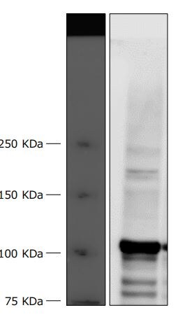
Western Blot: NFATC1/NFAT2 Antibody [NB100-56732] - Analysis of human (A)Ramos and (B) mouse RAW lysate using NFATc1 antibody at 1 ug/ml.![Western Blot: NFATC1/NFAT2 Antibody [NB100-56732]](https://resources.rndsystems.com/images/products/NFATC1-NFAT2-Antibody-Western-Blot-NB100-56732-img0001.jpg "Western Blot: NFATC1/NFAT2 Antibody [NB100-56732]")
Western Blot: NFATC1/NFAT2 Antibody [NB100-56732]
Western Blot: NFATC1/NFAT2 Antibody [NB100-56732] - Analysis of NFATc1 in Ramos cell lysate. (1) Without blocking peptide. (2) With blocking peptide.Applications for NFATC1/NFAT2 Antibody - BSA Free
Application
Recommended Usage
Cleavage Under Targets and Tagmentation
reported in scientific literature (PMID: 36073544)
Immunocytochemistry/ Immunofluorescence
1:20
Immunohistochemistry
reported in scientific literature (PMID 35167499)
Western Blot
1-3 ug/ml
Application Notes
Reviewed Applications
Read 2 reviews rated 4.5 using NB100-56732 in the following applications:
Formulation, Preparation, and Storage
Purification
Protein G purified
Formulation
PBS
Format
BSA Free
Preservative
0.05% Sodium Azide
Concentration
1.0 mg/ml
Shipping
The product is shipped with polar packs. Upon receipt, store it immediately at the temperature recommended below.
Stability & Storage
Store at 4C short term. Aliquot and store at -20C long term. Avoid freeze-thaw cycles.
Background: NFATC1
Long Name
Nuclear Factor of Activated T Cells C1
Alternate Names
NFAT2
Entrez Gene IDs
4772 (Human)
Gene Symbol
NFATC1
UniProt
Additional NFATC1 Products
Product Documents for NFATC1/NFAT2 Antibody - BSA Free
Certificate of Analysis
To download a Certificate of Analysis, please enter a lot or batch number in the search box below.
Product Specific Notices for NFATC1/NFAT2 Antibody - BSA Free
This product is for research use only and is not approved for use in humans or in clinical diagnosis. Primary Antibodies are guaranteed for 1 year from date of receipt.
Citations for NFATC1/NFAT2 Antibody - BSA Free
 Powered by Bioz
Customer Reviews for NFATC1/NFAT2 Antibody - BSA Free (2)
4.5 out of 5
2 Customer Ratings
Have you used NFATC1/NFAT2 Antibody - BSA Free?
Submit a review and receive an Amazon gift card!
$25/€18/£15/$25CAN/¥2500 Yen for a review with an image
$10/€7/£6/$10CAN/¥1110 Yen for a review without an image
Submit a review
Customer Images
Showing
1
-
2 of
2 reviews
Showing All
Filter By:
-
Application: Western BlotSample Tested: Immortalized podocytesSpecies: MouseVerified Customer | Posted 04/25/2018Anti-NFATc1 Ab: 1/500

-
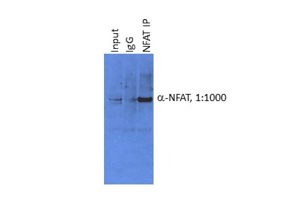
Application: Western BlotSample Tested:Species: MouseVerified Customer | Posted 02/12/2016Immunoblot analysis of NFATC (1:1000) following IgG or NFAT Immunoprecipitation
There are no reviews that match your criteria.
Protocols
Find general support by application which include: protocols, troubleshooting, illustrated assays, videos and webinars.
- Antigen Retrieval Protocol (PIER)
- Antigen Retrieval for Frozen Sections Protocol
- Appropriate Fixation of IHC/ICC Samples
- Cellular Response to Hypoxia Protocols
- Chromogenic IHC Staining of Formalin-Fixed Paraffin-Embedded (FFPE) Tissue Protocol
- Chromogenic Immunohistochemistry Staining of Frozen Tissue
- ClariTSA™ Fluorophore Kits
- Detection & Visualization of Antibody Binding
- Fluorescent IHC Staining of Frozen Tissue Protocol
- Graphic Protocol for Heat-induced Epitope Retrieval
- Graphic Protocol for the Preparation and Fluorescent IHC Staining of Frozen Tissue Sections
- Graphic Protocol for the Preparation and Fluorescent IHC Staining of Paraffin-embedded Tissue Sections
- Graphic Protocol for the Preparation of Gelatin-coated Slides for Histological Tissue Sections
- ICC Cell Smear Protocol for Suspension Cells
- ICC Immunocytochemistry Protocol Videos
- ICC for Adherent Cells
- IHC Sample Preparation (Frozen sections vs Paraffin)
- Immunocytochemistry (ICC) Protocol
- Immunocytochemistry Troubleshooting
- Immunofluorescence of Organoids Embedded in Cultrex Basement Membrane Extract
- Immunofluorescent IHC Staining of Formalin-Fixed Paraffin-Embedded (FFPE) Tissue Protocol
- Immunohistochemistry (IHC) and Immunocytochemistry (ICC) Protocols
- Immunohistochemistry Frozen Troubleshooting
- Immunohistochemistry Paraffin Troubleshooting
- Preparing Samples for IHC/ICC Experiments
- Preventing Non-Specific Staining (Non-Specific Binding)
- Primary Antibody Selection & Optimization
- Protocol for Heat-Induced Epitope Retrieval (HIER)
- Protocol for Making a 4% Formaldehyde Solution in PBS
- Protocol for VisUCyte™ HRP Polymer Detection Reagent
- Protocol for the Fluorescent ICC Staining of Cell Smears - Graphic
- Protocol for the Fluorescent ICC Staining of Cultured Cells on Coverslips - Graphic
- Protocol for the Preparation & Fixation of Cells on Coverslips
- Protocol for the Preparation and Chromogenic IHC Staining of Frozen Tissue Sections
- Protocol for the Preparation and Chromogenic IHC Staining of Frozen Tissue Sections - Graphic
- Protocol for the Preparation and Chromogenic IHC Staining of Paraffin-embedded Tissue Sections
- Protocol for the Preparation and Chromogenic IHC Staining of Paraffin-embedded Tissue Sections - Graphic
- Protocol for the Preparation and Fluorescent ICC Staining of Cells on Coverslips
- Protocol for the Preparation and Fluorescent ICC Staining of Non-adherent Cells
- Protocol for the Preparation and Fluorescent ICC Staining of Stem Cells on Coverslips
- Protocol for the Preparation and Fluorescent IHC Staining of Frozen Tissue Sections
- Protocol for the Preparation and Fluorescent IHC Staining of Paraffin-embedded Tissue Sections
- Protocol for the Preparation of Gelatin-coated Slides for Histological Tissue Sections
- Protocol for the Preparation of a Cell Smear for Non-adherent Cell ICC - Graphic
- R&D Systems Quality Control Western Blot Protocol
- TUNEL and Active Caspase-3 Detection by IHC/ICC Protocol
- The Importance of IHC/ICC Controls
- Troubleshooting Guide: Immunohistochemistry
- Troubleshooting Guide: Western Blot Figures
- Western Blot Conditions
- Western Blot Protocol
- Western Blot Protocol for Cell Lysates
- Western Blot Troubleshooting
- Western Blot Troubleshooting Guide
- View all Protocols, Troubleshooting, Illustrated assays and Webinars

