NHE3/SLC9A3 [p Ser605] Antibody (10A8) - BSA Free
Novus Biologicals | Catalog # NB110-74678
Key Product Details
Validated by
Species Reactivity
Validated:
Cited:
Applications
Validated:
Cited:
Label
Antibody Source
Format
Product Specifications
Immunogen
Reactivity Notes
Modification
Localization
Specificity
Clonality
Host
Isotype
Theoretical MW
Disclaimer note: The observed molecular weight of the protein may vary from the listed predicted molecular weight due to post translational modifications, post translation cleavages, relative charges, and other experimental factors.
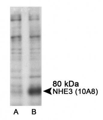
Scientific Data Images for NHE3/SLC9A3 [p Ser605] Antibody (10A8) - BSA Free
Applications for NHE3/SLC9A3 [p Ser605] Antibody (10A8) - BSA Free
Immunocytochemistry/ Immunofluorescence
Immunoprecipitation
Western Blot
Formulation, Preparation, and Storage
Purification
Formulation
Format
Preservative
Concentration
Shipping
Stability & Storage
Background: NHE3/SLC9A3
Long Name
Alternate Names
Gene Symbol
Additional NHE3/SLC9A3 Products
Product Documents for NHE3/SLC9A3 [p Ser605] Antibody (10A8) - BSA Free
Certificate of Analysis
To download a Certificate of Analysis, please enter a lot or batch number in the search box below.
Product Specific Notices for NHE3/SLC9A3 [p Ser605] Antibody (10A8) - BSA Free
This product is for research use only and is not approved for use in humans or in clinical diagnosis. Primary Antibodies are guaranteed for 1 year from date of receipt.
Related Research Areas
Citations for NHE3/SLC9A3 [p Ser605] Antibody (10A8) - BSA Free
 Powered by Bioz
Customer Reviews for NHE3/SLC9A3 [p Ser605] Antibody (10A8) - BSA Free
There are currently no reviews for this product. Be the first to review NHE3/SLC9A3 [p Ser605] Antibody (10A8) - BSA Free and earn rewards!
Have you used NHE3/SLC9A3 [p Ser605] Antibody (10A8) - BSA Free?
Submit a review and receive an Amazon gift card!
$25/€18/£15/$25CAN/¥2500 Yen for a review with an image
$10/€7/£6/$10CAN/¥1110 Yen for a review without an image
Submit a review
Protocols
View specific protocols for NHE3/SLC9A3 [p Ser605] Antibody (10A8) - BSA Free (NB110-74678):
Western Blot Protocol
1. Perform SDS-PAGE (4-12% MOPS) on samples to be analyzed, loading 30 ug of total protein per lane.
2. Transfer proteins to Nitrocellulose according to the instructions provided by the manufacturer of the transfer
apparatus.
3. Rinse membrane with dH2O and then stain the blot using Ponceau S for 1-2 minutes to access the transfer of proteins onto the nitrocellulose membrane. Rinse the blot in water to remove excess stain and mark the lane locations and locations of molecular weight markers using a pencil.
4. Rinse the blot in TBS for approximately 5 minutes.
5. Block the membrane using 5% NFDM + 1% BSA in TBS + Tween, 1 hour at RT.
6. Rinse the membrane in dH2O and then wash the membrane in wash buffer [TBS + 0.1% Tween] 3 times for 10 minutes each.
7. Dilute the mouse anti-NHE3 primary antibody (NB 110-74678) in blocking buffer and incubate 1 hour at room temperature.
8. Rinse the membrane in dH2O and then wash the membrane in wash buffer [TBS + 0.1% Tween] 3 times for 10 minutes each.
9. Apply the diluted mouse-IgG HRP-conjugated secondary antibody in blocking buffer (as per manufacturers
instructions) and incubate 1 hour at room temperature.
10. Wash the blot in wash buffer [TBS + 0.1% Tween] 3 times for 10 minutes each (this step can be repeated as required to reduce background).
11. Apply the detection reagent of choice in accordance with the manufacturers instructions (Pierce ECL).
Note: Tween-20 can be added to the blocking or antibody di
Find general support by application which include: protocols, troubleshooting, illustrated assays, videos and webinars.
- Appropriate Fixation of IHC/ICC Samples
- Cellular Response to Hypoxia Protocols
- ClariTSA™ Fluorophore Kits
- Detection & Visualization of Antibody Binding
- ICC Cell Smear Protocol for Suspension Cells
- ICC Immunocytochemistry Protocol Videos
- ICC for Adherent Cells
- Immunocytochemistry (ICC) Protocol
- Immunocytochemistry Troubleshooting
- Immunofluorescence of Organoids Embedded in Cultrex Basement Membrane Extract
- Immunohistochemistry (IHC) and Immunocytochemistry (ICC) Protocols
- Immunoprecipitation Protocol
- Preparing Samples for IHC/ICC Experiments
- Preventing Non-Specific Staining (Non-Specific Binding)
- Primary Antibody Selection & Optimization
- Protocol for VisUCyte™ HRP Polymer Detection Reagent
- Protocol for the Fluorescent ICC Staining of Cell Smears - Graphic
- Protocol for the Fluorescent ICC Staining of Cultured Cells on Coverslips - Graphic
- Protocol for the Preparation and Fluorescent ICC Staining of Cells on Coverslips
- Protocol for the Preparation and Fluorescent ICC Staining of Non-adherent Cells
- Protocol for the Preparation and Fluorescent ICC Staining of Stem Cells on Coverslips
- Protocol for the Preparation of a Cell Smear for Non-adherent Cell ICC - Graphic
- R&D Systems Quality Control Western Blot Protocol
- TUNEL and Active Caspase-3 Detection by IHC/ICC Protocol
- The Importance of IHC/ICC Controls
- Troubleshooting Guide: Western Blot Figures
- Western Blot Conditions
- Western Blot Protocol
- Western Blot Protocol for Cell Lysates
- Western Blot Troubleshooting
- Western Blot Troubleshooting Guide
- View all Protocols, Troubleshooting, Illustrated assays and Webinars
FAQs for NHE3/SLC9A3 [p Ser605] Antibody (10A8) - BSA Free
-
Q: The molecular weight of NHE3 is 92kDa but I see a band above 100 kDa. I am confused, because the size of these protein should not be greater than 100KD. Can you please help explain?
A: Higher than predicted molecular weight seen on WB is not uncommon if the protein is posttranslationally modified, especially glycosylated, and such is the case for NHE3. Please note that NHE3 gets phosphorylated and glycosylated (Uniprot ID P48764) and potentially, these modifications are adding ~10kDa extra to the predicted molecular weight. If you want to confirm it 100%, you could try de-glycosylating your protein and then performing a WB on it.