NK1R Antibody - Azide and BSA Free
Novus Biologicals | Catalog # NB300-101
![Immunocytochemistry/ Immunofluorescence: NK1R Antibody [NB300-101]](https://resources.rndsystems.com/images/products/Neurokinin-1-Receptor-Antibody-Immunocytochemistry-Immunofluorescence-NB300-101-img0007.jpg "Immunocytochemistry/ Immunofluorescence: NK1R Antibody [NB300-101]")
Key Product Details
Species Reactivity
Validated:
Cited:
Predicted:
Applications
Validated:
Cited:
Label
Antibody Source
Format
Product Specifications
Immunogen
Reactivity Notes
Localization
Specificity
Clonality
Host
Isotype
Scientific Data Images for NK1R Antibody - Azide and BSA Free
Immunocytochemistry/ Immunofluorescence: NK1R Antibody [NB300-101]
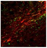
Immunocytochemistry/Immunofluorescence: Neurokinin 1 Receptor Antibody [NB300-101] - IF analysis of Neurokinin 1 Receptor in rat brain tissue. Image courtesy of anonymous customer product review.![Immunohistochemistry: NK1R Antibody [NB300-101]](https://resources.rndsystems.com/images/products/Neurokinin-1-Receptor-Antibody-Immunohistochemistry-NB300-101-img0005.jpg "Immunohistochemistry: NK1R Antibody [NB300-101]")
Immunohistochemistry: NK1R Antibody [NB300-101]
Immunohistochemistry: Neurokinin 1 Receptor Antibody [NB300-101] - Immunohistochemical analysis of Neurokinin 1 Receptor in mouse brain (selected cells, axon & dendrites) using NB300-101.![Immunocytochemistry/ Immunofluorescence: NK1R Antibody [NB300-101]](https://resources.rndsystems.com/images/products/Neurokinin-1-Receptor-Antibody-Immunocytochemistry-Immunofluorescence-NB300-101-img0006.jpg "Immunocytochemistry/ Immunofluorescence: NK1R Antibody [NB300-101]")
Immunocytochemistry/ Immunofluorescence: NK1R Antibody [NB300-101]
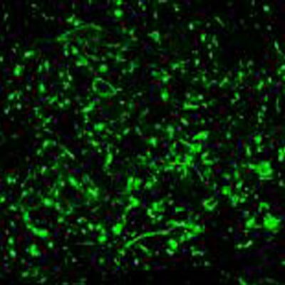
Immunocytochemistry/Immunofluorescence: Neurokinin 1 Receptor Antibody [NB300-101] - Confocal image of SPR reactivity using polyclonal anti-NK-1 Receptor (NB 300-101) in a lamina III neuron in the spinal cord of a rat.![Immunocytochemistry/ Immunofluorescence: NK1R Antibody [NB300-101]](https://resources.rndsystems.com/images/products/Neurokinin-1-Receptor-Antibody-Immunocytochemistry-Immunofluorescence-NB300-101-img0010.jpg "Immunocytochemistry/ Immunofluorescence: NK1R Antibody [NB300-101]")
Immunocytochemistry/ Immunofluorescence: NK1R Antibody [NB300-101]
Immunocytochemistry/Immunofluorescence: Neurokinin 1 Receptor Antibody [NB300-101] - IF analysis of Neurokinin 1 Receptor in rat brain tissue. Image courtesy of anonymous customer product review.Applications for NK1R Antibody - Azide and BSA Free
Immunocytochemistry/ Immunofluorescence
Immunohistochemistry
Immunohistochemistry-Frozen
Immunohistochemistry-Paraffin
Western Blot
Reviewed Applications
Read 2 reviews rated 4 using NB300-101 in the following applications:
Formulation, Preparation, and Storage
Purification
Formulation
Format
Preservative
Concentration
Shipping
Stability & Storage
Background: NK1R
Long Name
Alternate Names
Gene Symbol
UniProt
Additional NK1R Products
Product Documents for NK1R Antibody - Azide and BSA Free
Certificate of Analysis
To download a Certificate of Analysis, please enter a lot or batch number in the search box below.
Product Specific Notices for NK1R Antibody - Azide and BSA Free
This product is for research use only and is not approved for use in humans or in clinical diagnosis. Primary Antibodies are guaranteed for 1 year from date of receipt.
Citations for NK1R Antibody - Azide and BSA Free
 Powered by Bioz
Customer Reviews for NK1R Antibody - Azide and BSA Free (2)
Have you used NK1R Antibody - Azide and BSA Free?
Submit a review and receive an Amazon gift card!
$25/€18/£15/$25CAN/¥2500 Yen for a review with an image
$10/€7/£6/$10CAN/¥1110 Yen for a review without an image
Submit a review
Customer Images
-
Application: ImmunofluorescenceSample Tested: Rat brain tissueSpecies: RatVerified Customer | Posted 12/20/2010

-
Application: ImmunofluorescenceSample Tested: rat brain sectionsSpecies: RatVerified Customer | Posted 09/09/2010
There are no reviews that match your criteria.
Protocols
View specific protocols for NK1R Antibody - Azide and BSA Free (NB300-101):
Immunohistochemistry
1) Perfuse rat through the ascending aorta with 500 ml. of 0.1 M phosphate buffered saline (PBS) (pH 7.4 @ 4 degrees C), followed by 750 ml. of PBS containing 4% formaldehyde and 12.5% picric acid (pH 6.9, 4 degrees C). Tissue of interest is then dissected and post-fixed in PBS containing 4% formaldehyde and 12.5% picric acid (pH 6.9 at 4 degrees C) for at least 4 hours, followed by fixation in PBS containing 30% sucrose (pH 7.4, 4 degrees C) for at least 24 hours. Until the tissue sinks.
2) Cut fixed sections at 60 um using a sliding microtome.
3) If using culture cells, remove media and perform 2-3 washed in PBS (pH 7.4) -use same protocol without agitation.
4) Place tissue section in microcentrifuge tubes containing 1 ml. Of PBS, pH 7.4 as they are being cut. Wash sections (in PBS) on an upright rotator for 10-15 minutes.
5) Remove PBS and add 1 ml. of blocking solution (PBS + 1% normal donkey serum (NDS) + 0.3% triton X-100).
6) Incubate tissue sections in blocking solution for 30 minutes at room temperature (RT) on rotator.
7) Remove blocking solution and add anti-neurokinin-1 receptor or anti-Neurokinin-3 receptor(NB 300-101/NB 100-102) antibody +PBS + 1% NDS + 0.3% triton X-100 + NB 300-101/102 @ 1:100~1:1000
8) Incubate overnight on rotator at RT.
9) Remove primary antisera and perform 3 x 10 minute PBS washes (1ml volume) on rotator.
10) Add 1 ml. of secondary antibody and incubate for 2 hours at RT on rotator.
-PBS + 1% NDS + 0.3% triton X 100 + anti-rabbit Cy3 (Jackson Immunoresearch Labs) at 1:600
11) Perform 3 X 10 minute PBS washes (1 ml volume) on rotator
12) Mount tissue sections on gelatin coated slides and allow tissue to dry
13) Run slides through alcohol gradients (70%, 90%, 100%, xylene) leaving slide in each alcohol and xylene for 2 minutes
14) Coverslip with DPX mountant (FLUKA) ** Note: 24 well plates may be substituted for microcentrifuge
tubes. Use flat top bench rotator instead of upright rotator.
Find general support by application which include: protocols, troubleshooting, illustrated assays, videos and webinars.
- Antigen Retrieval Protocol (PIER)
- Antigen Retrieval for Frozen Sections Protocol
- Appropriate Fixation of IHC/ICC Samples
- Cellular Response to Hypoxia Protocols
- Chromogenic IHC Staining of Formalin-Fixed Paraffin-Embedded (FFPE) Tissue Protocol
- Chromogenic Immunohistochemistry Staining of Frozen Tissue
- ClariTSA™ Fluorophore Kits
- Detection & Visualization of Antibody Binding
- Fluorescent IHC Staining of Frozen Tissue Protocol
- Graphic Protocol for Heat-induced Epitope Retrieval
- Graphic Protocol for the Preparation and Fluorescent IHC Staining of Frozen Tissue Sections
- Graphic Protocol for the Preparation and Fluorescent IHC Staining of Paraffin-embedded Tissue Sections
- Graphic Protocol for the Preparation of Gelatin-coated Slides for Histological Tissue Sections
- ICC Cell Smear Protocol for Suspension Cells
- ICC Immunocytochemistry Protocol Videos
- ICC for Adherent Cells
- IHC Sample Preparation (Frozen sections vs Paraffin)
- Immunocytochemistry (ICC) Protocol
- Immunocytochemistry Troubleshooting
- Immunofluorescence of Organoids Embedded in Cultrex Basement Membrane Extract
- Immunofluorescent IHC Staining of Formalin-Fixed Paraffin-Embedded (FFPE) Tissue Protocol
- Immunohistochemistry (IHC) and Immunocytochemistry (ICC) Protocols
- Immunohistochemistry Frozen Troubleshooting
- Immunohistochemistry Paraffin Troubleshooting
- Preparing Samples for IHC/ICC Experiments
- Preventing Non-Specific Staining (Non-Specific Binding)
- Primary Antibody Selection & Optimization
- Protocol for Heat-Induced Epitope Retrieval (HIER)
- Protocol for Making a 4% Formaldehyde Solution in PBS
- Protocol for VisUCyte™ HRP Polymer Detection Reagent
- Protocol for the Fluorescent ICC Staining of Cell Smears - Graphic
- Protocol for the Fluorescent ICC Staining of Cultured Cells on Coverslips - Graphic
- Protocol for the Preparation & Fixation of Cells on Coverslips
- Protocol for the Preparation and Chromogenic IHC Staining of Frozen Tissue Sections
- Protocol for the Preparation and Chromogenic IHC Staining of Frozen Tissue Sections - Graphic
- Protocol for the Preparation and Chromogenic IHC Staining of Paraffin-embedded Tissue Sections
- Protocol for the Preparation and Chromogenic IHC Staining of Paraffin-embedded Tissue Sections - Graphic
- Protocol for the Preparation and Fluorescent ICC Staining of Cells on Coverslips
- Protocol for the Preparation and Fluorescent ICC Staining of Non-adherent Cells
- Protocol for the Preparation and Fluorescent ICC Staining of Stem Cells on Coverslips
- Protocol for the Preparation and Fluorescent IHC Staining of Frozen Tissue Sections
- Protocol for the Preparation and Fluorescent IHC Staining of Paraffin-embedded Tissue Sections
- Protocol for the Preparation of Gelatin-coated Slides for Histological Tissue Sections
- Protocol for the Preparation of a Cell Smear for Non-adherent Cell ICC - Graphic
- R&D Systems Quality Control Western Blot Protocol
- TUNEL and Active Caspase-3 Detection by IHC/ICC Protocol
- The Importance of IHC/ICC Controls
- Troubleshooting Guide: Immunohistochemistry
- Troubleshooting Guide: Western Blot Figures
- Western Blot Conditions
- Western Blot Protocol
- Western Blot Protocol for Cell Lysates
- Western Blot Troubleshooting
- Western Blot Troubleshooting Guide
- View all Protocols, Troubleshooting, Illustrated assays and Webinars
FAQs for NK1R Antibody - Azide and BSA Free
-
Q: Question about the Neurokinin 1 Receptor Antibody (NB300-101):why were the NK-1R in WB reported with different molecular mass(46kd, 50kd or about 70kd?)in publication 1,2 and 3? which is right?
A:
The theoretical MW of this protein is about 46KDa. Please see this reference for P25103 (https://www.uniprot.org/uniprotkb/P25103/entry). This is a protein database. The above MW weight is only theoretical and it can be expected that the MW differs between each experiment. The researcher who experienced this protein to be at 70KDa may not have reduced their protein enough which could have led to a protein complex, resulting in a larger MW.