NKX6.1 Antibody - BSA Free
Novus Biologicals | Catalog # NBP1-49672
![Western Blot: NKX6.1 AntibodyBSA Free [NBP1-49672]](https://resources.rndsystems.com/images/products/NKX6.1-Antibody-Western-Blot-NBP1-49672-img0004.jpg "Western Blot: NKX6.1 AntibodyBSA Free [NBP1-49672]")
Key Product Details
Species Reactivity
Validated:
Cited:
Applications
Validated:
Cited:
Label
Antibody Source
Format
Product Specifications
Immunogen
Reactivity Notes
Localization
Clonality
Host
Isotype
Scientific Data Images for NKX6.1 Antibody - BSA Free
Western Blot: NKX6.1 AntibodyBSA Free [NBP1-49672]
Western Blot: NKX6.1 Antibody [NBP1-49672] - Analysis of Nkx6.1 in human skeletal muscle.![Immunocytochemistry/ Immunofluorescence: NKX6.1 Antibody - BSA Free [NBP1-49672]](https://resources.rndsystems.com/images/products/NKX6.1-Antibody-Immunocytochemistry-Immunofluorescence-NBP1-49672-img0006.jpg "Immunocytochemistry/ Immunofluorescence: NKX6.1 Antibody - BSA Free [NBP1-49672]")
Immunocytochemistry/ Immunofluorescence: NKX6.1 Antibody - BSA Free [NBP1-49672]
Immunocytochemistry/Immunofluorescence: NKX6.1 Antibody [NBP1-49672] - Nkx6.1 antibody was tested at 1:250 in INS-1 cells with DyLight 488 (green). Nuclei and alpha-tubulin were counterstained with DAPI (blue) and DyLight 550 (red). Image objective 40X.![Immunohistochemistry: NKX6.1 Antibody - BSA Free [NBP1-49672]](https://resources.rndsystems.com/images/products/NKX6.1-Antibody---BSA-Free-Immunohistochemistry-NBP1-49672-img0012.jpg "Immunohistochemistry: NKX6.1 Antibody - BSA Free [NBP1-49672]")
Immunohistochemistry: NKX6.1 Antibody - BSA Free [NBP1-49672]
NKX6.1-Antibody---BSA-Free-Immunohistochemistry-NBP1-49672-img0012.jpg![Immunohistochemistry-Frozen: NKX6.1 Antibody - BSA Free [NBP1-49672]](https://resources.rndsystems.com/images/products/NKX6.1-Antibody-Immunohistochemistry-Frozen-NBP1-49672-img0009.jpg "Immunohistochemistry-Frozen: NKX6.1 Antibody - BSA Free [NBP1-49672]")
Immunohistochemistry-Frozen: NKX6.1 Antibody - BSA Free [NBP1-49672]
Immunohistochemistry-Frozen: NKX6.1 Antibody [NBP1-49672] - Analysis of NKX6.1 in mouse pancreas frozen tissue section using anti-NKX6.1 antibody. IHC-Fr image submitted by a verified customer review.![Immunohistochemistry: NKX6.1 Antibody - BSA Free [NBP1-49672]](https://resources.rndsystems.com/images/products/NKX6.1-Antibody-Immunohistochemistry-NBP1-49672-img0003.jpg "Immunohistochemistry: NKX6.1 Antibody - BSA Free [NBP1-49672]")
Immunohistochemistry: NKX6.1 Antibody - BSA Free [NBP1-49672]
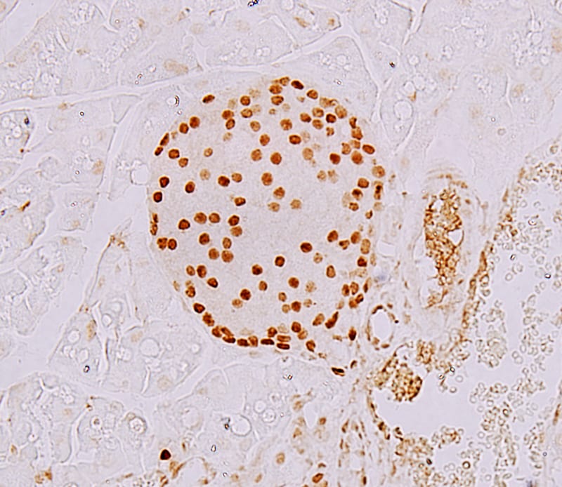
Immunohistochemistry: NKX6.1 Antibody [NBP1-49672] - Staining of Nkx6.1 in mouse intestine using DAB with hematoxylin counterstain.![Immunohistochemistry-Paraffin: NKX6.1 Antibody - BSA Free [NBP1-49672]](https://resources.rndsystems.com/images/products/NKX6.1-Antibody-Immunohistochemistry-Paraffin-NBP1-49672-img0007.jpg "Immunohistochemistry-Paraffin: NKX6.1 Antibody - BSA Free [NBP1-49672]")
Immunohistochemistry-Paraffin: NKX6.1 Antibody - BSA Free [NBP1-49672]
Immunohistochemistry-Paraffin: NKX6.1 Antibody [NBP1-49672] - Human fetal pancreas stained for Nkx6.1, green, and PDX1, grey. IHC-P image submitted by a verified customer review.![Immunohistochemistry-Paraffin: NKX6.1 Antibody - BSA Free [NBP1-49672]](https://resources.rndsystems.com/images/products/NKX6.1-Antibody-Immunohistochemistry-Paraffin-NBP1-49672-img0010.jpg "Immunohistochemistry-Paraffin: NKX6.1 Antibody - BSA Free [NBP1-49672]")
Immunohistochemistry-Paraffin: NKX6.1 Antibody - BSA Free [NBP1-49672]
Immunohistochemistry-Paraffin: NKX6.1 Antibody [NBP1-49672] - Mouse pancreas tissue. IHC-P image submitted by a verified customer review.![Immunohistochemistry-Paraffin: NKX6.1 Antibody - BSA Free [NBP1-49672]](https://resources.rndsystems.com/images/products/NKX6.1-Antibody-Immunohistochemistry-Paraffin-NBP1-49672-img0011.jpg "Immunohistochemistry-Paraffin: NKX6.1 Antibody - BSA Free [NBP1-49672]")
Immunohistochemistry-Paraffin: NKX6.1 Antibody - BSA Free [NBP1-49672]
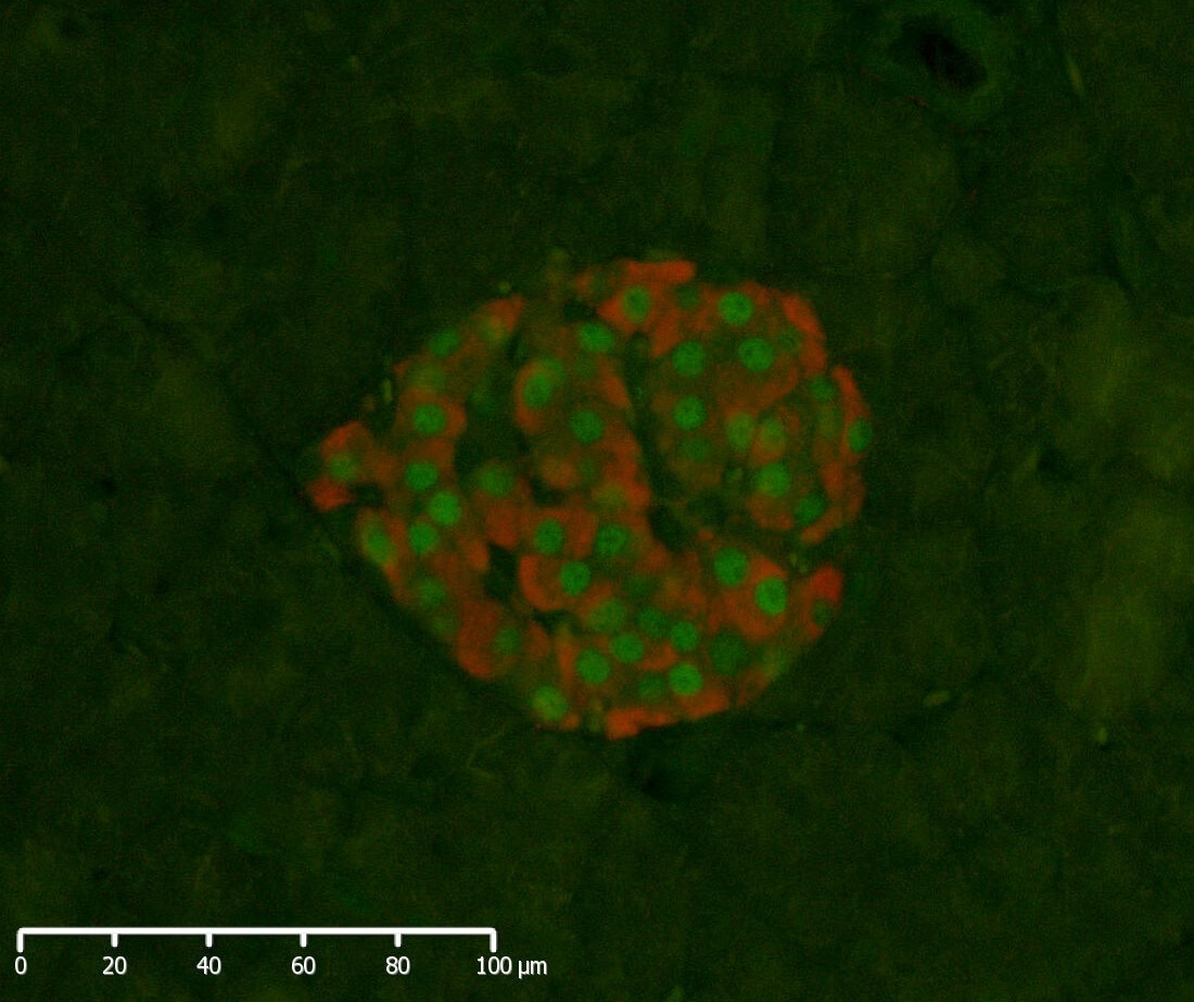
Immunohistochemistry-Paraffin: NKX6.1 Antibody [NBP1-49672] - Paraffin embeded rat pancreatic tissue at 40x resolution stained for insulin (red) and NKX6.1 (NBP1-49672, green). IHC-P image submitted by a verified customer review.![Simple Western: NKX6.1 AntibodyBSA Free [NBP1-49672]](https://resources.rndsystems.com/images/products/NKX6.1-Antibody-Simple-Western-NBP1-49672-img0008.jpg "Simple Western: NKX6.1 AntibodyBSA Free [NBP1-49672]")
Simple Western: NKX6.1 AntibodyBSA Free [NBP1-49672]
Simple Western: NKX6.1 Antibody [NBP1-49672] - Image shows a specific band for NKX6.1 in 0.5 mg/mL of BTC-6 lysate. This experiment was performed under reducing conditions using the 12-230 kDa separation system.
NKX6.1 in U-2 OS Human Cell Line.
NKX6.1 was detected in immersion fixed U-2 OS human osteosarcoma cell line using Rabbit anti-NKX6.1 Antigen Affinity Purified Polyclonal Antibody conjugated to Alexa Fluor® 488 (Catalog # NBP1-49672AF488) (green) at 10 µg/mL overnight at 4C. Cells were counterstained with DAPI (blue). Cells were imaged using a 100X objective and digitally deconvolved.
Immunocytochemistry/ Immunofluorescence: NKX6.1 Antibody - BSA Free [NBP1-49672] -
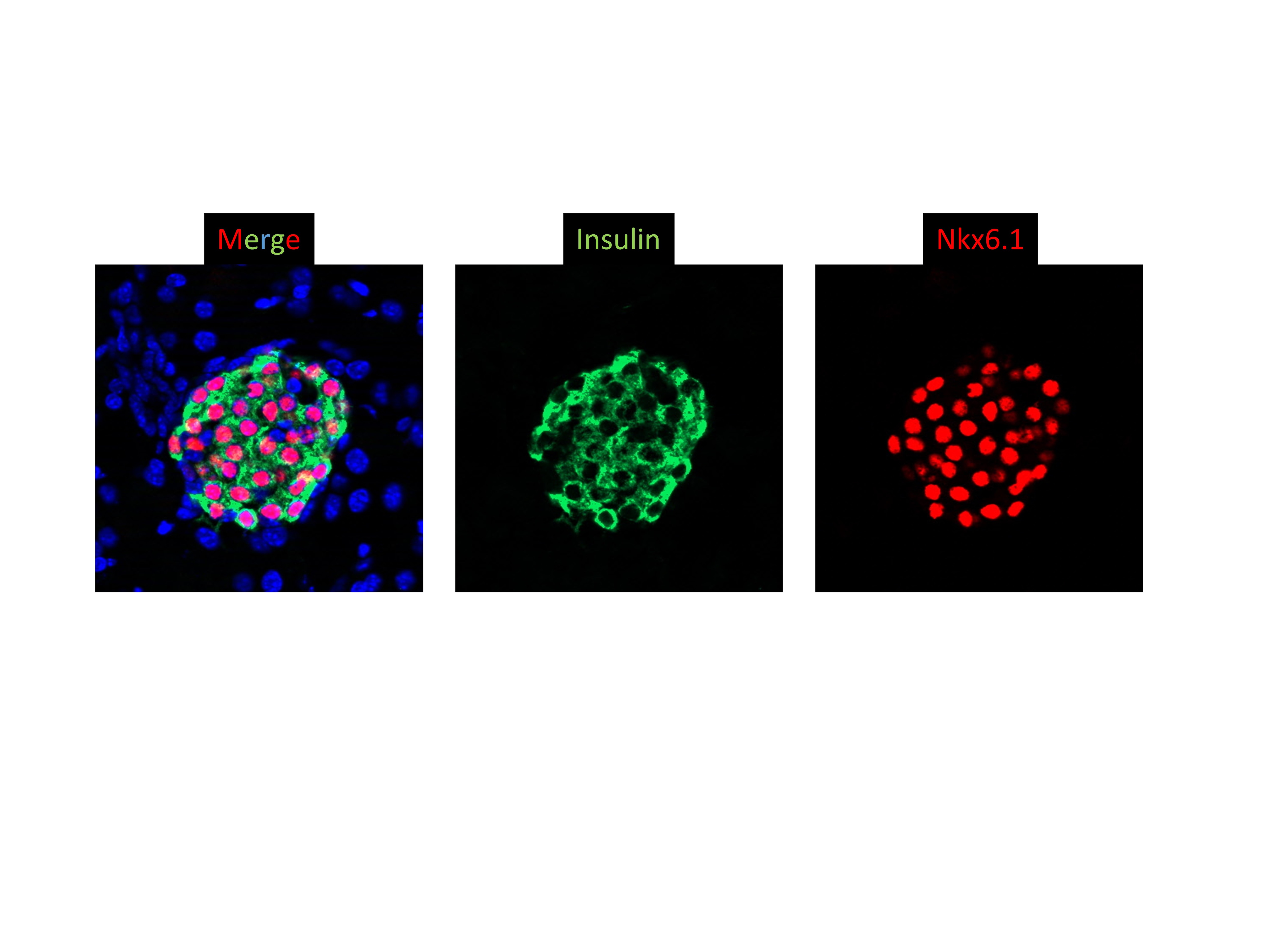
Characteristics of dedifferentiated beta cells identified by NKX6.1. a Representative immunofluorescence images of pancreatic sections with NKX6.1 and Insulin (Ins). b Relationships among different dedifferentiated beta cell labeled by white boxes in (a). Scale bars: 20 μm, Red: NKX6.1, Green: Ins, Blue: DAPI Image collected and cropped by CiteAb from the following open publication (https://pubmed.ncbi.nlm.nih.gov/33711989), licensed under a CC-BY license. Not internally tested by Novus Biologicals.
Immunocytochemistry/ Immunofluorescence: NKX6.1 Antibody - BSA Free [NBP1-49672] -
Representative immunofluorescence images of pancreatic sections of non-diabetic (ND) subjects and T2DM subjects. a Representative images of immunostaining with Insulin and NKX6.1 in the pancreatic sections of non-diabetic (ND) subjects and T2DM subjects. White arrows marked dedifferentiated beta cells with NKX6.1 dislocation. Scale bar: 20 μm, Red: NKX6.1, Green: Ins, Blue: DAPI. b Quantification of NKX6.1Nuc-Ins+ cell count and percentage in beta cells. c Quantification of NKX6.1cytIns− cell count and percentage per islet. d Correlation between NKX6.1Nuc-Ins+ cells per islet with HbA1c in T2DM subjects (Simple linear regression). n = 40 for non-diabetic (ND) subjects; n = 20 for T2DM subjects. Data were shown as mean +/- SEM. *P < 0.05. ***P < 0.001 Image collected and cropped by CiteAb from the following open publication (https://pubmed.ncbi.nlm.nih.gov/33711989), licensed under a CC-BY license. Not internally tested by Novus Biologicals.
Immunocytochemistry/ Immunofluorescence: NKX6.1 Antibody - BSA Free [NBP1-49672] -
Characteristics of dedifferentiated beta cells identified by NKX6.1. a Representative immunofluorescence images of pancreatic sections with NKX6.1 and Insulin (Ins). b Relationships among different dedifferentiated beta cell labeled by white boxes in (a). Scale bars: 20 μm, Red: NKX6.1, Green: Ins, Blue: DAPI Image collected and cropped by CiteAb from the following open publication (https://pubmed.ncbi.nlm.nih.gov/33711989), licensed under a CC-BY license. Not internally tested by Novus Biologicals.Applications for NKX6.1 Antibody - BSA Free
Immunocytochemistry/ Immunofluorescence
Immunohistochemistry
Immunohistochemistry-Paraffin
In vitro assay
Simple Western
Western Blot
In Simple Western only 10 - 15 uL of the recommended dilution is used per data point.
See Simple Western Antibody Database for Simple Western validation: Tested in BTC-6 lysate 0.5 mg/mL, separated by Size, antibody dilution of 1:100. Separated by Size-Wes, Sally Sue/Peggy Sue.
Reviewed Applications
Read 4 reviews rated 4.8 using NBP1-49672 in the following applications:
Formulation, Preparation, and Storage
Purification
Formulation
Format
Preservative
Concentration
Shipping
Stability & Storage
Background: NKX6.1
Long Name
Alternate Names
Gene Symbol
UniProt
Additional NKX6.1 Products
Product Documents for NKX6.1 Antibody - BSA Free
Certificate of Analysis
To download a Certificate of Analysis, please enter a lot or batch number in the search box below.
Product Specific Notices for NKX6.1 Antibody - BSA Free
This product is for research use only and is not approved for use in humans or in clinical diagnosis. Primary Antibodies are guaranteed for 1 year from date of receipt.
Related Research Areas
Citations for NKX6.1 Antibody - BSA Free
 Powered by Bioz
Customer Reviews for NKX6.1 Antibody - BSA Free (4)
Have you used NKX6.1 Antibody - BSA Free?
Submit a review and receive an Amazon gift card!
$25/€18/£15/$25CAN/¥2500 Yen for a review with an image
$10/€7/£6/$10CAN/¥1110 Yen for a review without an image
Submit a review
Customer Images
-(01-ml)_NBP1-49672_10356.jpg)
-
Application: Immunohistochemistry-ParaffinSample Tested: Pancreas tissueSpecies: RatVerified Customer | Posted 04/29/2020Paraffin embeded rat pancreatic tissue at 40x resolution stained for insulin (red) and NKX6.1 (NBP1-49672, green).We used a 1:1600 dilution for our assays.

-
Application: Immunohistochemistry-ParaffinSample Tested: Mouse PancreasSpecies: MouseVerified Customer | Posted 01/08/2019Very good antibody. Highly recommended.
-
Application: ImmunofluorescenceSample Tested:Species: MouseVerified Customer | Posted 10/30/2015
-
Application: Immunohistochemistry-ParaffinSample Tested: Human fetal pancreasSpecies: HumanVerified Customer | Posted 09/24/2014Paraffin embedded section of human fetal pancreas stained for Nkx6.1 (NBP1-49672), green, and PDX1, grey.
There are no reviews that match your criteria.
Protocols
View specific protocols for NKX6.1 Antibody - BSA Free (NBP1-49672):
Culture cells to appropriate density in 35 mm culture dishes or 6-well plates.
1. Remove culture medium and wash the cells briefly in PBS. Add 4% paraformaldehyde to the dish and fix at room temperature for 10 minutes.
2. Remove the paraformaldehyde and wash the cells in PBS.
3. Permeabilize the cells with 0.1% Triton X100 or other suitable detergent for 2 min.
4. Remove the permeabilization buffer and wash three times for 5 minutes each in PBS. Be sure to not let the specimen dry out.
5. To block nonspecific antibody binding, incubate in 10% normal goat serum from 1 hour to overnight at room temperature.
6. Add primary antibody at appropriate dilution and incubate overnight at 4C.
7. Remove primary antibody and replace with PBS. Wash three times for 5 minutes each.
8. Add secondary antibody at appropriate dilution. Incubate for 1 hour at room temperature.
9. Remove secondary antibody and replace with PBS. Wash three times for 5 minutes each.
10. Counter stain DNA with DAPI if required.
Antigen Unmasking:
Bring slides to a boil in 10 mM sodium citrate buffer (pH 6.0) then maintain at a sub-boiling temperature for 10 minutes. Cool slides on bench-top for 30 minutes (keep slides in the sodium citrate buffer at all times).
Staining:
1. Wash sections in deionized water three times for 5 minutes each.
2. Wash sections in PBS for 5 minutes.
3. Block each section with 100-400 ul blocking solution (1% BSA in PBS) for 1 hour at room temperature.
4. Remove blocking solution and add 100-400 ul diluted primary antibody. Incubate overnight at 4 C.
5. Remove antibody solution and wash sections in wash buffer three times for 5 minutes each.
6. Add 100-400 ul HRP polymer conjugated secondary antibody. Incubate 30 minutes at room temperature.
7. Wash sections three times in wash buffer for 5 minutes each.
8. Add 100-400 ul DAB substrate to each section and monitor staining closely.
9. As soon as the sections develop, immerse slides in deionized water.
10. Counterstain sections in hematoxylin.
11. Wash sections in deionized water two times for 5 minutes each.
12. Dehydrate sections.
13. Mount coverslips.
1. Perform SDS-PAGE on samples to be analyzed, loading 10-25 ug of total protein per lane.
2. Transfer proteins to PVDF membrane according to the instructions provided by the manufacturer of the membrane and transfer apparatus.
3. Stain the membrane with Ponceau S (or similar product) to assess transfer success, and mark molecular weight standards where appropriate.
4. Rinse the blot TBS -0.05% Tween 20 (TBST).
5. Block the membrane in 5% Non-fat milk in TBST (blocking buffer) for at least 1 hour.
6. Wash the membrane in TBST three times for 10 minutes each.
7. Dilute primary antibody in 1% Non-fat milk and incubate overnight at 4C with gentle rocking.
8. Wash the membrane in TBST three times for 10 minutes each.
9. Incubate the membrane in diluted HRP conjugated secondary antibody in blocking buffer (as per manufacturer's instructions) for 1 hour at room temperature.
10. Wash the blot in TBST three times for 10 minutes each (this step can be repeated as required to reduce background).
11. Apply the detection reagent of choice in accordance with the manufacturer's instructions.
Find general support by application which include: protocols, troubleshooting, illustrated assays, videos and webinars.
- Antigen Retrieval Protocol (PIER)
- Antigen Retrieval for Frozen Sections Protocol
- Appropriate Fixation of IHC/ICC Samples
- Cellular Response to Hypoxia Protocols
- Chromogenic IHC Staining of Formalin-Fixed Paraffin-Embedded (FFPE) Tissue Protocol
- Chromogenic Immunohistochemistry Staining of Frozen Tissue
- ClariTSA™ Fluorophore Kits
- Detection & Visualization of Antibody Binding
- Fluorescent IHC Staining of Frozen Tissue Protocol
- Graphic Protocol for Heat-induced Epitope Retrieval
- Graphic Protocol for the Preparation and Fluorescent IHC Staining of Frozen Tissue Sections
- Graphic Protocol for the Preparation and Fluorescent IHC Staining of Paraffin-embedded Tissue Sections
- Graphic Protocol for the Preparation of Gelatin-coated Slides for Histological Tissue Sections
- ICC Cell Smear Protocol for Suspension Cells
- ICC Immunocytochemistry Protocol Videos
- ICC for Adherent Cells
- IHC Sample Preparation (Frozen sections vs Paraffin)
- Immunocytochemistry (ICC) Protocol
- Immunocytochemistry Troubleshooting
- Immunofluorescence of Organoids Embedded in Cultrex Basement Membrane Extract
- Immunofluorescent IHC Staining of Formalin-Fixed Paraffin-Embedded (FFPE) Tissue Protocol
- Immunohistochemistry (IHC) and Immunocytochemistry (ICC) Protocols
- Immunohistochemistry Frozen Troubleshooting
- Immunohistochemistry Paraffin Troubleshooting
- Preparing Samples for IHC/ICC Experiments
- Preventing Non-Specific Staining (Non-Specific Binding)
- Primary Antibody Selection & Optimization
- Protocol for Heat-Induced Epitope Retrieval (HIER)
- Protocol for Making a 4% Formaldehyde Solution in PBS
- Protocol for VisUCyte™ HRP Polymer Detection Reagent
- Protocol for the Fluorescent ICC Staining of Cell Smears - Graphic
- Protocol for the Fluorescent ICC Staining of Cultured Cells on Coverslips - Graphic
- Protocol for the Preparation & Fixation of Cells on Coverslips
- Protocol for the Preparation and Chromogenic IHC Staining of Frozen Tissue Sections
- Protocol for the Preparation and Chromogenic IHC Staining of Frozen Tissue Sections - Graphic
- Protocol for the Preparation and Chromogenic IHC Staining of Paraffin-embedded Tissue Sections
- Protocol for the Preparation and Chromogenic IHC Staining of Paraffin-embedded Tissue Sections - Graphic
- Protocol for the Preparation and Fluorescent ICC Staining of Cells on Coverslips
- Protocol for the Preparation and Fluorescent ICC Staining of Non-adherent Cells
- Protocol for the Preparation and Fluorescent ICC Staining of Stem Cells on Coverslips
- Protocol for the Preparation and Fluorescent IHC Staining of Frozen Tissue Sections
- Protocol for the Preparation and Fluorescent IHC Staining of Paraffin-embedded Tissue Sections
- Protocol for the Preparation of Gelatin-coated Slides for Histological Tissue Sections
- Protocol for the Preparation of a Cell Smear for Non-adherent Cell ICC - Graphic
- R&D Systems Quality Control Western Blot Protocol
- TUNEL and Active Caspase-3 Detection by IHC/ICC Protocol
- The Importance of IHC/ICC Controls
- Troubleshooting Guide: Immunohistochemistry
- Troubleshooting Guide: Western Blot Figures
- Western Blot Conditions
- Western Blot Protocol
- Western Blot Protocol for Cell Lysates
- Western Blot Troubleshooting
- Western Blot Troubleshooting Guide
- View all Protocols, Troubleshooting, Illustrated assays and Webinars
FAQs for NKX6.1 Antibody - BSA Free
-
Q: Has this antibody been tested on cryo sections for immunohistochemistry?
A: Unfortunately the antibody has never been tested with any frozen section (so called IHC-Fr). This antibody had been tested, in lab, for using in WB, ICC/IF, and IHC-P as these were already posted on the web. Our company use "IHC" just for a faster indexing/searching or browsing the webpages. In theory, when an antibody product can be used in IHC-P, usually it can be used in IHC-Fr with the same dilutions; but not the way around. Therefore you could consider to include NBP1-49672 for using in your ongoing IHC-Fr experiments.
Associated Pathways
