![Western Blot: NONO Antibody [NB100-1556]](https://resources.rndsystems.com/images/products/NONO-Antibody-Western-Blot-NB100-1556-img0023.jpg "Western Blot: NONO Antibody [NB100-1556]")
Loading...
Key Product Details
Validated by
Independent Antibodies, Biological Validation
Species Reactivity
Validated:
Human, Mouse, Rat
Cited:
Human, Mouse
Predicted:
Orangutan (100%). Backed by our 100% Guarantee.
Applications
Validated:
Immunohistochemistry, Immunohistochemistry-Paraffin, Western Blot, Immunocytochemistry/ Immunofluorescence, Immunoprecipitation
Cited:
Western Blot
Label
Unconjugated
Antibody Source
Polyclonal Rabbit IgG
Loading...
Product Specifications
Immunogen
The immunogen recognized by this antibody maps to a region between residues 350 and 400 of human Non-POU Domain Containing, Octamer-binding using the numbering given in entry NP_031389.3 (GeneID 4841).
Clonality
Polyclonal
Host
Rabbit
Isotype
IgG
Theoretical MW
54 kDa.
Disclaimer note: The observed molecular weight of the protein may vary from the listed predicted molecular weight due to post translational modifications, post translation cleavages, relative charges, and other experimental factors.
Disclaimer note: The observed molecular weight of the protein may vary from the listed predicted molecular weight due to post translational modifications, post translation cleavages, relative charges, and other experimental factors.
Scientific Data Images for NONO Antibody
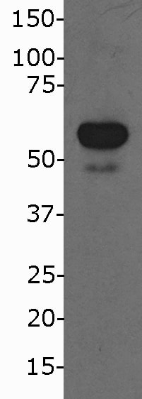
Western Blot: NONO Antibody [NB100-1556]
Western Blot: NONO Antibody [NB100-1556] - B-cell whole cell lysate 40ug. Image submitted by a verified customer review![Immunocytochemistry: NONO Antibody [NB100-1556]](https://resources.rndsystems.com/images/products/NONO-Antibody-Immunocytochemistry-NB100-1556-img0024.jpg "Immunocytochemistry: NONO Antibody [NB100-1556]")
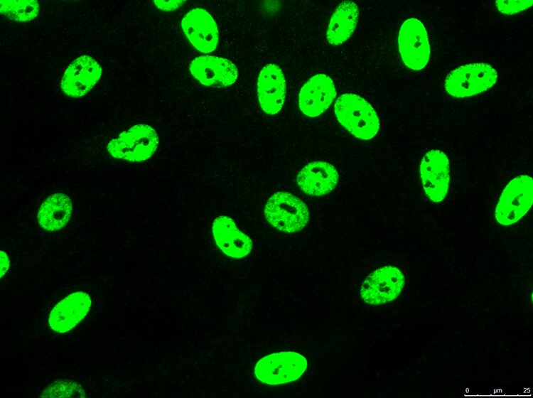
Immunocytochemistry: NONO Antibody [NB100-1556]
Immunocytochemistry: NONO Antibody [NB100-1556] - NONO in human fibroblasts at 1:200 dilution in 1X PBS/1% BSA/0.3% Triton X-100, 16 hours 4C. Image submitted by a verified customer review.![Immunohistochemistry: NONO Antibody [NB100-1556]](https://resources.rndsystems.com/images/products/NONO-Antibody-Immunohistochemistry-NB100-1556-img0026.jpg "Immunohistochemistry: NONO Antibody [NB100-1556]")
Immunohistochemistry: NONO Antibody [NB100-1556]
Immunohistochemistry: NONO Antibody [NB100-1556]![Western Blot: NONO Antibody [NB100-1556]](https://resources.rndsystems.com/images/products/NONO-Antibody-Western-Blot-NB100-1556-img0016.jpg "Western Blot: NONO Antibody [NB100-1556]")
Western Blot: NONO Antibody [NB100-1556]
Western Blot: NONO Antibody [NB100-1556] - Detection of Human NONO on HeLa whole cell lysate (5, 15, and 50 microgram) and 293T cell lystate (50 microgram) using NB100-1556. NONO was also immunoprecipitated using anti-NONO antibody NB100-1554 or NB100-1555 at 3 microg/mg lysate.![Western Blot: NONO Antibody [NB100-1556]](https://resources.rndsystems.com/images/products/NONO-Antibody-Western-Blot-NB100-1556-img0018.jpg "Western Blot: NONO Antibody [NB100-1556]")
![Immunohistochemistry-Paraffin: NONO Antibody [NB100-1556]](https://resources.rndsystems.com/images/products/NONO-Antibody-Immunohistochemistry-Paraffin-NB100-1556-img0020.jpg "Immunohistochemistry-Paraffin: NONO Antibody [NB100-1556]")
Immunohistochemistry-Paraffin: NONO Antibody [NB100-1556]
Immunohistochemistry-Paraffin: NONO Antibody [NB100-1556] - Sample: FFPE section of human breast carcinoma (left) and mouse squamous cell carcinoma (right). Antibody: Affinity purified rabbit anti- NONO used at a dilution of 1:1,000 (0.2 ug/ml) and 1:200 (1ug/ml). Detection: DAB![Immunohistochemistry-Paraffin: NONO Antibody [NB100-1556]](https://resources.rndsystems.com/images/products/NONO-Antibody-Immunohistochemistry-Paraffin-NB100-1556-img0025.jpg "Immunohistochemistry-Paraffin: NONO Antibody [NB100-1556]")
Immunohistochemistry-Paraffin: NONO Antibody [NB100-1556]
Immunohistochemistry-Paraffin: NONO Antibody [NB100-1556] - Affinity purified rabbit anti-NONO used at a dilution of 1:1,000 (0.2 ug/ml).Applications for NONO Antibody
Application
Recommended Usage
Immunohistochemistry
1:200 - 1:1:000
Immunohistochemistry-Paraffin
1:200 - 1:1:000
Immunoprecipitation
1-4 ug/mg lysate
Western Blot
1:2000-1:10000
Application Notes
NONO antibody validated for ICC/IF, WB from verified customer reviews. Epitope retrieval with citrate buffer pH 6.0 is recommended for FFPE tissue sections.
Reviewed Applications
Read 2 reviews rated 4.5 using NB100-1556 in the following applications:
Formulation, Preparation, and Storage
Purification
Immunogen affinity purified
Formulation
TBS and 0.1% BSA
Preservative
0.09% Sodium Azide
Concentration
0.2 mg/ml
Shipping
The product is shipped with polar packs. Upon receipt, store it immediately at the temperature recommended below.
Stability & Storage
Store at 4C. Do not freeze.
Background: NONO
Alternate Names
NMT5552 kDa subunit, non-POU domain containing, octamer-binding, NRB54non-POU-domain-containing, octamer-binding, p54(nrb)
Entrez Gene IDs
4841 (Human)
Gene Symbol
NONO
UniProt
Additional NONO Products
Product Documents for NONO Antibody
Certificate of Analysis
To download a Certificate of Analysis, please enter a lot or batch number in the search box below.
Product Specific Notices for NONO Antibody
This product is for research use only and is not approved for use in humans or in clinical diagnosis. Primary Antibodies are guaranteed for 1 year from date of receipt.
Citations for NONO Antibody
 Powered by Bioz
Customer Reviews for NONO Antibody (2)
4.5 out of 5
2 Customer Ratings
Have you used NONO Antibody?
Submit a review and receive an Amazon gift card!
$25/€18/£15/$25CAN/¥2500 Yen for a review with an image
$10/€7/£6/$10CAN/¥1110 Yen for a review without an image
Submit a review
Customer Images
Showing
1
-
2 of
2 reviews
Showing All
Filter By:
-
Application: Western BlotSample Tested: B-cell whole cell lysate 40ugSpecies: HumanVerified Customer | Posted 03/09/2018BIOSCARF

-
Application: ImmunocytochemistrySample Tested: fibroblastsSpecies: HumanVerified Customer | Posted 01/11/20181:200 in 1X PBS/1% BSA/0.3% Triton X-100, 16 hours 4C.
There are no reviews that match your criteria.
Protocols
Find general support by application which include: protocols, troubleshooting, illustrated assays, videos and webinars.
- Antigen Retrieval Protocol (PIER)
- Antigen Retrieval for Frozen Sections Protocol
- Appropriate Fixation of IHC/ICC Samples
- Cellular Response to Hypoxia Protocols
- Chromogenic IHC Staining of Formalin-Fixed Paraffin-Embedded (FFPE) Tissue Protocol
- Chromogenic Immunohistochemistry Staining of Frozen Tissue
- ClariTSA™ Fluorophore Kits
- Detection & Visualization of Antibody Binding
- Fluorescent IHC Staining of Frozen Tissue Protocol
- Graphic Protocol for Heat-induced Epitope Retrieval
- Graphic Protocol for the Preparation and Fluorescent IHC Staining of Frozen Tissue Sections
- Graphic Protocol for the Preparation and Fluorescent IHC Staining of Paraffin-embedded Tissue Sections
- Graphic Protocol for the Preparation of Gelatin-coated Slides for Histological Tissue Sections
- ICC Cell Smear Protocol for Suspension Cells
- ICC Immunocytochemistry Protocol Videos
- ICC for Adherent Cells
- IHC Sample Preparation (Frozen sections vs Paraffin)
- Immunocytochemistry (ICC) Protocol
- Immunocytochemistry Troubleshooting
- Immunofluorescence of Organoids Embedded in Cultrex Basement Membrane Extract
- Immunofluorescent IHC Staining of Formalin-Fixed Paraffin-Embedded (FFPE) Tissue Protocol
- Immunohistochemistry (IHC) and Immunocytochemistry (ICC) Protocols
- Immunohistochemistry Frozen Troubleshooting
- Immunohistochemistry Paraffin Troubleshooting
- Immunoprecipitation Protocol
- Preparing Samples for IHC/ICC Experiments
- Preventing Non-Specific Staining (Non-Specific Binding)
- Primary Antibody Selection & Optimization
- Protocol for Heat-Induced Epitope Retrieval (HIER)
- Protocol for Making a 4% Formaldehyde Solution in PBS
- Protocol for VisUCyte™ HRP Polymer Detection Reagent
- Protocol for the Fluorescent ICC Staining of Cell Smears - Graphic
- Protocol for the Fluorescent ICC Staining of Cultured Cells on Coverslips - Graphic
- Protocol for the Preparation & Fixation of Cells on Coverslips
- Protocol for the Preparation and Chromogenic IHC Staining of Frozen Tissue Sections
- Protocol for the Preparation and Chromogenic IHC Staining of Frozen Tissue Sections - Graphic
- Protocol for the Preparation and Chromogenic IHC Staining of Paraffin-embedded Tissue Sections
- Protocol for the Preparation and Chromogenic IHC Staining of Paraffin-embedded Tissue Sections - Graphic
- Protocol for the Preparation and Fluorescent ICC Staining of Cells on Coverslips
- Protocol for the Preparation and Fluorescent ICC Staining of Non-adherent Cells
- Protocol for the Preparation and Fluorescent ICC Staining of Stem Cells on Coverslips
- Protocol for the Preparation and Fluorescent IHC Staining of Frozen Tissue Sections
- Protocol for the Preparation and Fluorescent IHC Staining of Paraffin-embedded Tissue Sections
- Protocol for the Preparation of Gelatin-coated Slides for Histological Tissue Sections
- Protocol for the Preparation of a Cell Smear for Non-adherent Cell ICC - Graphic
- R&D Systems Quality Control Western Blot Protocol
- TUNEL and Active Caspase-3 Detection by IHC/ICC Protocol
- The Importance of IHC/ICC Controls
- Troubleshooting Guide: Immunohistochemistry
- Troubleshooting Guide: Western Blot Figures
- Western Blot Conditions
- Western Blot Protocol
- Western Blot Protocol for Cell Lysates
- Western Blot Troubleshooting
- Western Blot Troubleshooting Guide
- View all Protocols, Troubleshooting, Illustrated assays and Webinars
Loading...