NOX1 Antibody - Azide Free
Novus Biologicals | Catalog # NBP1-31546
![Western Blot: NOX1 Antibody [NBP1-31546]](https://resources.rndsystems.com/images/products/NOX1-Antibody-Western-Blot-NBP1-31546-img0008.jpg "Western Blot: NOX1 Antibody [NBP1-31546]")
Loading...
Key Product Details
Validated by
Orthogonal Validation, Biological Validation
Species Reactivity
Validated:
Human, Mouse, Rat
Cited:
Human, Mouse, Rat
Applications
Validated:
Immunohistochemistry, Immunohistochemistry-Paraffin, Western Blot, Flow Cytometry, Immunocytochemistry/ Immunofluorescence, Immunoprecipitation
Cited:
Immunohistochemistry-Paraffin, Western Blot, Immunocytochemistry/ Immunofluorescence, Immunoprecipitation, IHC-P WB
Label
Unconjugated
Antibody Source
Polyclonal Rabbit IgG
Format
Azide Free
Loading...
Product Specifications
Immunogen
Recombinant protein encompassing a sequence within the center region of human NOX1. The exact sequence is proprietary.
Reactivity Notes
Immunogen displays the following percentage of sequence identity for non-tested species: Bovine (85%).
Localization
Membrane, Multi-pass membrane protein
Clonality
Polyclonal
Host
Rabbit
Isotype
IgG
Theoretical MW
65 kDa.
Disclaimer note: The observed molecular weight of the protein may vary from the listed predicted molecular weight due to post translational modifications, post translation cleavages, relative charges, and other experimental factors.
Disclaimer note: The observed molecular weight of the protein may vary from the listed predicted molecular weight due to post translational modifications, post translation cleavages, relative charges, and other experimental factors.
Scientific Data Images for NOX1 Antibody - Azide Free
![Immunohistochemistry-Paraffin: NOX1 Antibody [NBP1-31546]](https://resources.rndsystems.com/images/products/NOX1-Antibody-Immunohistochemistry-Paraffin-NBP1-31546-img0013.jpg "Immunohistochemistry-Paraffin: NOX1 Antibody [NBP1-31546]")
Immunohistochemistry-Paraffin: NOX1 Antibody [NBP1-31546]
Immunohistochemistry-Paraffin: NOX1 Antibody [NBP1-31546] - Mouse kidney. NOX1 stained by NOX1 antibody diluted at 1:500. Antigen Retrieval: Citrate buffer, pH 6.0, 15 min.![Flow Cytometry: NOX1 Antibody [NBP1-31546]](https://resources.rndsystems.com/images/products/NOX1-Antibody-Flow-Cytometry-NBP1-31546-img0006.jpg "Flow Cytometry: NOX1 Antibody [NBP1-31546]")
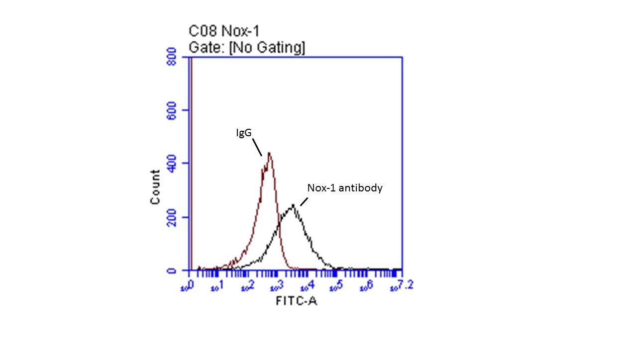
Flow Cytometry: NOX1 Antibody [NBP1-31546]
Flow Cytometry: NOX1 Antibody [NBP1-31546] - Nox-1 is present at the surface of resting platelets. Human platelets (4 x 10^8 cells/mL) were isolated from whole blood and incubated with the Nox-1 antibody or Rabbit IgG control at 1:250 dilution for 30 minutes at room temperature. FITC-tagged anti-rabbit (secondary antibody) was added at 1:100 dilution and incubated for an additional 30 minutes. Platelets were then diluted 1:25 v/v in Tyrodes buffer and read using a BD Accuri flow cytometer. Image from verified customer review.![Western Blot: NOX1 Antibody [NBP1-31546]](https://resources.rndsystems.com/images/products/NOX1-Antibody-Western-Blot-NBP1-31546-img0002.jpg "Western Blot: NOX1 Antibody [NBP1-31546]")
Western Blot: NOX1 Antibody [NBP1-31546]
Western Blot: NOX1 Antibody [NBP1-31546] - Sample (30 ug of whole cell lysate) A: JC B: BCL-1 7. 5% SDS PAGE; antibody diluted at 1:5000.![Western Blot: NOX1 Antibody [NBP1-31546]](https://resources.rndsystems.com/images/products/NOX1-Antibody-Western-Blot-NBP1-31546-img0003.jpg "Western Blot: NOX1 Antibody [NBP1-31546]")
Western Blot: NOX1 Antibody [NBP1-31546]
Western Blot: NOX1 Antibody [NBP1-31546] - Sample (30 ug of whole cell lysate) A: 293T B: A431 C: HeLa 7. 5% SDS PAGE, antibody diluted at 1:2000.![Western Blot: NOX1 Antibody [NBP1-31546]](https://resources.rndsystems.com/images/products/NOX1-Antibody-Western-Blot-NBP1-31546-img0010.jpg "Western Blot: NOX1 Antibody [NBP1-31546]")
Western Blot: NOX1 Antibody [NBP1-31546]
Western Blot: NOX1 Antibody [NBP1-31546] - Mouse tissue extract (50 ug) was separated by 7.5% SDS-PAGE, and the membrane was blotted with NOX1 antibody diluted at 1:2000. The HRP-conjugated anti-rabbit IgG antibody (NBP2-19301) was used to detect the primary antibody.![Immunohistochemistry-Paraffin: NOX1 Antibody [NBP1-31546]](https://resources.rndsystems.com/images/products/NOX1-Antibody-Immunohistochemistry-Paraffin-NBP1-31546-img0005.jpg "Immunohistochemistry-Paraffin: NOX1 Antibody [NBP1-31546]")
Immunohistochemistry-Paraffin: NOX1 Antibody [NBP1-31546]
Immunohistochemistry-Paraffin: NOX1 Antibody [NBP1-31546] - NOX1 antibody detects NOX1 protein at cytoplasm on human lung carcinoma by immunohistochemical analysis.Sample: Paraffin-embedded human lung carcinoma.NOX1 antibody diluted at 1:500.
Antigen Retrieval: Trilogy™ (EDTA based, pH 8.0) buffer, 15min

Western Blot: NOX1 Antibody [NBP1-31546] -
Western Blot: NOX1 Antibody [NBP1-31546] - Blueberry polyphenol extract (BPE) did not affect the expression of NADPH oxidases (NOX) in angiotensin (Ang) II-treated human aortic endothelial cells (HAECs). HAECs were treated with 200 µg/mL of BPE for 1 h then treated with 200 nM of Ang II for 12 h. Protein expression of NOX1 (A,B), NOX2 (C,D), NOX4 (E,F), & NOX5 (G,H) were determined by Western blot. Quantification was performed using Image Lab (Bio-Rad Laboratories, Inc.). Data are expressed as mean ± SD from three (NOX1 & NOX4) & five (NOX2 & NOX5) independent experiments. Values that do not share the same letter are significantly different from each other (p ≤ 0.05). Image collected & cropped by CiteAb from the following publication (https://pubmed.ncbi.nlm.nih.gov/35453301), licensed under a CC-BY license. Not internally tested by Novus Biologicals.
Western Blot: NOX1 Antibody [NBP1-31546] -
Untreated (-) and treated (+) HCT116 whole cell extracts (30 ug) were separated by 7.5% SDS-PAGE, and the membrane was blotted with NOX1 antibody (NBP1-31546) diluted at 1:2000. The HRP-conjugated anti-rabbit IgG antibody was used to detect the primary antibody.
Immunocytochemistry/ Immunofluorescence: NOX1 Antibody [NBP1-31546] -
NOX1 antibody detects NOX1 protein at cytoplasm by immunofluorescent analysis.Sample: HepG2 cells were fixed in 4% paraformaldehyde at RT for 15 min.
Green: NOX1 protein stained by NOX1 antibody (NBP1-31546) diluted at 1:500.
Blue: Hoechst 33342 staining.
Applications for NOX1 Antibody - Azide Free
Application
Recommended Usage
Flow Cytometry
Validated from a verified customer review.
Immunocytochemistry/ Immunofluorescence
1:100-1:1000
Immunohistochemistry
1:100-1:1000
Immunohistochemistry-Paraffin
1:100-1:1000
Immunoprecipitation
Reported in scientific literature (PMID: 27094494)
Western Blot
1:500-1:3000
Reviewed Applications
Read 1 review rated 5 using NBP1-31546 in the following applications:
Flow Cytometry Panel Builder
Bio-Techne Knows Flow Cytometry
Save time and reduce costly mistakes by quickly finding compatible reagents using the Panel Builder Tool.
Advanced Features
- Spectra Viewer - Custom analysis of spectra from multiple fluorochromes
- Spillover Popups - Visualize the spectra of individual fluorochromes
- Antigen Density Selector - Match fluorochrome brightness with antigen density

Formulation, Preparation, and Storage
Purification
Antigen Affinity-purified
Formulation
PBS, 1% BSA, 20% Glycerol
Format
Azide Free
Preservative
0.025% Proclin 300
Concentration
Concentrations vary lot to lot. See vial label for concentration. If unlisted please contact technical services.
Shipping
The product is shipped with polar packs. Upon receipt, store it immediately at the temperature recommended below.
Stability & Storage
Aliquot and store at -20C or -80C. Avoid freeze-thaw cycles.
Background: NOX1
Long Name
NADPH Oxidase 1
Alternate Names
MOX-1, NOH1
Entrez Gene IDs
27305 (Human)
Gene Symbol
NOX1
UniProt
Additional NOX1 Products
Product Documents for NOX1 Antibody - Azide Free
Certificate of Analysis
To download a Certificate of Analysis, please enter a lot or batch number in the search box below.
Product Specific Notices for NOX1 Antibody - Azide Free
This product is for research use only and is not approved for use in humans or in clinical diagnosis. Primary Antibodies are guaranteed for 1 year from date of receipt.
Related Research Areas
Citations for NOX1 Antibody - Azide Free
 Powered by Bioz
Customer Reviews for NOX1 Antibody - Azide Free (1)
5 out of 5
1 Customer Rating
Have you used NOX1 Antibody - Azide Free?
Submit a review and receive an Amazon gift card!
$25/€18/£15/$25CAN/¥2500 Yen for a review with an image
$10/€7/£6/$10CAN/¥1110 Yen for a review without an image
Submit a review
Customer Images
Showing
1
-
1 of
1 review
Showing All
Filter By:
-
Application: Flow CytometrySample Tested: PlateletsSpecies: HumanVerified Customer | Posted 01/24/2019Nox-1 is present at the surface of resting platelets.Platelets (4 x 10^8 cells/mL) were isolated from whole blood and incubated with the Nox-1 antibody or Rabbit IgG control at 1:250 dilution for 30 minutes at room temperature. FITC-tagged anti-rabbit (secondary antibody) was added at 1:100 dilution and incubated for an additional 30 minutes. Platelets were then diluted 1:25 v/v in Tyrodes buffer and read using a BD Accuri flow cytometer.

There are no reviews that match your criteria.
Protocols
Find general support by application which include: protocols, troubleshooting, illustrated assays, videos and webinars.
- 7-Amino Actinomycin D (7-AAD) Cell Viability Flow Cytometry Protocol
- Antigen Retrieval Protocol (PIER)
- Antigen Retrieval for Frozen Sections Protocol
- Appropriate Fixation of IHC/ICC Samples
- Cellular Response to Hypoxia Protocols
- Chromogenic IHC Staining of Formalin-Fixed Paraffin-Embedded (FFPE) Tissue Protocol
- Chromogenic Immunohistochemistry Staining of Frozen Tissue
- ClariTSA™ Fluorophore Kits
- Detection & Visualization of Antibody Binding
- Extracellular Membrane Flow Cytometry Protocol
- Flow Cytometry Protocol for Cell Surface Markers
- Flow Cytometry Protocol for Staining Membrane Associated Proteins
- Flow Cytometry Staining Protocols
- Flow Cytometry Troubleshooting Guide
- Fluorescent IHC Staining of Frozen Tissue Protocol
- Graphic Protocol for Heat-induced Epitope Retrieval
- Graphic Protocol for the Preparation and Fluorescent IHC Staining of Frozen Tissue Sections
- Graphic Protocol for the Preparation and Fluorescent IHC Staining of Paraffin-embedded Tissue Sections
- Graphic Protocol for the Preparation of Gelatin-coated Slides for Histological Tissue Sections
- ICC Cell Smear Protocol for Suspension Cells
- ICC Immunocytochemistry Protocol Videos
- ICC for Adherent Cells
- IHC Sample Preparation (Frozen sections vs Paraffin)
- Immunocytochemistry (ICC) Protocol
- Immunocytochemistry Troubleshooting
- Immunofluorescence of Organoids Embedded in Cultrex Basement Membrane Extract
- Immunofluorescent IHC Staining of Formalin-Fixed Paraffin-Embedded (FFPE) Tissue Protocol
- Immunohistochemistry (IHC) and Immunocytochemistry (ICC) Protocols
- Immunohistochemistry Frozen Troubleshooting
- Immunohistochemistry Paraffin Troubleshooting
- Immunoprecipitation Protocol
- Intracellular Flow Cytometry Protocol Using Alcohol (Methanol)
- Intracellular Flow Cytometry Protocol Using Detergents
- Intracellular Nuclear Staining Flow Cytometry Protocol Using Detergents
- Intracellular Staining Flow Cytometry Protocol Using Alcohol Permeabilization
- Intracellular Staining Flow Cytometry Protocol Using Detergents to Permeabilize Cells
- Preparing Samples for IHC/ICC Experiments
- Preventing Non-Specific Staining (Non-Specific Binding)
- Primary Antibody Selection & Optimization
- Propidium Iodide Cell Viability Flow Cytometry Protocol
- Protocol for Heat-Induced Epitope Retrieval (HIER)
- Protocol for Liperfluo
- Protocol for Making a 4% Formaldehyde Solution in PBS
- Protocol for VisUCyte™ HRP Polymer Detection Reagent
- Protocol for the Characterization of Human Th22 Cells
- Protocol for the Characterization of Human Th9 Cells
- Protocol for the Fluorescent ICC Staining of Cell Smears - Graphic
- Protocol for the Fluorescent ICC Staining of Cultured Cells on Coverslips - Graphic
- Protocol for the Preparation & Fixation of Cells on Coverslips
- Protocol for the Preparation and Chromogenic IHC Staining of Frozen Tissue Sections
- Protocol for the Preparation and Chromogenic IHC Staining of Frozen Tissue Sections - Graphic
- Protocol for the Preparation and Chromogenic IHC Staining of Paraffin-embedded Tissue Sections
- Protocol for the Preparation and Chromogenic IHC Staining of Paraffin-embedded Tissue Sections - Graphic
- Protocol for the Preparation and Fluorescent ICC Staining of Cells on Coverslips
- Protocol for the Preparation and Fluorescent ICC Staining of Non-adherent Cells
- Protocol for the Preparation and Fluorescent ICC Staining of Stem Cells on Coverslips
- Protocol for the Preparation and Fluorescent IHC Staining of Frozen Tissue Sections
- Protocol for the Preparation and Fluorescent IHC Staining of Paraffin-embedded Tissue Sections
- Protocol for the Preparation of Gelatin-coated Slides for Histological Tissue Sections
- Protocol for the Preparation of a Cell Smear for Non-adherent Cell ICC - Graphic
- Protocol: Annexin V and PI Staining by Flow Cytometry
- Protocol: Annexin V and PI Staining for Apoptosis by Flow Cytometry
- R&D Systems Quality Control Western Blot Protocol
- TUNEL and Active Caspase-3 Detection by IHC/ICC Protocol
- The Importance of IHC/ICC Controls
- Troubleshooting Guide: Fluorokine Flow Cytometry Kits
- Troubleshooting Guide: Immunohistochemistry
- Troubleshooting Guide: Western Blot Figures
- Western Blot Conditions
- Western Blot Protocol
- Western Blot Protocol for Cell Lysates
- Western Blot Troubleshooting
- Western Blot Troubleshooting Guide
- View all Protocols, Troubleshooting, Illustrated assays and Webinars
Loading...