Nox4 Antibody - BSA Free
Novus Biologicals | Catalog # NB110-58849
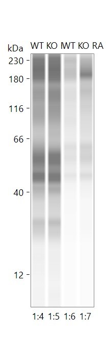
![Western Blot: Nox4 AntibodyBSA Free [NB110-58849]](https://resources.rndsystems.com/images/products/Nox4-Antibody---BSA-Free-Western-Blot-NB110-58849-img0013.jpg "Western Blot: Nox4 AntibodyBSA Free [NB110-58849]")
Key Product Details
Validated by
Species Reactivity
Validated:
Cited:
Applications
Validated:
Cited:
Label
Antibody Source
Format
Product Specifications
Immunogen
Reactivity Notes
Localization
Clonality
Host
Isotype
Scientific Data Images for Nox4 Antibody - BSA Free
Western Blot: Nox4 AntibodyBSA Free [NB110-58849]
Western Blot: Nox4 Antibody - BSA Free [NB110-58849] - Detection of NOX4 on IMR90 lysate. Image courtesy of Naomi Logsdon, University of Alabama at Birmingham.![Immunocytochemistry/ Immunofluorescence: Nox4 Antibody - BSA Free [NB110-58849]](https://resources.rndsystems.com/images/products/Nox4-Antibody---BSA-Free-Immunocytochemistry-Immunofluorescence-NB110-58849-img0019.jpg "Immunocytochemistry/ Immunofluorescence: Nox4 Antibody - BSA Free [NB110-58849]")
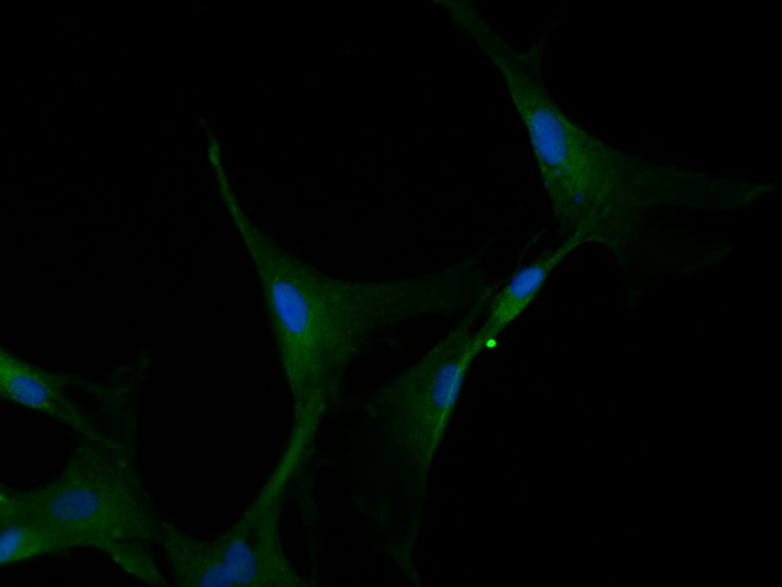
Immunocytochemistry/ Immunofluorescence: Nox4 Antibody - BSA Free [NB110-58849]
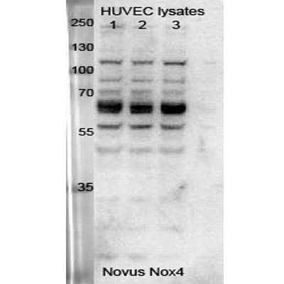
Immunocytochemistry/Immunofluorescence: Nox4 Antibody - BSA Free [NB110-58849] - Porcine aortic endothelial cells stained with NB110-58849 (green). Nuclei were counterstained with DAPI (blue). Image from verified customer review.![Western Blot: Nox4 AntibodyBSA Free [NB110-58849]](https://resources.rndsystems.com/images/products/Nox4-Antibody---BSA-Free-Western-Blot-NB110-58849-img0012.jpg "Western Blot: Nox4 AntibodyBSA Free [NB110-58849]")
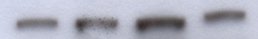
Western Blot: Nox4 AntibodyBSA Free [NB110-58849]
Western Blot: Nox4 Antibody - BSA Free [NB110-58849] - Detection of NOX4 in HUVEC whole cell lysate. Lanes 1 and 2: serum-starved HUVEC lysate denatured at 95C for 5 minutes. Lanes 3: serum-starved HUVEC lysate denatured at room temperature for 10 minutes. Image from verified customer review.![Immunocytochemistry/ Immunofluorescence: Nox4 Antibody - BSA Free [NB110-58849]](https://resources.rndsystems.com/images/products/Nox4-Antibody---BSA-Free-Immunocytochemistry-Immunofluorescence-NB110-58849-img0011.jpg "Immunocytochemistry/ Immunofluorescence: Nox4 Antibody - BSA Free [NB110-58849]")
Immunocytochemistry/ Immunofluorescence: Nox4 Antibody - BSA Free [NB110-58849]
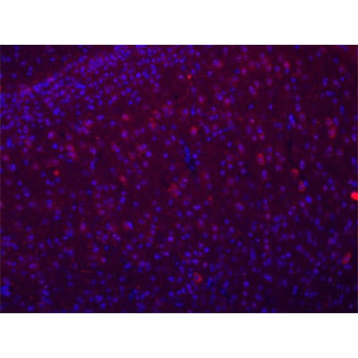
Immunocytochemistry/Immunofluorescence: Nox4 Antibody - BSA Free [NB110-58849] - Detection of NOX4 in mouse brain showing positive staining in neurons in the cortex. Image provided by Rachel Reith.![Immunohistochemistry: Nox4 Antibody - BSA Free [NB110-58849]](https://resources.rndsystems.com/images/products/Nox4-Antibody---BSA-Free-Immunohistochemistry-NB110-58849-img0009.jpg "Immunohistochemistry: Nox4 Antibody - BSA Free [NB110-58849]")
Immunohistochemistry: Nox4 Antibody - BSA Free [NB110-58849]
Immunohistochemistry: Nox4 Antibody - BSA Free [NB110-58849] - Detection of NOX4 in proximal convoluted tubules of the kidney using NB110-58849 at 5 ug/mL.![Immunohistochemistry: Nox4 Antibody - BSA Free [NB110-58849]](https://resources.rndsystems.com/images/products/Nox4-Antibody---BSA-Free-Immunohistochemistry-NB110-58849-img0022.jpg "Immunohistochemistry: Nox4 Antibody - BSA Free [NB110-58849]")
Immunohistochemistry: Nox4 Antibody - BSA Free [NB110-58849]
Nox4-Antibody---BSA-Free-Immunohistochemistry-NB110-58849-img0022.jpg![Knockdown Validated: Nox4 Antibody - BSA Free [NB110-58849]](https://resources.rndsystems.com/images/products/Nox4%20Antibody%20-%20BSA%20Free-Knockdown%20Validated-NB110-58849-img0023.jpg "Western Blot: Nox4 Antibody - BSA Free [NB110-58849]")
![Knockdown Validated: Nox4 Antibody - BSA Free [NB110-58849]](https://resources.rndsystems.com/images/products/Nox4-Antibody---BSA-Free-Knockdown-Validated-NB110-58849-img0020.jpg "Western Blot: Nox4 Antibody - BSA Free [NB110-58849]")

Immunohistochemistry: Nox4 Antibody - BSA Free [NB110-58849] -
Presence of NOX4 in human ovarian tissue. Immunohistochemistry using human ovarian sections and an anti-NOX4 antibody from ProSci showed positive staining for NOX4 in granulosa (GC) and theca cells (TC) of a secondary follicle (A), of a small antral follicle (B), of a large antral follicle (C) as well as in luteinized GCs (LGC) and luteinized TCs (LTC) of the corpus luteum (E). Serum controls lacked first antibody (D and F). Scale bars: A–E = 30 µm, F = 50 µm.
Immunohistochemistry: Nox4 Antibody - BSA Free [NB110-58849] -
AT1-KO mice are resistant to alcohol-induced oxidative stress. Oxidative stress was examined by immunohistochemical staining of 3-NT (A) and 4-HNE (B). NOX2 (C) and NOX4 (D) expression was examined by immunohistochemical staining. Data are presented as means ± SD (the animal number for each group is indicated in Figure 1). *, P < 0.05 vs corresponding control; #, P < 0.05 vs WT alcohol group. Bar = 50 μM.
Western Blot: Nox4 Antibody - BSA Free [NB110-58849] -
Western Blot: Nox4 Antibody - BSA Free [NB110-58849] - Effect of metformin on bleomycin-induced lung fibrosis development in mice. f WB using anti-NOX4, & anti-beta -actin of cell lysates from normal LF (lane 1, 2, 3) & IPF LF (lane 4, 5, 6). Lower panel is average (±SEM) taken from 3patients shown as relative expression. Open bar is normal LF & filled bar is IPF LF. *p < 0.05 Image collected & cropped by CiteAb from following publication (http://respiratory-research.biomedcentral.com/articles/10.1186/s12931-0…), licensed under a CC-BY license. Not internally tested by Novus Biologicals.
Western Blot: Nox4 Antibody - BSA Free [NB110-58849] -
Western Blot: Nox4 Antibody - BSA Free [NB110-58849] - Fasting protects against oxidative stress resulting after two weeks of unilateral ischemia-reperfusion (IR) injury. (A) Representative kidney sections (10×) immunostained for 8-hydroxy-2′-deoxyguanosine (8-OHdG) in sham, IR, & IR + Fasting experimental groups at day 14 post-IR or sham surgery. (B) Immunoblots (left) & quantification (right) of catalase (CAT), glutamate-cysteine ligase modifier subunit (GCLM), & nicotinamide adenine dinucleotide phosphate oxidase 4 (NOX4) in the kidney cortex of rats from sham, IR, & IR + Fasting experimental groups. GAPDH, glyceraldehyde-3-phosphate dehydrogenase. Data are expressed as mean ± SEM. n = 3 animals per group. * p < 0.05. Image collected & cropped by CiteAb from the following publication (https://pubmed.ncbi.nlm.nih.gov/31443530), licensed under a CC-BY license. Not internally tested by Novus Biologicals.
Western Blot: Nox4 Antibody - BSA Free [NB110-58849] -
Western Blot: Nox4 Antibody - BSA Free [NB110-58849] - SPA0355 decreased renal oxidative stress & regulated levels of pro-oxidant & antioxidant enzymes in LPS-treated mice. (A) Representative images of IHC of 4-hydroxynonenal (4-HNE) in kidneys. Bar = 20 μm. (B) Percentage of 4-HNE-positive area per field. (C) Renal levels of malondialdehyde (MDA). (D) Representative images of Western blotting of nicotinamide adenine dinucleotide phosphate oxidase 4 (NOX4) & GAPDH. (E) The mRNA levels of manganese superoxide dismutase (MnSOD) & catalase. Results are from 8 mice per group (biological replicates) & 2 or 3 technical replicates per mouse. *** p < 0.001 vs. vehicle-treated mice (Veh). ###p <0.001 vs. LPS-injected mice (LPS). Image collected & cropped by CiteAb from the following publication (https://pubmed.ncbi.nlm.nih.gov/32635491), licensed under a CC-BY license. Not internally tested by Novus Biologicals.
Immunohistochemistry: Nox4 Antibody - BSA Free [NB110-58849] -
Immunohistochemistry: Nox4 Antibody - BSA Free [NB110-58849] - Presence of NOX4 in human ovarian tissue. Immunohistochemistry using human ovarian sections & an anti-NOX4 antibody from ProSci showed positive staining for NOX4 in granulosa (GC) & theca cells (TC) of a secondary follicle (A), of a small antral follicle (B), of a large antral follicle (C) as well as in luteinized GCs (LGC) & luteinized TCs (LTC) of the corpus luteum (E). Serum controls lacked first antibody (D & F). Scale bars: A–E = 30 µm, F = 50 µm. Image collected & cropped by CiteAb from the following publication (https://pubmed.ncbi.nlm.nih.gov/30837663), licensed under a CC-BY license. Not internally tested by Novus Biologicals.
Western Blot: Nox4 Antibody - BSA Free [NB110-58849] -
Western Blot: Nox4 Antibody - BSA Free [NB110-58849] - Role of NADPH oxidases in IH-induced HIF-2 alpha degradation. A. Effect of NADPH oxidase (Nox) inhibitors Apocynin (Apo, 1 mM) & AEBSF (15 µM) on HIF-2 alpha protein following exposure to IH. B. HIF-2 alpha expression in PC12 cells transfected with Nox2 & Nox4 siRNA & exposed to normoxia (N) or IH. Tubulin expression was monitored as control for protein loading. Bottom panels of A & B represent average data of densitometric analysis of the immunoblots presented as mean ± S.E.M from three independent experiments. *p<0.05; n.s. not significant, p>0.05. Image collected & cropped by CiteAb from the following publication (https://pubmed.ncbi.nlm.nih.gov/24124516), licensed under a CC-BY license. Not internally tested by Novus Biologicals.
Western Blot: Nox4 Antibody - BSA Free [NB110-58849] -
Western Blot: Nox4 Antibody - BSA Free [NB110-58849] - The representative images of immunohistochemical staining (A–D; sections were counterstained with hematoxylin; original magnification, × 20), & western blot assays (E,F) showed that FLL treatment decreased Nox4 expression in tibias & uteri of OVX rats (n = 9). In addition, FLL treatment also decreased cytochrome C (Cyto-C; G) & increased Bcl-2 expression (H) in the tibias of OVX rats (n = 9). Data are presented as mean ± SD. IOD denotes integrated optical density of interested areas. #p < 0.05 with Sham group, *p < 0.05 compared with OVX group. Image collected & cropped by CiteAb from the following publication (https://pubmed.ncbi.nlm.nih.gov/28588482), licensed under a CC-BY license. Not internally tested by Novus Biologicals.
Western Blot: Nox4 Antibody - BSA Free [NB110-58849] -
Western Blot: Nox4 Antibody - BSA Free [NB110-58849] - Blockade of LTCC suppressed CaMKII-NF-kB pathway in DOX-treated hearts. (a) Representative immunoblots & quantitative analysis of CaMKII, phosphorylated CaMKII, & GAPDH in DOX (3 doses of DOX at 6 mg/kg body weight every third day for 1 week) or control vehicle (phosphate-buffered saline: PBS) treated-C57B/6 J mouse hearts subjected to either nifedipine (Nif, 10 mg/day/day) or saline for 9 days (n = 5). (b) Representative immunoblots & quantitative analysis of NF-kappa B, phosphorylated NF-kappa B, cleaved caspase 3, & GAPDH in each group (n = 5). The experiment was conducted 3 times. (c–g) Representative immunoblotsa & quantitative analysis of ERK, phosphorylated ERK, JNK, phosphorylated JNK, Nox4, p53, & GAPDH in each group (n = 5). Image collected & cropped by CiteAb from the following publication (https://pubmed.ncbi.nlm.nih.gov/31285514), licensed under a CC-BY license. Not internally tested by Novus Biologicals.
Western Blot: Nox4 Antibody - BSA Free [NB110-58849] -
Western Blot: Nox4 Antibody - BSA Free [NB110-58849] - Diabetic atrophied muscles exhibited a state of heightened oxidative stress (HSOS). (a & b) Superoxide generation was measured in frozen muscle sections of control & diabetic using dihydroethidium-based confocal microscopic staining technique. (c & d) NADPH oxidase in a membrane fraction was assessed according to procedure involving the substrate NADPH & lucigenin chemiluminescence or the Amplex Red/horseradish peroxidase fluorescence-based assays. (e & f) Muscle NADPH oxidase-related isoforms including NOX2 & NOX4 were determined at the mRNA (e) & protein levels (f) using RT-PCR & Western blotting-based techniques. (g) Mitochondrial H2O2 generation at the steady state level & in the presence of added glutamate/malate substrates was measured using the Amplex Red/horseradish peroxidase fluorescence-based assay (g). Activities of complexes I (h) & III (i) of the electron transport chain were measured using spectrophotometric-based assay. Abbreviation: C: control; D: diabetic. Values are means ± SEM for at least 6 animals/group. ∗Significantly different from corresponding control values at P ≤ 0.05. Image collected & cropped by CiteAb from the following publication (https://pubmed.ncbi.nlm.nih.gov/30510624), licensed under a CC-BY license. Not internally tested by Novus Biologicals.
Western Blot: Nox4 Antibody - BSA Free [NB110-58849] -
Western Blot: Nox4 Antibody - BSA Free [NB110-58849] - Characterization of NOX4 & PKM2 in human RCC tumors & adjacent tissue. a Mitochondrial fractions were prepared from human tumors (T) or uninvolved adjacent tissue (N). NOX4 expression was examined by western blot analysis. Prohibitin was probed as a mitochondrial marker & loading control. b Quantitation of NOX4 distribution in the mitochondrial fraction from a. The results are expressed as the means using one-way ANOVA with Tukey’s post hoc test where ± S.E.M. *p < 0.05 compared to normal (N). c Mitochondria fractions were prepared from RCC tumors & NADPH-dependent superoxide generation was examined in the presence (+) or absence (−) of ATP. The results are from eight tumors & are expressed as the means using one-way ANOVA with Tukey’s post hoc test where ±S.E.M. **p < 0.01 is compared to without (−) ATP. d PKM2 & PKM1 expression was examined by western blot analysis in lysates prepared from human tumors (T) or uninvolved adjacent tissue (N) from the same patient. Actin as loading control Image collected & cropped by CiteAb from the following publication (https://pubmed.ncbi.nlm.nih.gov/29051480), licensed under a CC-BY license. Not internally tested by Novus Biologicals.
Western Blot: Nox4 Antibody - BSA Free [NB110-58849] -
Western Blot: Nox4 Antibody - BSA Free [NB110-58849] - Fbs stimulated by BMM differentiate into myofibroblasts via the Cyr61/Nox4 pathway. (A) Western blotting for Fb differentiation, involving analysis of specific factors such as FSP-1, NOX2, NOX4, collagen-1, & Cyr61. The expression of these factors was normalized to that of GAPDH; (B) Cells were treated with BMM for 3 or 6 h or DPI (5 μM; NOX inhibitor) for 1 h or were co-treated with DPI & BMM. ROS production was measured using the DCF-DA assay & calculated as a percentage of the mean fluorescence intensity compared with that of the control. * p < 0.05 as compared to the control; # p < 0.05 as compared to the BMM (6 h)-treated group; (C) Cells were treated with BMM for 3 h or DPI (5 μM, NOX inhibitor) for 1 h or were co-treated with DPI & BMM. Cyr61 expression was normalized to that of GAPDH after western blotting. The values indicate intensities of protein expression with respect to that of the loading control; (D) Secretion of MMPs from BMM-treated-Fbs by using conditioned medium, followed by western blotting analysis. The expression levels were normalized to PonceauS, used as a loading control. *** p < 0.001 as compared to the control. Image collected & cropped by CiteAb from the following publication (http://www.mdpi.com/1422-0067/19/4/1164), licensed under a CC-BY license. Not internally tested by Novus Biologicals.
Western Blot: Nox4 Antibody - BSA Free [NB110-58849] -
Western Blot: Nox4 Antibody - BSA Free [NB110-58849] - DSC restores redox balance in colonic tissues. A, Colitis was induced as described in Materials & Methods & treated with or without DSC (50 mg/kg). Representative bands & densitometry analysis of Nrf2 nuclear translocation & HO‐1 expression in colonic tissues. Histone H3 & beta ‐actin were used as loading control, respectively. B, Colitis was induced as described in Materials & Methods & treated with lentiviral Nox4 shRNA or lentiviral scrambled shRNA. Representative bands & densitometry analysis of Nrf2 nuclear translocation & HO‐1 expression in colonic tissues. Histone H3 & beta ‐actin were used as loading control, respectively. BMDM was stimulated with or without DSC (50 μmol/L) as described in Materials & Methods. C, Representative bands & densitometry analysis of Nox4. beta ‐actin was used as loading control. D, Representative bands & densitometry analysis of Nrf2 nuclear translocation & cytoplasmic HO‐1 expression in BMDM. beta ‐actin or histone H3 was used as loading control. Data shown are means ± SEM of n = 8 in each group. *P < 0.05 vs Control cell or mice (CTL), #P < 0.05 vs DSS‐treated mice or LPS‐stimulated cells Image collected & cropped by CiteAb from the following publication (https://pubmed.ncbi.nlm.nih.gov/32945118), licensed under a CC-BY license. Not internally tested by Novus Biologicals.
Western Blot: Nox4 Antibody - BSA Free [NB110-58849] -
Western Blot: Nox4 Antibody - BSA Free [NB110-58849] - DSC ameliorates Nox4 expression & ROS production. A, Colitis was induced as described in Materials & Methods, & the colonic tissues were collected as indicated periods. Representative bands & quantitative analysis of Nox4 in colonic tissues were shown. B‐F, Colitis was induced as described in Materials & Methods & treated with or without DSC (50 mg/kg). B, Representative bands & quantitative analysis of Nox4 in colonic tissues. C, Representative images & quantitative analysis of ROS production by DHE staining in colonic tissues, scale bar = 100 μm. D, Quantitative analysis of GSH/GSSG ratio. E, Quantitative analysis of tissue H2O2 production. Colitis was induced as described in Materials & Methods & treated with or lentiviral Nox4 shRNA. F, Representative bands & quantitative analysis of Nox4 in colonic tissues. G, Representative images & quantitative analysis of colon length. H, Representative images of H&E staining & histological score. I, Representative bands & quantitative analysis of ZO‐2 & claudin‐1 expression in colonic tissues. beta ‐actin was used as loading control. Data shown are means ± SEM of n = 8 in each group. *P < 0.05 vs Control (CTL), #P < 0.05 vs DSS‐treated mice Image collected & cropped by CiteAb from the following publication (https://pubmed.ncbi.nlm.nih.gov/32945118), licensed under a CC-BY license. Not internally tested by Novus Biologicals.Applications for Nox4 Antibody - BSA Free
Immunoblotting
Immunocytochemistry/ Immunofluorescence
Immunohistochemistry
Immunohistochemistry-Frozen
Immunohistochemistry-Paraffin
Western Blot
Reviewed Applications
Read 9 reviews rated 4.1 using NB110-58849 in the following applications:
Formulation, Preparation, and Storage
Purification
Formulation
Format
Preservative
Concentration
Shipping
Stability & Storage
Background: Nox4
Long Name
Alternate Names
Gene Symbol
Additional Nox4 Products
Product Documents for Nox4 Antibody - BSA Free
Certificate of Analysis
To download a Certificate of Analysis, please enter a lot or batch number in the search box below.
Product Specific Notices for Nox4 Antibody - BSA Free
This product is for research use only and is not approved for use in humans or in clinical diagnosis. Primary Antibodies are guaranteed for 1 year from date of receipt.
Related Research Areas
Citations for Nox4 Antibody - BSA Free
 Powered by Bioz
Customer Reviews for Nox4 Antibody - BSA Free (9)
Have you used Nox4 Antibody - BSA Free?
Submit a review and receive an Amazon gift card!
$25/€18/£15/$25CAN/¥2500 Yen for a review with an image
$10/€7/£6/$10CAN/¥1110 Yen for a review without an image
Submit a review
Customer Images

-
Application: Simple WesternSample Tested: Lung tissueSpecies: MouseVerified Customer | Posted 09/08/2025Nox4 protein expression in mouse lung homogenates using JESSTested the Nox4 protein expression in mouse lung homogenates
 Bio-Techne ResponseThis review reflects a new species or application tested on a primary antibody.
 -
Application: Western BlotSample Tested: H4-II-E-C3 rat hepatoma cell lineSpecies: RatVerified Customer | Posted 03/01/2018silence of NOX4 in H4-II-E-C3 cells.
-
Application: ImmunofluorescenceSample Tested: porcine aortic endothelial cellsSpecies: OtherVerified Customer | Posted 07/25/2016cells stained with nox-4 antibody (gree); blue=dapi
-
Application: Western BlotSample Tested: PAOEC cell lysateSpecies: OtherVerified Customer | Posted 07/22/2016
-
Application: Western BlotSample Tested: MouseSpecies: MouseVerified Customer | Posted 07/01/2014
-
Application: Western BlotSample Tested: SH-SY5Y whole cell lysteSpecies: OtherVerified Customer | Posted 06/13/2014
-
Application: Immunohistochemistry-ParaffinSample Tested: mouse brainSpecies: MouseVerified Customer | Posted 01/30/2013
-
Application: Western BlotSample Tested: HUVEC whole cell lysateSpecies: HumanVerified Customer | Posted 12/10/2012
-
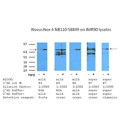
Application: Western BlotSample Tested: IMR90 fibroblast, +/- TGF beta, whole cell lysateSpecies: HumanVerified Customer | Posted 11/15/2012
There are no reviews that match your criteria.
Protocols
View specific protocols for Nox4 Antibody - BSA Free (NB110-58849):
Culture cells to appropriate density in 35 mm culture dishes or 6-well plates.
1. Remove culture medium and wash the cells briefly in PBS. Add 10% formalin to the dish and fix at room temperature for 10 minutes.
2. Remove the formalin and wash the cells in PBS.
3. Permeablize the cells with 0.1% Triton X100 or other suitable detergent for 10 min.
4. Remove the permeablization buffer and wash three times for 10 minutes each in PBS. Be sure to not let the specimen dry out.
5. To block nonspecific antibody binding, incubate in 10% normal goat serum from 1 hour to overnight at room temperature.
6. Add primary antibody at appropriate dilution and incubate overnight at 4C.
7. Remove primary antibody and replace with PBS. Wash three times for 10 minutes each.
8. Add secondary antibody at appropriate dilution. Incubate for 1 hour at room temperature.
9. Remove secondary antibody and replace with PBS. Wash three times for 10 minutes each.
10. Counter stain DNA with DAPi if required.
Antigen Unmasking:
Bring slides to a boil in 10 mM sodium citrate buffer (pH 6.0) then maintain at a sub-boiling temperature for 10 minutes. Cool slides on bench-top for 30 minutes (keep slides in the sodium citrate buffer at all times).
Staining:
1. Wash sections in deionized water three times for 5 minutes each.
2. Wash sections in PBS for 5 minutes.
3. Block each section with 100-400 ul blocking solution (1% BSA in PBS) for 1 hour at room temperature.
4. Remove blocking solution and add 100-400 ul diluted primary antibody. Incubate overnight at 4 C.
5. Remove antibody solution and wash sections in wash buffer three times for 5 minutes each.
6. Add 100-400 ul HRP polymer conjugated secondary antibody. Incubate 30 minutes at room temperature.
7. Wash sections three times in wash buffer for 5 minutes each.
8. Add 100-400 ul DAB substrate to each section and monitor staining closely.
9. As soon as the sections develop, immerse slides in deionized water.
10. Counterstain sections in hematoxylin.
11. Wash sections in deionized water two times for 5 minutes each.
12. Dehydrate sections.
13. Mount coverslips.
1. Perform SDS-PAGE on samples to be analyzed, loading 10-25 ug of total protein per lane.
2. Transfer proteins to PVDF membrane according to the instructions provided by the manufacturer of the membrane and transfer apparatus.
3. Stain the membrane with Ponceau S (or similar product) to assess transfer success, and mark molecular weight standards where appropriate.
4. Rinse the blot TBS -0.05% Tween 20 (TBST).
5. Block the membrane in 5% Non-fat milk in TBST (blocking buffer) for at least 1 hour.
6. Wash the membrane in TBST three times for 10 minutes each.
7. Dilute primary antibody in blocking buffer and incubate overnight at 4C with gentle rocking.
8. Wash the membrane in TBST three times for 10 minutes each.
9. Incubate the membrane in diluted HRP conjugated secondary antibody in blocking buffer (as per manufacturer's instructions) for 1 hour at room temperature.
10. Wash the blot in TBST three times for 10 minutes each (this step can be repeated as required to reduce background).
11. Apply the detection reagent of choice in accordance with the manufacturer's instructions.
Find general support by application which include: protocols, troubleshooting, illustrated assays, videos and webinars.
- Antigen Retrieval Protocol (PIER)
- Antigen Retrieval for Frozen Sections Protocol
- Appropriate Fixation of IHC/ICC Samples
- Cellular Response to Hypoxia Protocols
- Chromogenic IHC Staining of Formalin-Fixed Paraffin-Embedded (FFPE) Tissue Protocol
- Chromogenic Immunohistochemistry Staining of Frozen Tissue
- ClariTSA™ Fluorophore Kits
- Detection & Visualization of Antibody Binding
- Fluorescent IHC Staining of Frozen Tissue Protocol
- Graphic Protocol for Heat-induced Epitope Retrieval
- Graphic Protocol for the Preparation and Fluorescent IHC Staining of Frozen Tissue Sections
- Graphic Protocol for the Preparation and Fluorescent IHC Staining of Paraffin-embedded Tissue Sections
- Graphic Protocol for the Preparation of Gelatin-coated Slides for Histological Tissue Sections
- ICC Cell Smear Protocol for Suspension Cells
- ICC Immunocytochemistry Protocol Videos
- ICC for Adherent Cells
- IHC Sample Preparation (Frozen sections vs Paraffin)
- Immunocytochemistry (ICC) Protocol
- Immunocytochemistry Troubleshooting
- Immunofluorescence of Organoids Embedded in Cultrex Basement Membrane Extract
- Immunofluorescent IHC Staining of Formalin-Fixed Paraffin-Embedded (FFPE) Tissue Protocol
- Immunohistochemistry (IHC) and Immunocytochemistry (ICC) Protocols
- Immunohistochemistry Frozen Troubleshooting
- Immunohistochemistry Paraffin Troubleshooting
- Preparing Samples for IHC/ICC Experiments
- Preventing Non-Specific Staining (Non-Specific Binding)
- Primary Antibody Selection & Optimization
- Protocol for Heat-Induced Epitope Retrieval (HIER)
- Protocol for Making a 4% Formaldehyde Solution in PBS
- Protocol for VisUCyte™ HRP Polymer Detection Reagent
- Protocol for the Fluorescent ICC Staining of Cell Smears - Graphic
- Protocol for the Fluorescent ICC Staining of Cultured Cells on Coverslips - Graphic
- Protocol for the Preparation & Fixation of Cells on Coverslips
- Protocol for the Preparation and Chromogenic IHC Staining of Frozen Tissue Sections
- Protocol for the Preparation and Chromogenic IHC Staining of Frozen Tissue Sections - Graphic
- Protocol for the Preparation and Chromogenic IHC Staining of Paraffin-embedded Tissue Sections
- Protocol for the Preparation and Chromogenic IHC Staining of Paraffin-embedded Tissue Sections - Graphic
- Protocol for the Preparation and Fluorescent ICC Staining of Cells on Coverslips
- Protocol for the Preparation and Fluorescent ICC Staining of Non-adherent Cells
- Protocol for the Preparation and Fluorescent ICC Staining of Stem Cells on Coverslips
- Protocol for the Preparation and Fluorescent IHC Staining of Frozen Tissue Sections
- Protocol for the Preparation and Fluorescent IHC Staining of Paraffin-embedded Tissue Sections
- Protocol for the Preparation of Gelatin-coated Slides for Histological Tissue Sections
- Protocol for the Preparation of a Cell Smear for Non-adherent Cell ICC - Graphic
- R&D Systems Quality Control Western Blot Protocol
- TUNEL and Active Caspase-3 Detection by IHC/ICC Protocol
- The Importance of IHC/ICC Controls
- Troubleshooting Guide: Immunohistochemistry
- Troubleshooting Guide: Western Blot Figures
- Western Blot Conditions
- Western Blot Protocol
- Western Blot Protocol for Cell Lysates
- Western Blot Troubleshooting
- Western Blot Troubleshooting Guide
- View all Protocols, Troubleshooting, Illustrated assays and Webinars
FAQs for Nox4 Antibody - BSA Free
-
Q: Do you have an alternative product for NOX4 Antibody (NB110-58851). I will be using it for porcine.
A:
NB110-58849 is validated in the same species (apart from porcine) and applications as NB110-58851 and should be a suitable alternative for you. NB110-58851 was raised to a synthetic peptide made to a C-terminal region (within residues 500-578) of the human NOX4 protein, whilst NB110-58849 was raised to a synthetic peptide made to an internal region (between residues 100-200) of the human NOX4 protein. Both antibodies are rabbit polyclonals. As NB110-58849 has not yet been tested in porcine samples, you may be interested in our Innovator's Reward programme. This allows you to try our primary antibodies in an untested species or application, without the financial risk of failure. To participate you simply need to go to the antibody’s webpage and complete an online review with an image, detailing the positive or negative results of your study. In return you will receive a discount voucher for 100% of the purchase price of the reviewed product. More details of this programme can be found here: Innovator's Reward Program. I ran an alignment for you between the human NOX4 sequence and an unreviewed porcine NOX4 sequence from Uniprot (accession number F1STQ7.). The homology is high between the two proteins. Please do, however, be aware that this porcine sequence is unreviewed.
-
Q: Is NB110-58849PEP the exact peptide sequence you raised your polyclonal antibody against? And, do you have any Westerns or IHC images with your NOX4 antibody combined with this as a blocking peptide and showing very little background?
A: We have used this blocking peptide in our testing in the past; however, I am unable to find any available images on our system. NOX4 does tend to be difficult to work with in WB, as there are 9 different isoforms of the protein. However, we will guarantee that the blocking peptide and the antibody will perform together.
-
Q: What is the molecular weight of NOX4 (for western blot)?
A:
The MW of this protein is Length:578 Mass (Da):66,932 on Uniprot. There are Application Notes for this antibody: In Western blot this antibody recognizes a band at ~67 kDa or larger representing isoform 1 of NOX4, and what appears to be a non-specific band ~48 kDa. The observed band size may vary depending on sample type and glycosylation.
-
Q: Do you have an alternative product for NOX4 Antibody (NB110-58851). I will be using it for porcine.
A:
NB110-58849 is validated in the same species (apart from porcine) and applications as NB110-58851 and should be a suitable alternative for you. NB110-58851 was raised to a synthetic peptide made to a C-terminal region (within residues 500-578) of the human NOX4 protein, whilst NB110-58849 was raised to a synthetic peptide made to an internal region (between residues 100-200) of the human NOX4 protein. Both antibodies are rabbit polyclonals. As NB110-58849 has not yet been tested in porcine samples, you may be interested in our Innovator's Reward programme. This allows you to try our primary antibodies in an untested species or application, without the financial risk of failure. To participate you simply need to go to the antibody’s webpage and complete an online review with an image, detailing the positive or negative results of your study. In return you will receive a discount voucher for 100% of the purchase price of the reviewed product. More details of this programme can be found here: Innovator's Reward Program. I ran an alignment for you between the human NOX4 sequence and an unreviewed porcine NOX4 sequence from Uniprot (accession number F1STQ7.). The homology is high between the two proteins. Please do, however, be aware that this porcine sequence is unreviewed.
-
Q: Is NB110-58849PEP the exact peptide sequence you raised your polyclonal antibody against? And, do you have any Westerns or IHC images with your NOX4 antibody combined with this as a blocking peptide and showing very little background?
A: We have used this blocking peptide in our testing in the past; however, I am unable to find any available images on our system. NOX4 does tend to be difficult to work with in WB, as there are 9 different isoforms of the protein. However, we will guarantee that the blocking peptide and the antibody will perform together.
-
Q: What is the molecular weight of NOX4 (for western blot)?
A:
The MW of this protein is Length:578 Mass (Da):66,932 on Uniprot. There are Application Notes for this antibody: In Western blot this antibody recognizes a band at ~67 kDa or larger representing isoform 1 of NOX4, and what appears to be a non-specific band ~48 kDa. The observed band size may vary depending on sample type and glycosylation.

