Noxa Antibody (114C307.1) - Non-Recombinant Monoclonal
Novus Biologicals | Catalog # NB600-1159
Key Product Details
Validated by
Biological Validation
Species Reactivity
Validated:
Human, Mouse, Rat
Cited:
Human, Mouse, Rat
Applications
Validated:
Immunohistochemistry, Immunohistochemistry-Paraffin, Western Blot, Flow Cytometry, Immunocytochemistry/ Immunofluorescence
Cited:
Immunohistochemistry-Paraffin, Western Blot, Immunocytochemistry/ Immunofluorescence, IF/IHC
Label
Unconjugated
Antibody Source
Monoclonal Mouse IgG1 kappa Clone # 114C307.1
Format
Non-Recombinant Monoclonal
Loading...
Product Specifications
Immunogen
This Noxa antibody was developed by immunizing mice with a fusion protein containing human Noxa.
Reactivity Notes
Reactivity in rat is reported in scientific literature (Seda et al Hepatol Res. 2010, 40: 701-710). Rat reactivity reported in scientific literature (PMID:33031904).
Localization
Mitochondrion, also reported in Cytosol (PMID 21145489) & Nucleus (PMID 12879012).
Specificity
An ~80 kD background band may also be seen in Jurkat cell lysate.
Clonality
Monoclonal
Host
Mouse
Isotype
IgG1 kappa
Theoretical MW
14.96 kDa.
Disclaimer note: The observed molecular weight of the protein may vary from the listed predicted molecular weight due to post translational modifications, post translation cleavages, relative charges, and other experimental factors.
Disclaimer note: The observed molecular weight of the protein may vary from the listed predicted molecular weight due to post translational modifications, post translation cleavages, relative charges, and other experimental factors.
Scientific Data Images for Noxa Antibody (114C307.1) - Non-Recombinant Monoclonal
![Western Blot: Noxa Antibody (114C307.1)Non-Recombinant Monoclonal [NB600-1159]](https://resources.rndsystems.com/images/products/Noxa-Antibody-114C307-1-Non-Recombinant-Monoclonal-Western-Blot-NB600-1159-img0020.jpg "Western Blot: Noxa Antibody (114C307.1)Non-Recombinant Monoclonal [NB600-1159]")
Western Blot: Noxa Antibody (114C307.1)Non-Recombinant Monoclonal [NB600-1159]
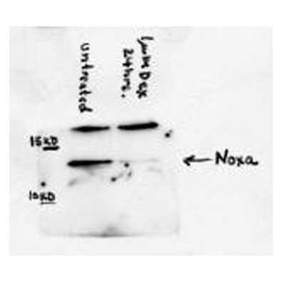
Western Blot: Noxa Antibody (114C307.1) - Non-Recombinant Monoclonal [NB600-1159] - Western blot analysis in RL-7 cells (a follicular lymphoma).![Immunocytochemistry/ Immunofluorescence: Noxa Antibody (114C307.1) - Non-Recombinant Monoclonal [NB600-1159]](https://resources.rndsystems.com/images/products/Noxa-Antibody-114C307-1-Non-Recombinant-Monoclonal-Immunocytochemistry-Immunofluorescence-NB600-1159-img0011.jpg "Immunocytochemistry/ Immunofluorescence: Noxa Antibody (114C307.1) - Non-Recombinant Monoclonal [NB600-1159]")
Immunocytochemistry/ Immunofluorescence: Noxa Antibody (114C307.1) - Non-Recombinant Monoclonal [NB600-1159]
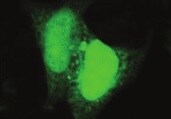
Immunocytochemistry/Immunofluorescence: Noxa Antibody (114C307.1) - Non-Recombinant Monoclonal [NB600-1159] - ICC/IF analysis using the azide-free version of NB600-1159 at 1:10. HeLa cells with DyLight 488 (green). Nuclei and alpha-tubulin were counterstained with DAPI (blue) and DyLight 550 (red). Image objective 40X.![Immunohistochemistry-Paraffin: Noxa Antibody (114C307.1) - Non-Recombinant Monoclonal [NB600-1159]](https://resources.rndsystems.com/images/products/Noxa-Antibody-114C307-1-Non-Recombinant-Monoclonal-Immunohistochemistry-Paraffin-NB600-1159-img0018.jpg "Immunohistochemistry-Paraffin: Noxa Antibody (114C307.1) - Non-Recombinant Monoclonal [NB600-1159]")
Immunohistochemistry-Paraffin: Noxa Antibody (114C307.1) - Non-Recombinant Monoclonal [NB600-1159]
Immunohistochemistry-Paraffin: Noxa Antibody (114C307.1) - Non-Recombinant Monoclonal [NB600-1159] - Bouin-fixed, paraffin-embedded Chronic Lymphocytic Leukemia (CLL) xenograft stained with Noxa antibody (1:2000), peroxidase-conjugate and DAB chromogen. Staining for 2 hr at RT. Image using the azide-free form of this antibody.![Flow Cytometry: Noxa Antibody (114C307.1) - Non-Recombinant Monoclonal [NB600-1159]](https://resources.rndsystems.com/images/products/Noxa-Antibody-114C307-1-Non-Recombinant-Monoclonal-Flow-Cytometry-NB600-1159-img0022.jpg "Flow Cytometry: Noxa Antibody (114C307.1) - Non-Recombinant Monoclonal [NB600-1159]")
Flow Cytometry: Noxa Antibody (114C307.1) - Non-Recombinant Monoclonal [NB600-1159]
Flow Cytometry: Noxa Antibody (114C307.1) - Non-Recombinant Monoclonal [NB600-1159] - Flow cytometry after an intracellular stain was performed on MCF7 cells with Noxa [114C307.1] antibody NB600-1159AF700 (blue) and a matched isotype control (orange). Cells were fixed with 4% PFA and then permeabilized with 0.1% saponin. Cells were incubated in an antibody dilution of 5 ug/mL for 30 minutes at room temperature. Both antibodies were conjugated to Alexa Fluor 700.![Western Blot: Noxa Antibody (114C307.1)Non-Recombinant Monoclonal [NB600-1159]](https://resources.rndsystems.com/images/products/Noxa-Antibody-114C307-1-Non-Recombinant-Monoclonal-Western-Blot-NB600-1159-img0017.jpg "Western Blot: Noxa Antibody (114C307.1)Non-Recombinant Monoclonal [NB600-1159]")
![Flow Cytometry: Noxa Antibody (114C307.1) - Non-Recombinant Monoclonal [NB600-1159]](https://resources.rndsystems.com/images/products/Noxa-Antibody-114C307-1-Non-Recombinant-Monoclonal-Flow-Cytometry-NB600-1159-img0013.jpg "Flow Cytometry: Noxa Antibody (114C307.1) - Non-Recombinant Monoclonal [NB600-1159]")
Flow Cytometry: Noxa Antibody (114C307.1) - Non-Recombinant Monoclonal [NB600-1159]
Flow Cytometry: Noxa Antibody (114C307.1) - Non-Recombinant Monoclonal [NB600-1159] - Flow cytometry after an intracellular stain was performed on HeLa cells with Noxa [114C307.1] antibody NB600-1159AF700 (blue) and a matched isotype control (orange). Cells were fixed with 4% PFA and then permeabilized with 0.1% saponin. Cells were incubated in an antibody dilution of 5 ug/mL for 30 minutes at room temperature. Both antibodies were conjugated to Alexa Fluor 700.![Flow Cytometry: Noxa Antibody (114C307.1) - Non-Recombinant Monoclonal [NB600-1159]](https://resources.rndsystems.com/images/products/Noxa-Antibody-114C307-1-Non-Recombinant-Monoclonal-Flow-Cytometry-NB600-1159-img0015.jpg "Flow Cytometry: Noxa Antibody (114C307.1) - Non-Recombinant Monoclonal [NB600-1159]")
Flow Cytometry: Noxa Antibody (114C307.1) - Non-Recombinant Monoclonal [NB600-1159]
Flow Cytometry: Noxa Antibody (114C307.1) - Non-Recombinant Monoclonal [NB600-1159] - Flow cytometry after an intracellular stain was performed on MCF7 cells with Noxa [114C307.1] antibody NB600-1159AF700 (blue) and a matched isotype control (orange). Cells were fixed with 4% PFA and then permeabilized with 0.1% saponin. Cells were incubated in an antibody dilution of 5 ug/mL for 30 minutes at room temperature. Both antibodies were conjugated to Alexa Fluor 700.![Flow Cytometry: Noxa Antibody (114C307.1) - Non-Recombinant Monoclonal [NB600-1159]](https://resources.rndsystems.com/images/products/Noxa-Antibody-114C307-1-Non-Recombinant-Monoclonal-Flow-Cytometry-NB600-1159-img0016.jpg "Flow Cytometry: Noxa Antibody (114C307.1) - Non-Recombinant Monoclonal [NB600-1159]")
Flow Cytometry: Noxa Antibody (114C307.1) - Non-Recombinant Monoclonal [NB600-1159]
Flow Cytometry: Noxa Antibody (114C307.1) - Non-Recombinant Monoclonal [NB600-1159] - An intracellular stain was performed on Ntera2 cells with Noxa Antibody [114C307.1] NB600-1159 (blue) and a matched isotype control (orange). Cells were fixed with 4% PFA and then permeabilized with 0.1% saponin. Cells were incubated in an antibody dilution of 1.0 ug/mL for 30 minutes at room temperature, followed by Mouse IgG (H+L) Cross-Adsorbed Secondary Antibody, Dylight 550 (35503, Thermo Fisher). - Non-Recombinant Monoclonal [NB600-1159] -")
Western Blot: Noxa Antibody (114C307.1) - Non-Recombinant Monoclonal [NB600-1159] -
Doc counteracts the ABT-737-mediated increase in Mcl-1 protein.Western blot showing that treatment of LNCaP and PC3 cells with 1 µM ABT-737 (A) increases Mcl-1 but treatment with 1 nM Doc (D) decreases Mcl-1. Combination of Doc + ABT-737 (DA) decreases Mcl-1. In LNCaP, there is less Bcl-2 (D, 24 h) and Bak (DA, 48 h) but few differences in Bcl-xL, Bax, Bid, and Noxa. In PC3, there are few differences in Bcl-2, Bcl-xL, Bax, Bak, Bid, and Noxa. - Non-Recombinant Monoclonal [NB600-1159] -")
Western Blot: Noxa Antibody (114C307.1) - Non-Recombinant Monoclonal [NB600-1159] -
Western Blot: Noxa Antibody (114C307.1) - Non-Recombinant Monoclonal [NB600-1159] - EGb761 regulates Bcl-2 family proteins expression in melanoma cells.(A) EGb761 alters the expression levels of anti- & pro-apoptotic Bcl-2 family proteins in melanoma cell lines. Whole cell lysates from Mel-RM & Mel-AT cells treated with EGb761 (400 μg/ml) for indicated time periods were subjected to Western blot analysis. The data shown are representative of three individual experiments. (B) 5% ethanol as control vehicle did not alter the expression levels of Mcl-1. Mel-AT cells with 5% ethanol for increasing periods. Whole cell lysates from Mel-AT cells treated were subjected to Western blot analysis. The data shown are representative of three individual experiments.(C) Mel-RM & Mel-AT cells were treated with EGb761 (400 μg/ml) or 5% ethanol for the indicated periods. Total RNA was isolated & subjected to real-time PCR analysis for Mcl-1. The relative abundance of mRNA expression treated with 5% ethanol was arbitrarily designated as 1. Columns, mean of three individual experiments; bars, SEM. * Present p<0.05 vs control.(D) Relative expression of anti-apoptosis Bcl-2 family proteins in melanoma cell lines Mel-RM & Mel-AT without treatment. Quantitative expression levels of Mcl-1, Bcl-2 & Bcl-XL were normalized to GAPDH.(E) Relative expression of pro-apoptosis Bcl-2 family proteins in melanoma cell lines Mel-RM & Mel-AT without treatment. Quantitative expression levels of Bax, Bid, Noxa, PUMA, Bim, Bad & Bak were normalized to GAPDH. Image collected & cropped by CiteAb from the following publication (https://pubmed.ncbi.nlm.nih.gov/25860257), licensed under a CC-BY license. Not internally tested by Novus Biologicals.Applications for Noxa Antibody (114C307.1) - Non-Recombinant Monoclonal
Application
Recommended Usage
Flow Cytometry
1 ug / million cells
Immunocytochemistry/ Immunofluorescence
1:20-1:1000
Immunohistochemistry
1:10-1:500
Immunohistochemistry-Paraffin
5 ug/ml
Western Blot
1-2 ug/ml
Reviewed Applications
Read 2 reviews rated 4.5 using NB600-1159 in the following applications:
Flow Cytometry Panel Builder
Bio-Techne Knows Flow Cytometry
Save time and reduce costly mistakes by quickly finding compatible reagents using the Panel Builder Tool.
Advanced Features
- Spectra Viewer - Custom analysis of spectra from multiple fluorochromes
- Spillover Popups - Visualize the spectra of individual fluorochromes
- Antigen Density Selector - Match fluorochrome brightness with antigen density

Formulation, Preparation, and Storage
Purification
Protein G purified
Formulation
PBS
Format
Non-Recombinant Monoclonal
Preservative
0.05% Sodium Azide
Concentration
1.0 mg/ml
Shipping
The product is shipped with polar packs. Upon receipt, store it immediately at the temperature recommended below.
Stability & Storage
Store at 4C short term. Aliquot and store at -20C long term. Avoid freeze-thaw cycles.
Background: Noxa
Noxa mediated apoptosis may follow its transcriptional activation by p53 as part of the DNA damage response. However, Noxa expression may be induced by HIF-1 alpha under hypoxia, promoting apoptosis independently of p53 (1). Inhibition of the anti-apoptotic Bcl2 family member Myeloid cell leukemia-1 (Mcl-1) by Noxa, leads to the activation of pro-apoptotic Bcl2 homologous antagonist killer (Bak) and Bcl2 associated X (Bax) proteins, and apoptosis through the intrinsic mitochondrial pathway (2). Other BH3 only proteins such as Bim, Puma, Bad and Bid act via the same mechanism, targeting Mcl-1 and inducing Bak/Bax mediated mitochondrial outer membrane permeabilization (MOMP), cytochrome c release and caspase 9 activation (3). Overexpression of antiapoptotic Bcl2 family members is common in various types of cancer including prostate cancer. A recent study identified antiapoptotic Bcl2 proteins involved in the development of resistance towards the androgen receptor antagonist enzalutamide, which is used for the treatment of metastatic castration-resistant prostate cancer (mCRPC) (3).
References
1.Ploner, C., Kofler, R., & Villunger, A. (2008). Noxa: At the tip of the balance between life and death. Oncogene. https://doi.org/10.1038/onc.2009.46
2.Xiang, W., Yang, C. Y., & Bai, L. (2018). MCL-1 inhibition in cancer treatment. OncoTargets and Therapy. https://doi.org/10.2147/OTT.S146228
3.Pilling, A. B., & Hwang, C. (2019). Targeting prosurvival BCL2 signaling through Akt blockade sensitizes castration-resistant prostate cancer cells to enzalutamide. Prostate. https://doi.org/10.1002/pros.23843
Alternate Names
adult T cell leukemia-derived PMA-responsive, APR, Immediate-early-response protein APR, NOXAPMA-induced protein 1, phorbol-12-myristate-13-acetate-induced protein 1, PMA-Induced Protein 1, Protein Noxa
Gene Symbol
PMAIP1
UniProt
Additional Noxa Products
Product Documents for Noxa Antibody (114C307.1) - Non-Recombinant Monoclonal
Certificate of Analysis
To download a Certificate of Analysis, please enter a lot or batch number in the search box below.
Product Specific Notices for Noxa Antibody (114C307.1) - Non-Recombinant Monoclonal
This product is for research use only and is not approved for use in humans or in clinical diagnosis. Primary Antibodies are guaranteed for 1 year from date of receipt.
Citations for Noxa Antibody (114C307.1) - Non-Recombinant Monoclonal
 Powered by Bioz
Customer Reviews for Noxa Antibody (114C307.1) - Non-Recombinant Monoclonal (2)
4.5 out of 5
2 Customer Ratings
Have you used Noxa Antibody (114C307.1) - Non-Recombinant Monoclonal?
Submit a review and receive an Amazon gift card!
$25/€18/£15/$25CAN/¥2500 Yen for a review with an image
$10/€7/£6/$10CAN/¥1110 Yen for a review without an image
Submit a review
Customer Images
Showing
1
-
2 of
2 reviews
Showing All
Filter By:
-
Application: ImmunocytochemistrySample Tested: HeLa cellsSpecies: HumanVerified Customer | Posted 09/10/2021HeLa cells

-
Application: Western BlotSample Tested: WEHI 7.2 mouse T cell lymphoma cell lysate, Sample Amount: 100ugSpecies: MouseVerified Customer | Posted 10/04/2011
There are no reviews that match your criteria.
Protocols
Find general support by application which include: protocols, troubleshooting, illustrated assays, videos and webinars.
- 7-Amino Actinomycin D (7-AAD) Cell Viability Flow Cytometry Protocol
- Antigen Retrieval Protocol (PIER)
- Antigen Retrieval for Frozen Sections Protocol
- Appropriate Fixation of IHC/ICC Samples
- Cellular Response to Hypoxia Protocols
- Chromogenic IHC Staining of Formalin-Fixed Paraffin-Embedded (FFPE) Tissue Protocol
- Chromogenic Immunohistochemistry Staining of Frozen Tissue
- ClariTSA™ Fluorophore Kits
- Detection & Visualization of Antibody Binding
- Extracellular Membrane Flow Cytometry Protocol
- Flow Cytometry Protocol for Cell Surface Markers
- Flow Cytometry Protocol for Staining Membrane Associated Proteins
- Flow Cytometry Staining Protocols
- Flow Cytometry Troubleshooting Guide
- Fluorescent IHC Staining of Frozen Tissue Protocol
- Graphic Protocol for Heat-induced Epitope Retrieval
- Graphic Protocol for the Preparation and Fluorescent IHC Staining of Frozen Tissue Sections
- Graphic Protocol for the Preparation and Fluorescent IHC Staining of Paraffin-embedded Tissue Sections
- Graphic Protocol for the Preparation of Gelatin-coated Slides for Histological Tissue Sections
- ICC Cell Smear Protocol for Suspension Cells
- ICC Immunocytochemistry Protocol Videos
- ICC for Adherent Cells
- IHC Sample Preparation (Frozen sections vs Paraffin)
- Immunocytochemistry (ICC) Protocol
- Immunocytochemistry Troubleshooting
- Immunofluorescence of Organoids Embedded in Cultrex Basement Membrane Extract
- Immunofluorescent IHC Staining of Formalin-Fixed Paraffin-Embedded (FFPE) Tissue Protocol
- Immunohistochemistry (IHC) and Immunocytochemistry (ICC) Protocols
- Immunohistochemistry Frozen Troubleshooting
- Immunohistochemistry Paraffin Troubleshooting
- Intracellular Flow Cytometry Protocol Using Alcohol (Methanol)
- Intracellular Flow Cytometry Protocol Using Detergents
- Intracellular Nuclear Staining Flow Cytometry Protocol Using Detergents
- Intracellular Staining Flow Cytometry Protocol Using Alcohol Permeabilization
- Intracellular Staining Flow Cytometry Protocol Using Detergents to Permeabilize Cells
- Preparing Samples for IHC/ICC Experiments
- Preventing Non-Specific Staining (Non-Specific Binding)
- Primary Antibody Selection & Optimization
- Propidium Iodide Cell Viability Flow Cytometry Protocol
- Protocol for Heat-Induced Epitope Retrieval (HIER)
- Protocol for Liperfluo
- Protocol for Making a 4% Formaldehyde Solution in PBS
- Protocol for VisUCyte™ HRP Polymer Detection Reagent
- Protocol for the Characterization of Human Th22 Cells
- Protocol for the Characterization of Human Th9 Cells
- Protocol for the Fluorescent ICC Staining of Cell Smears - Graphic
- Protocol for the Fluorescent ICC Staining of Cultured Cells on Coverslips - Graphic
- Protocol for the Preparation & Fixation of Cells on Coverslips
- Protocol for the Preparation and Chromogenic IHC Staining of Frozen Tissue Sections
- Protocol for the Preparation and Chromogenic IHC Staining of Frozen Tissue Sections - Graphic
- Protocol for the Preparation and Chromogenic IHC Staining of Paraffin-embedded Tissue Sections
- Protocol for the Preparation and Chromogenic IHC Staining of Paraffin-embedded Tissue Sections - Graphic
- Protocol for the Preparation and Fluorescent ICC Staining of Cells on Coverslips
- Protocol for the Preparation and Fluorescent ICC Staining of Non-adherent Cells
- Protocol for the Preparation and Fluorescent ICC Staining of Stem Cells on Coverslips
- Protocol for the Preparation and Fluorescent IHC Staining of Frozen Tissue Sections
- Protocol for the Preparation and Fluorescent IHC Staining of Paraffin-embedded Tissue Sections
- Protocol for the Preparation of Gelatin-coated Slides for Histological Tissue Sections
- Protocol for the Preparation of a Cell Smear for Non-adherent Cell ICC - Graphic
- Protocol: Annexin V and PI Staining by Flow Cytometry
- Protocol: Annexin V and PI Staining for Apoptosis by Flow Cytometry
- R&D Systems Quality Control Western Blot Protocol
- TUNEL and Active Caspase-3 Detection by IHC/ICC Protocol
- The Importance of IHC/ICC Controls
- Troubleshooting Guide: Fluorokine Flow Cytometry Kits
- Troubleshooting Guide: Immunohistochemistry
- Troubleshooting Guide: Western Blot Figures
- Western Blot Conditions
- Western Blot Protocol
- Western Blot Protocol for Cell Lysates
- Western Blot Troubleshooting
- Western Blot Troubleshooting Guide
- View all Protocols, Troubleshooting, Illustrated assays and Webinars
Loading...