Nrf2 Antibody - BSA Free
Novus Biologicals | Catalog # NBP1-32822

Key Product Details
Validated by
Species Reactivity
Validated:
Cited:
Predicted:
Applications
Validated:
Cited:
Label
Antibody Source
Format
Product Specifications
Immunogen
Localization
Clonality
Host
Isotype
Theoretical MW
Disclaimer note: The observed molecular weight of the protein may vary from the listed predicted molecular weight due to post translational modifications, post translation cleavages, relative charges, and other experimental factors.
Scientific Data Images for Nrf2 Antibody - BSA Free
Western Blot Analysis of Nrf2 in SH-SY5Y Cells
Nrf2-Antibody-Western-Blot-NBP1-32822-img0046.jpg
Chromatin Immunoprecipitation Performed Using Anti-Nrf2 Antibody
ChIP was performed with HepG2 chromatin extract and 5 ug of either normal rabbit IgG or anti-NRF2 antibody. The precipitated DNA was detected by PCR with primer set targeting to GCLC gene locus.
Western Blot Detection of Nrf2 in NRF2-Transfected and Non-Transfected 293T Whole Cell Extracts
Non-transfected (-) and NRF2-transfected (+, including 3xFlag-tag) 293T whole cell extracts (30ug) were separated by 5% SDS-PAGE, and the membrane was blotted with NRF2 antibody diluted by 1:1000.
Western Blot Analysis of Nrf2 in Treated and Untreated RAW264.7 Whole Cell Extracts
Untreated (-) and treated (+) RAW264.7 whole cell extracts (30 ug) were separated by 5% SDS-PAGE, and the membrane was blotted with NRF2 antibody [N2C2], Internal diluted at 1:500. The HRP-conjugated anti-rabbit IgG antibody (NBP2-19301) was used to detect the primary antibody.
Western Blotting of Nrf2 in Treated and Untreated Neuro2A Whole Cell Extracts
Untreated (-) and treated (+) Neuro2A whole cell extracts (30 ug) were separated by 5% SDS-PAGE, and the membrane was blotted with NRF2 antibody [N2C2], Internal (NBP1-32822) diluted at 1:500. The HRP-conjugated anti-rabbit IgG antibody (NBP2-19301) was used to detect the primary antibody.
Western Blotting of Nrf2 in Treated and Untreated HepG2 hole Cell Extracts
Untreated (-) and treated (+) HepG2 whole cell extracts (30 ug) were separated by 5% SDS-PAGE, and the membrane was blotted with NRF2 antibody [N2C2], Internal (NBP1-32822) diluted at 1:500. The HRP-conjugated anti-rabbit IgG antibody (NBP2-19301) was used to detect the primary antibody.


Immunocytochemistry/Immunofluorescence Analysis of Nrf2 in NIH/3T3 Cells
NIH/3T3 cells were fixed in 4% paraformaldehyde at RT for 15 min. Green: NRF2 protein stained by NRF2 antibody [N2C2], Internal diluted at 1:500. Blue: Hoechst 33342 staining. Scale bar = 10 um.
Immunoprecipitation of Nrf2 from HepG2 Whole Cell Extracts Using Nrf2 Antibody
Immunoprecipitation of NRF2 protein from HepG2 whole cell extracts using 5 ug of NRF2 antibody [N2C2], Internal Western blot analysis was performed using NRF2 antibody [N2C2], Internal.. EasyBlot anti-Rabbit IgG was used as a secondary reagent.
Western Blot: Nrf2 Antibody [NBP1-32822] -
Western Blot: Nrf2 Antibody [NBP1-32822] - Blueberry polyphenol extract (BPE) increases the expression of NRF2 & HO-1 while reducing NF-kappa B p65 phosphorylation in angiotensin (Ang) II-treated human aortic endothelial cells (HAECs). HAECs were treated with 200 µg/mL of BPE for 1 h then treated with 200 nM of Ang II for 12 h. Protein expression of NRF2 (A,B), HO-1 (C,D), & NF-kappa B p65 (E,F) were determined by Western blot. Quantification was performed using Image Lab (Bio-Rad Laboratories, Inc.). Data are expressed as mean ± SD from nine (HO-1), & three (NRF2 & NF-kappa B) independent experiments. Values that do not share the same letter are significantly different from each other (p ≤ 0.05). Image collected & cropped by CiteAb from the following publication (https://pubmed.ncbi.nlm.nih.gov/35453301), licensed under a CC-BY license. Not internally tested by Novus Biologicals.
Western Blot: Nrf2 Antibody [NBP1-32822] -
Western Blot: Nrf2 Antibody [NBP1-32822] - NRF2 antibody validation. (A) NRF2 expression was silenced in MCF7 cells by transiently transfecting NRF2-specific siRNAs or a negative control siRNA for 48 h, then NRF2 protein expression was determined using two different antibodies (Abcam: ab31163; NOVUS: NBP1-32822). (B–D) 4T1 cells were treated with NRF2 activators, RA839 or tBHQ, or MG-132, a proteasome inhibitor, in the concentrations indicated for 48 h, then NRF2 protein expression was determined by western blotting using two different antibodies (Abcam: ab31163; NOVUS: NBP1-32822). Image collected & cropped by CiteAb from the following publication (https://pubmed.ncbi.nlm.nih.gov/31461945), licensed under a CC-BY license. Not internally tested by Novus Biologicals.
Western Blot: Nrf2 Antibody [NBP1-32822] -
Western Blot: Nrf2 Antibody [NBP1-32822] - Maternal exercise during pregnancy on mitochondrial biogenesis in the fetal hearts. (A) Levels of relative mRNA expression measured by qRT‐PCR. n = 9–12/group. Maternal exercise during pregnancy did not alter levels of mRNA in Ppargc1a & Tfam, while it significantly upregulated the levels of mRNA in Nrf1 & Nrf2. (B–D) Densitometric analyses of protein expression levels relative to the sedentary group with representative images of western blots were shown. No significant differences in PGC‐1 alpha, NRF1, & NRF2 (P > 0.05). n = 5–6/group. * P < 0.05, significantly different from the sedentary group. Black bar: fetal hearts from sedentary dams; gray bar: fetal hearts from exercised dams. Image collected & cropped by CiteAb from the following publication (https://pubmed.ncbi.nlm.nih.gov/28292876), licensed under a CC-BY license. Not internally tested by Novus Biologicals.
Western Blot: Nrf2 Antibody [NBP1-32822] -
Western Blot: Nrf2 Antibody [NBP1-32822] - Nrf2 is required for UroA/UAS03 mediated upregulation of tight junction proteins. a Nrf2 levels were determined by immunoblots in HT29 cells treated with vehicle/UroA/UAS03 (50 μM) for 24 h. b Nrf2 expression in cytosolic & nuclear fractions of HT29 cells treated with Veh/UroA/UAS03 (50 μM) for 6 h. c Immunofluorescence confocal images of HT29 cells treated with vehicle/UroA/UAS03 (50 μM) for 6 h. The cells were stained with anti-Nrf2 antibody & DAPI. Relative green fluorescence (n = ~20 cells) intensity was measured. Scale bars indicate 25 μm. d Expression of Cldn4 & NQO1 in colon explants from WT, Nrf2−/−, & AhR−/− mice treated with vehicle/UroA/UAS03 (50 μM) for 24 h. Immunoblots were quantified using Image J software. e mRNA levels of Cldn4, Nrf2, & HO1 from colon explant cultures was measured by real-time PCR using SyBr green method. f C57BL/6, Nrf2−/−, & AhR−/− mice (n = 3) treated orally daily with veh or UroA/UAS03 (20 mg/kg) for 1 week. Cldn4 & NQO1 protein levels in colons were measured by immunoblots & quantified by Image J software. All in vitro studies were performed in triplicates. The immunoblots of colon explants & colon tissues were quantified from at least 6 independent runs. The levels of proteins were normalized to beta -actin & Wild type vehicle treatment was set to 1 & calculated the fold changes. Statistics performed using unpaired t-test using Graphpad Prism software. Error bars, ±SEM; *p < 0.05; **p < 0.01; ***p < 0.001. Source Data are provided as a Source Data File Image collected & cropped by CiteAb from the following publication (https://pubmed.ncbi.nlm.nih.gov/30626868), licensed under a CC-BY license. Not internally tested by Novus Biologicals.
Western Blot: Nrf2 Antibody [NBP1-32822] -
Western Blot: Nrf2 Antibody [NBP1-32822] - Modulation of Nrf2 & Keap1 mRNA & protein levels by compounds 1–6, curcumin (CURC), & dimethyl fumarate (DMF). (A–B) RNA from total cellular extracts of SH-SY5Y cells treated for 24 hours with 5 μM compounds or 20 μM DMF were analyzed for Nrf2 (A) & Keap1 (B) mRNA expression by RT-qPCR. GAPDH was used as housekeeping gene. Results are shown as mean ± SEM; no statistically significant data with Dunnett’s multiple comparison test (A, n = 3, F ratio = 1.249; B, n = 3, F ratio = 1.671). (C–D) Cellular extracts of SH-SY5Y cells treated for 24 hours with compounds at 5 μM or 20 μM DMF were analyzed for Nrf2 (C) & Keap1 (D) protein levels by Western blot. Anti-tubulin was used as protein loading control. Results are shown as ratio (% of CTR) ± SEM; **p < 0.01, versus CTR; Dunnett’s multiple comparison test (C, n ≥ 5, F ratio = 3.981; D, n = 3, F ratio = 0.4049). Image collected & cropped by CiteAb from the following publication (https://pubmed.ncbi.nlm.nih.gov/32047434), licensed under a CC-BY license. Not internally tested by Novus Biologicals.
Western Blot: Nrf2 Antibody [NBP1-32822] -
Western Blot: Nrf2 Antibody [NBP1-32822] - CT activated AMPK/SIRT1 signaling. (A,B) HepG2 cells were treated with 2.5 μM CT for indicated times. Western blot analysis of phosphorylated AMPK, ACC, SIRT1, & Nrf2. (C) C57BL/6 mice were pair-fed either control or ethanol-containing diet with or without CT (20 or 40 mg/kg) for four weeks. Western blot analysis of phosphorylated AMPK, SIRT1. CYP2E1, & Nrf2. (D) HepG2 cells were incubated with 50 mM ethanol & treated with CT (2.5 or 5 μM) for 24 h. Western blot analysis of phosphorylated AMPK, SIRT1, CYP2E1, & Nrf2. (E) AML-12 cells were incubated with 50 mM ethanol & treated with CT (2.5 or 5 μM) for 24 h. Western blot analysis of phosphorylated AMPK & SIRT1. The images are representative (F) HepG2 cells were pretreated with CT (2.5 μM) for 3 h or with compound C (comp C) (10 μM) for 6 h, followed by ethanol (100 μM) treatment. Measurement of intracellular TG levels. Data are shown as mean ± SD of three independent experiments. #p < 0.05 vs. untreated control, ** p < 0.01 vs. ethanol-treated group. §§p < 0.01 vs. ethanol & CT-treated group. Densitometric analysis of western blots are given in Supplementary Figures S2 & S3A–G. Image collected & cropped by CiteAb from the following publication (https://pubmed.ncbi.nlm.nih.gov/31906014), licensed under a CC-BY license. Not internally tested by Novus Biologicals.
Western Blot: Nrf2 Antibody [NBP1-32822] -
Western Blot: Nrf2 Antibody [NBP1-32822] - MPE & MSE exert anti-oxidant effects in 3T3-L1 adipocytes. 3T3-L1 cells were treated with pro-differentiative agents for 8 days in the presence or absence of 100 µg/mL MPE or MSE, as reported in Methods. (A) Intracellular ROS were detected using the redox-sensitive fluorochrome H2-DCFDA. After differentiation, the medium was replaced with 10 µM H2DCFDA solution & the incubation was protracted for 30 min at 37 °C. The oxidation of the fluorochrome generates green fluorescence, which was visualized by a Leica microscope equipped with a DC300F camera using a FITC filter. Representative micrographs of fluorescence microscopy were taken at 200× magnification. (B). Western blotting analysis of Nrf2, MnSOD & HO-1 in 3T3-L1 cells differentiated without or with 100 µg/mL MPE or MSE. Equal loading of proteins was verified by immunoblotting for beta -actin & showed values were assigned in relation to undifferentiated cells (Undif.). The bar graphs represent the mean of three independent experiments ± SD. * p < 0.05, ** p < 0.01 with respect to the undifferentiated 3T3-L1 cells, # p < 0.05, ## p < 0.01 & ### p < 0.001 with respect to the differentiated untreated 3T3-L1 cells (Dif.). Image collected & cropped by CiteAb from the following publication (https://pubmed.ncbi.nlm.nih.gov/35204243), licensed under a CC-BY license. Not internally tested by Novus Biologicals.
Western Blot: Nrf2 Antibody [NBP1-32822] -
Western Blot: Nrf2 Antibody [NBP1-32822] - NRF2 modulates epirubicin resistance in breast cancer cells. (A) MCF-7 & MCF-7 EpiR cells were treated for 24 h with epirubicin at 1 µM. MCF-7 & MCF-7 EpiR cells were stained with CellROX Deep Red reagent & analyzed for ROS levels by flow cytometry. Data were analyzed by FlowJo software. The mean fluorescence values were presented as relative ROS level compared to untreated cells (0 µM). Data presented as mean ± SD. Student’s t-test was used to compare the means: ** p < 0.01; *** p < 0.001; n.s. nonsignificant. (N = 4) (B) Knockdown of NRF2 was achieved by transfecting 4 specific siRNA against NRF2 (siNRF2, 150pmol) to MCF-7 EpiR cells in a 6-well plate. Non-targeting siRNAs were used as control (NSC). At 24 h post-transfection, these cells were seeded in 6-well plates & treated with increasing doses of epirubicin for 14 days. Their sensitivity to epirubicin was assessed by clonogenic assay. Their clonogenicity in response to epirubicin was analyzed by two-way ANOVA & found to be significantly different (** p < 0.01) from one another. (N = 3) (C) Expression of NRF2 in MCF-7 cells & MCF-7 EpiR cells was detected by Western blot. beta -Tubulin served as the loading control. (N = 3). Image collected & cropped by CiteAb from the following publication (https://pubmed.ncbi.nlm.nih.gov/32110852), licensed under a CC-BY license. Not internally tested by Novus Biologicals.
Immunocytochemistry/ Immunofluorescence: Nrf2 Antibody [NBP1-32822] -
Immunocytochemistry/ Immunofluorescence: Nrf2 Antibody [NBP1-32822] - Nrf2 antibody [N2C2], Internal detects Nrf2 protein at nucleus by immunofluorescent analysis.Sample: Neuro2A cells were fixed in 4% paraformaldehyde at RT for 15 min.
Green: Nrf2 stained by Nrf2 antibody [N2C2], Internal (NBP1-32822) diluted at 1:1000.

Immunohistochemistry-Paraffin: Nrf2 Antibody [NBP1-32822] -
Immunohistochemistry-Paraffin: Nrf2 Antibody [NBP1-32822] - Nrf2 antibody [N2C2], Internal detects Nrf2 protein at cytoplasm and nucleus by immunohistochemical analysis.Sample: Paraffin-embedded human breast carcinoma.
Nrf2 stained by Nrf2 antibody [N2C2], Internal (NBP1-32822) diluted at 1:500.
Antigen Retrieval: Citrate buffer, pH 6.0, 15 min

Immunocytochemistry/ Immunofluorescence: Nrf2 Antibody [NBP1-32822] -
Immunocytochemistry/ Immunofluorescence: Nrf2 Antibody [NBP1-32822] - Nrf2 antibody [N2C2], Internal detects Nrf2 protein at nucleus by immunofluorescent analysis.Sample: HeLa cells were fixed in 4% paraformaldehyde at RT for 15 min.
Green: Nrf2 stained by Nrf2 antibody [N2C2], Internal (NBP1-32822) diluted at 1:1000.
Red: phalloidin, a cytoskeleton marker, diluted at 1:200.
Scale bar= 10 um.

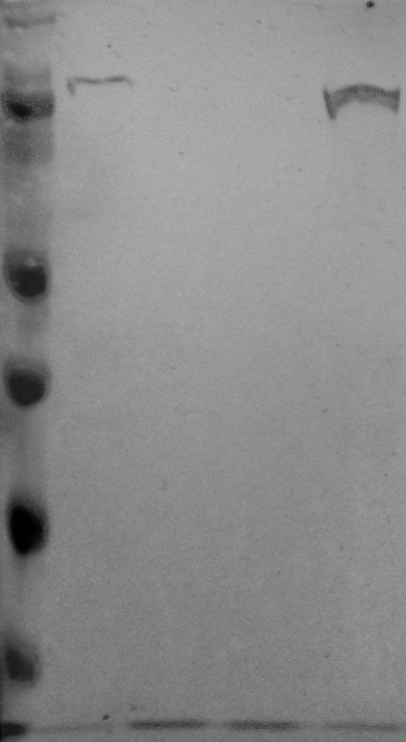
Western Blot: Nrf2 Antibody [NBP1-32822] -
Untreated (-) and treated (+) HepG2 whole cell extracts (30 ug) were separated by 5% SDS-PAGE, and the membrane was blotted with Nrf2 antibody [N2C2], Internal (NBP1-32822) diluted at 1:2000. The HRP-conjugated anti-rabbit IgG antibody was used to detect the primary antibody.
Immunocytochemistry/ Immunofluorescence: Nrf2 Antibody [NBP1-32822] -
NRF2 antibody [N2C2], Internal detects NRF2 protein at nucleus by immunofluorescent analysis.Sample: Mock and treated HeLa cells were fixed in 4% paraformaldehyde at RT for 15 min.
Green: NRF2 stained by NRF2 antibody [N2C2], Internal (NBP1-32822) diluted at 1:500.
Blue: Fluoroshield with DAPI.

Western Blot: Nrf2 Antibody [NBP1-32822] -
Untreated (-) and treated (+) HepG2 whole cell extracts (30 ug) were separated by 5% SDS-PAGE, and the membranes were blotted with NRF2 antibody [N2C2], Internal (NBP1-32822) diluted at 1:500 and competitor's antibody diluted at 1:500. The HRP-conjugated anti-rabbit IgG antibody was used to detect the primary antibody.*The competitor is not affiliated with Novus and does not endorse this product.
Applications for Nrf2 Antibody - BSA Free
Chromatin Immunoprecipitation (ChIP)
Flow Cytometry
Immunocytochemistry/ Immunofluorescence
Immunohistochemistry
Immunohistochemistry-Paraffin
Immunoprecipitation
Western Blot
Reviewed Applications
Read 6 reviews rated 4 using NBP1-32822 in the following applications:
Flow Cytometry Panel Builder
Bio-Techne Knows Flow Cytometry
Save time and reduce costly mistakes by quickly finding compatible reagents using the Panel Builder Tool.
Advanced Features
- Spectra Viewer - Custom analysis of spectra from multiple fluorochromes
- Spillover Popups - Visualize the spectra of individual fluorochromes
- Antigen Density Selector - Match fluorochrome brightness with antigen density

Formulation, Preparation, and Storage
Purification
Formulation
Format
Preservative
Concentration
Shipping
Stability & Storage
Background: Nrf2
Long Name
Alternate Names
Gene Symbol
UniProt
Additional Nrf2 Products
Product Documents for Nrf2 Antibody - BSA Free
Certificate of Analysis
To download a Certificate of Analysis, please enter a lot or batch number in the search box below.
Product Specific Notices for Nrf2 Antibody - BSA Free
This product is for research use only and is not approved for use in humans or in clinical diagnosis. Primary Antibodies are guaranteed for 1 year from date of receipt.
Citations for Nrf2 Antibody - BSA Free
 Powered by Bioz
Customer Reviews for Nrf2 Antibody - BSA Free (6)
Have you used Nrf2 Antibody - BSA Free?
Submit a review and receive an Amazon gift card!
$25/€18/£15/$25CAN/¥2500 Yen for a review with an image
$10/€7/£6/$10CAN/¥1110 Yen for a review without an image
Submit a review
Customer Images
-
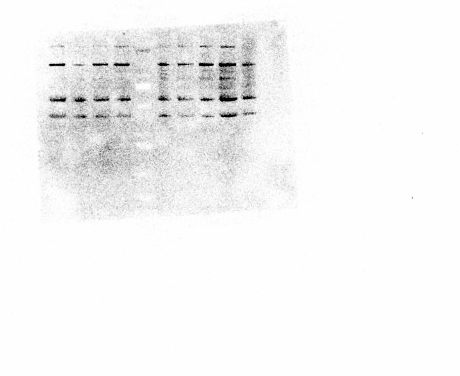
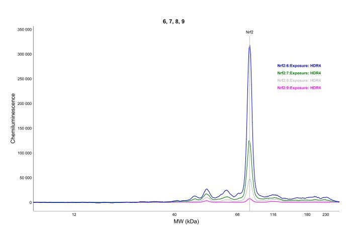
Application: Western BlotSample Tested: Bone Marrrow Derived MacrophagesSpecies: MouseVerified Customer | Posted 04/03/2024NRF2 Western blot BMDM

-
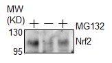
Application: Western BlotSample Tested: Rat CardiomyocyteSpecies: RatVerified Customer | Posted 01/26/2023MG132, a proteasome inhibitor, induces Nrf2 in rat cardiomyocytes
-
Application: Simple WesternSample Tested: skin woundSpecies: rat skin wound and RatVerified Customer | Posted 02/07/2020
-
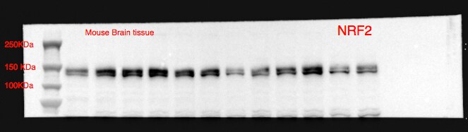
Application: Western BlotSample Tested: Brain (substantia nigra)Species: MouseVerified Customer | Posted 10/05/2017
-
Application: Western BlotSample Tested: BxPC3, GH3 and SH-SY5Y whole cell lysatesSpecies: HumanVerified Customer | Posted 04/28/2017
-
Application: Western BlotSample Tested: HepG2 (liver) cell lysateSpecies: HumanVerified Customer | Posted 03/01/2017Marker, HepG2 (endogenous), blank, blank, HepG2 (over-expressed) (30ug protein per well). Band observed is about 90kDa.Primary rabbit anti NRF2 antibody diluted 1:500 in TBS. Membrane incubation for 2 hours at RT. Secondary IRDye 800CW goat anti rabbit antibody diluted 1:10000 in TBS. Membrane incubation for 1 hour at RT. Detection by Li-Cor Odyssey
There are no reviews that match your criteria.
Protocols
Find general support by application which include: protocols, troubleshooting, illustrated assays, videos and webinars.
- 7-Amino Actinomycin D (7-AAD) Cell Viability Flow Cytometry Protocol
- Antigen Retrieval Protocol (PIER)
- Antigen Retrieval for Frozen Sections Protocol
- Appropriate Fixation of IHC/ICC Samples
- Cellular Response to Hypoxia Protocols
- ChIP Protocol Video
- Chromatin Immunoprecipitation (ChIP) Protocol
- Chromatin Immunoprecipitation Protocol
- Chromogenic IHC Staining of Formalin-Fixed Paraffin-Embedded (FFPE) Tissue Protocol
- Chromogenic Immunohistochemistry Staining of Frozen Tissue
- ClariTSA™ Fluorophore Kits
- Detection & Visualization of Antibody Binding
- Extracellular Membrane Flow Cytometry Protocol
- Flow Cytometry Protocol for Cell Surface Markers
- Flow Cytometry Protocol for Staining Membrane Associated Proteins
- Flow Cytometry Staining Protocols
- Flow Cytometry Troubleshooting Guide
- Fluorescent IHC Staining of Frozen Tissue Protocol
- Graphic Protocol for Heat-induced Epitope Retrieval
- Graphic Protocol for the Preparation and Fluorescent IHC Staining of Frozen Tissue Sections
- Graphic Protocol for the Preparation and Fluorescent IHC Staining of Paraffin-embedded Tissue Sections
- Graphic Protocol for the Preparation of Gelatin-coated Slides for Histological Tissue Sections
- ICC Cell Smear Protocol for Suspension Cells
- ICC Immunocytochemistry Protocol Videos
- ICC for Adherent Cells
- IHC Sample Preparation (Frozen sections vs Paraffin)
- Immunocytochemistry (ICC) Protocol
- Immunocytochemistry Troubleshooting
- Immunofluorescence of Organoids Embedded in Cultrex Basement Membrane Extract
- Immunofluorescent IHC Staining of Formalin-Fixed Paraffin-Embedded (FFPE) Tissue Protocol
- Immunohistochemistry (IHC) and Immunocytochemistry (ICC) Protocols
- Immunohistochemistry Frozen Troubleshooting
- Immunohistochemistry Paraffin Troubleshooting
- Immunoprecipitation Protocol
- Intracellular Flow Cytometry Protocol Using Alcohol (Methanol)
- Intracellular Flow Cytometry Protocol Using Detergents
- Intracellular Nuclear Staining Flow Cytometry Protocol Using Detergents
- Intracellular Staining Flow Cytometry Protocol Using Alcohol Permeabilization
- Intracellular Staining Flow Cytometry Protocol Using Detergents to Permeabilize Cells
- Preparing Samples for IHC/ICC Experiments
- Preventing Non-Specific Staining (Non-Specific Binding)
- Primary Antibody Selection & Optimization
- Propidium Iodide Cell Viability Flow Cytometry Protocol
- Protocol for Heat-Induced Epitope Retrieval (HIER)
- Protocol for Liperfluo
- Protocol for Making a 4% Formaldehyde Solution in PBS
- Protocol for VisUCyte™ HRP Polymer Detection Reagent
- Protocol for the Characterization of Human Th22 Cells
- Protocol for the Characterization of Human Th9 Cells
- Protocol for the Fluorescent ICC Staining of Cell Smears - Graphic
- Protocol for the Fluorescent ICC Staining of Cultured Cells on Coverslips - Graphic
- Protocol for the Preparation & Fixation of Cells on Coverslips
- Protocol for the Preparation and Chromogenic IHC Staining of Frozen Tissue Sections
- Protocol for the Preparation and Chromogenic IHC Staining of Frozen Tissue Sections - Graphic
- Protocol for the Preparation and Chromogenic IHC Staining of Paraffin-embedded Tissue Sections
- Protocol for the Preparation and Chromogenic IHC Staining of Paraffin-embedded Tissue Sections - Graphic
- Protocol for the Preparation and Fluorescent ICC Staining of Cells on Coverslips
- Protocol for the Preparation and Fluorescent ICC Staining of Non-adherent Cells
- Protocol for the Preparation and Fluorescent ICC Staining of Stem Cells on Coverslips
- Protocol for the Preparation and Fluorescent IHC Staining of Frozen Tissue Sections
- Protocol for the Preparation and Fluorescent IHC Staining of Paraffin-embedded Tissue Sections
- Protocol for the Preparation of Gelatin-coated Slides for Histological Tissue Sections
- Protocol for the Preparation of a Cell Smear for Non-adherent Cell ICC - Graphic
- Protocol: Annexin V and PI Staining by Flow Cytometry
- Protocol: Annexin V and PI Staining for Apoptosis by Flow Cytometry
- R&D Systems Quality Control Western Blot Protocol
- TUNEL and Active Caspase-3 Detection by IHC/ICC Protocol
- The Importance of IHC/ICC Controls
- Troubleshooting Guide: Fluorokine Flow Cytometry Kits
- Troubleshooting Guide: Immunohistochemistry
- Troubleshooting Guide: Western Blot Figures
- Western Blot Conditions
- Western Blot Protocol
- Western Blot Protocol for Cell Lysates
- Western Blot Troubleshooting
- Western Blot Troubleshooting Guide
- View all Protocols, Troubleshooting, Illustrated assays and Webinars
FAQs for Nrf2 Antibody - BSA Free
-
Q: Would you please check if thimerosal is contained or not contained in the current lot of this item?
A: I can confirm that our Nrf2 antibody with catalogue number NBP1-32822 is supplied in Phosphate Buffered Saline (PBS) with 20% glycerol and 0.025% Proclin. The change to the preservative was made at the beginning of April 2015.
Associated Pathways
