Nucleoli Marker Antibody (NM95) [Biotin]
Novus Biologicals | Catalog # NBP2-34695B
Key Product Details
Species Reactivity
Human, Bovine (Negative), Mouse (Negative), Rat (Negative)
Applications
Immunohistochemistry-Paraffin, Flow Cytometry, Immunocytochemistry/ Immunofluorescence
Label
Biotin
Antibody Source
Monoclonal Mouse IgG1 kappa Clone # NM95
Loading...
Product Specifications
Immunogen
Nuclei of myeloid leukemia biopsy cells
Reactivity Notes
Does not react with Mouse, Rat or Bovine.
Localization
Nucleoli
Marker
Marker For Human Cells
Specificity
This monoclonal antibody is an excellent marker for human cells in xenographic model research. It reacts specifically with human cells. This monoclonal antibody is part of a new panel of reagents, which recognizes subcellular organelles or compartments of human cells. These markers may be useful in identification of these organelles in cells, tissues, and biochemical preparations. monoclonal antibody NM95 recognizes an antigen associated with the nucleoli in human cells. It can be used to stain the nucleoli in cell or tissue preparations and can be used as a marker of the nucleoli in subcellular fractions. It produces a speckled pattern in the nuclei of cells of normal and malignant cells and may be used to stain the nucleoli of cells in fixed or frozen tissue sections. It can be used with paraformaldehyde fixed frozen tissue or cell preparations and formalin fixed, paraffin-embedded tissue sections.
Clonality
Monoclonal
Host
Mouse
Isotype
IgG1 kappa
Description
This conjugate is made on demand. Actual recovery may vary from the stated volume of this product. The volume will be greater than or equal to the unit size stated on the datasheet.
Scientific Data Images for Nucleoli Marker Antibody (NM95) [Biotin]
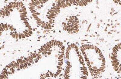
Immunohistochemistry-Paraffin: Nucleoli Marker Antibody (NM95) [Biotin] [NBP2-34695B] - Human ovarian cancer cell line, staining of nucleoli. Antibody at 1:150. Image from verified customer review.
![Nucleoli Marker Antibody (NM95) [Biotin]](https://resources.rndsystems.com/images/products/nbp2-34695b_mouse-monoclonal-nucleoli-marker-antibody-nm95-biotin-immunohistochemistry-paraffin-213202411392..jpg "Immunohistochemistry-Paraffin: Mouse Monoclonal Nucleoli Marker Antibody (NM95) [Biotin] [NBP2-34695B] -")
Immunohistochemistry-Paraffin: Mouse Monoclonal Nucleoli Marker Antibody (NM95) [Biotin] [NBP2-34695B] -
Immunohistochemistry-Paraffin: Mouse Monoclonal Nucleoli Marker Antibody (NM95) [Biotin] [NBP2-34695B] - Nucleoli Marker Antibody (NM95) immunoreactivity in adipose surrounding mouse kidney. The antibody was diluted 1 to 100 and left on tissue sections for 16h at 4 degrees Celsius. Image from a verified customer review.Applications for Nucleoli Marker Antibody (NM95) [Biotin]
Application
Recommended Usage
Flow Cytometry
Optimal dilutions of this antibody should be experimentally determined.
Immunocytochemistry/ Immunofluorescence
Optimal dilutions of this antibody should be experimentally determined.
Immunohistochemistry-Paraffin
Optimal dilutions of this antibody should be experimentally determined.
Reviewed Applications
Read 2 reviews rated 5 using NBP2-34695B in the following applications:
Flow Cytometry Panel Builder
Bio-Techne Knows Flow Cytometry
Save time and reduce costly mistakes by quickly finding compatible reagents using the Panel Builder Tool.
Advanced Features
- Spectra Viewer - Custom analysis of spectra from multiple fluorochromes
- Spillover Popups - Visualize the spectra of individual fluorochromes
- Antigen Density Selector - Match fluorochrome brightness with antigen density

Formulation, Preparation, and Storage
Purification
Protein A or G purified
Formulation
PBS
Preservative
0.05% Sodium Azide
Concentration
Please see the vial label for concentration. If unlisted please contact technical services.
Shipping
The product is shipped with polar packs. Upon receipt, store it immediately at the temperature recommended below.
Stability & Storage
Store at 4C in the dark.
Product Documents for Nucleoli Marker Antibody (NM95) [Biotin]
Certificate of Analysis
To download a Certificate of Analysis, please enter a lot or batch number in the search box below.
Product Specific Notices for Nucleoli Marker Antibody (NM95) [Biotin]
This product is for research use only and is not approved for use in humans or in clinical diagnosis. Primary Antibodies are guaranteed for 1 year from date of receipt.
Customer Reviews for Nucleoli Marker Antibody (NM95) [Biotin] (2)
5 out of 5
2 Customer Ratings
Have you used Nucleoli Marker Antibody (NM95) [Biotin]?
Submit a review and receive an Amazon gift card!
$25/€18/£15/$25CAN/¥2500 Yen for a review with an image
$10/€7/£6/$10CAN/¥1110 Yen for a review without an image
Submit a review
Customer Images
![Nucleoli Marker Antibody (NM95) [Biotin] NBP2-34695B](https://resources.rndsystems.com/images/reviews/review_nbp2-34695b_63231.jpg)
![Nucleoli Marker Antibody (NM95) [Biotin] NBP2-34695B](https://resources.rndsystems.com/images/reviews/review_nbp2-34695b_48616_0_0.jpg)
Showing
1
-
2 of
2 reviews
Showing All
Filter By:
-
Application: Immunohistochemistry-ParaffinSample Tested: PBMCSpecies: HumanVerified Customer | Posted 03/18/2024Nucleoli Marker Antibody NM95 immunoreactivity in adipose surrounding mouse kidney. The antibody was diluted 1 to 100 and left on tissue sections for 16h at 4 degrees Celsius.Used Target Retrieval Solution preheated to 95 degrees Celsius. Total retrieval time was 20m in a vegetable steamer followed by a 15m cooldown period.
![Nucleoli Marker Antibody (NM95) [Biotin] NBP2-34695B](data:image/png;base64,R0lGODlhAQABAAD/ACwAAAAAAQABAAACADs=)
-
Application: Immunohistochemistry-ParaffinSample Tested: Human ovarian cancer cell linesSpecies: HumanVerified Customer | Posted 07/01/2019Human Nucleoli Ovarian Cancer StainTested in IHC application, turned out excellent without any background noise using 1:150 dilution.
There are no reviews that match your criteria.
Protocols
Find general support by application which include: protocols, troubleshooting, illustrated assays, videos and webinars.
- 7-Amino Actinomycin D (7-AAD) Cell Viability Flow Cytometry Protocol
- Antigen Retrieval Protocol (PIER)
- Antigen Retrieval for Frozen Sections Protocol
- Appropriate Fixation of IHC/ICC Samples
- Cellular Response to Hypoxia Protocols
- Chromogenic IHC Staining of Formalin-Fixed Paraffin-Embedded (FFPE) Tissue Protocol
- Chromogenic Immunohistochemistry Staining of Frozen Tissue
- ClariTSA™ Fluorophore Kits
- Detection & Visualization of Antibody Binding
- Extracellular Membrane Flow Cytometry Protocol
- Flow Cytometry Protocol for Cell Surface Markers
- Flow Cytometry Protocol for Staining Membrane Associated Proteins
- Flow Cytometry Staining Protocols
- Flow Cytometry Troubleshooting Guide
- Fluorescent IHC Staining of Frozen Tissue Protocol
- Graphic Protocol for Heat-induced Epitope Retrieval
- Graphic Protocol for the Preparation and Fluorescent IHC Staining of Frozen Tissue Sections
- Graphic Protocol for the Preparation and Fluorescent IHC Staining of Paraffin-embedded Tissue Sections
- Graphic Protocol for the Preparation of Gelatin-coated Slides for Histological Tissue Sections
- ICC Cell Smear Protocol for Suspension Cells
- ICC Immunocytochemistry Protocol Videos
- ICC for Adherent Cells
- IHC Sample Preparation (Frozen sections vs Paraffin)
- Immunocytochemistry (ICC) Protocol
- Immunocytochemistry Troubleshooting
- Immunofluorescence of Organoids Embedded in Cultrex Basement Membrane Extract
- Immunofluorescent IHC Staining of Formalin-Fixed Paraffin-Embedded (FFPE) Tissue Protocol
- Immunohistochemistry (IHC) and Immunocytochemistry (ICC) Protocols
- Immunohistochemistry Frozen Troubleshooting
- Immunohistochemistry Paraffin Troubleshooting
- Intracellular Flow Cytometry Protocol Using Alcohol (Methanol)
- Intracellular Flow Cytometry Protocol Using Detergents
- Intracellular Nuclear Staining Flow Cytometry Protocol Using Detergents
- Intracellular Staining Flow Cytometry Protocol Using Alcohol Permeabilization
- Intracellular Staining Flow Cytometry Protocol Using Detergents to Permeabilize Cells
- Preparing Samples for IHC/ICC Experiments
- Preventing Non-Specific Staining (Non-Specific Binding)
- Primary Antibody Selection & Optimization
- Propidium Iodide Cell Viability Flow Cytometry Protocol
- Protocol for Heat-Induced Epitope Retrieval (HIER)
- Protocol for Liperfluo
- Protocol for Making a 4% Formaldehyde Solution in PBS
- Protocol for VisUCyte™ HRP Polymer Detection Reagent
- Protocol for the Characterization of Human Th22 Cells
- Protocol for the Characterization of Human Th9 Cells
- Protocol for the Fluorescent ICC Staining of Cell Smears - Graphic
- Protocol for the Fluorescent ICC Staining of Cultured Cells on Coverslips - Graphic
- Protocol for the Preparation & Fixation of Cells on Coverslips
- Protocol for the Preparation and Chromogenic IHC Staining of Frozen Tissue Sections
- Protocol for the Preparation and Chromogenic IHC Staining of Frozen Tissue Sections - Graphic
- Protocol for the Preparation and Chromogenic IHC Staining of Paraffin-embedded Tissue Sections
- Protocol for the Preparation and Chromogenic IHC Staining of Paraffin-embedded Tissue Sections - Graphic
- Protocol for the Preparation and Fluorescent ICC Staining of Cells on Coverslips
- Protocol for the Preparation and Fluorescent ICC Staining of Non-adherent Cells
- Protocol for the Preparation and Fluorescent ICC Staining of Stem Cells on Coverslips
- Protocol for the Preparation and Fluorescent IHC Staining of Frozen Tissue Sections
- Protocol for the Preparation and Fluorescent IHC Staining of Paraffin-embedded Tissue Sections
- Protocol for the Preparation of Gelatin-coated Slides for Histological Tissue Sections
- Protocol for the Preparation of a Cell Smear for Non-adherent Cell ICC - Graphic
- Protocol: Annexin V and PI Staining by Flow Cytometry
- Protocol: Annexin V and PI Staining for Apoptosis by Flow Cytometry
- TUNEL and Active Caspase-3 Detection by IHC/ICC Protocol
- The Importance of IHC/ICC Controls
- Troubleshooting Guide: Fluorokine Flow Cytometry Kits
- Troubleshooting Guide: Immunohistochemistry
- View all Protocols, Troubleshooting, Illustrated assays and Webinars
Loading...