Opsin 1 (Medium Wave) Antibody
Novus Biologicals | Catalog # NB110-74730
![Immunohistochemistry: Opsin 1 (Medium Wave) Antibody [NB110-74730]](https://resources.rndsystems.com/images/products/Opsin-1-Medium-Wave-Antibody-Immunohistochemistry-NB110-74730-img0011.jpg "Immunohistochemistry: Opsin 1 (Medium Wave) Antibody [NB110-74730]")
Key Product Details
Validated by
Species Reactivity
Validated:
Cited:
Applications
Validated:
Cited:
Label
Antibody Source
Product Specifications
Immunogen
Reactivity Notes
Clonality
Host
Isotype
Scientific Data Images for Opsin 1 (Medium Wave) Antibody
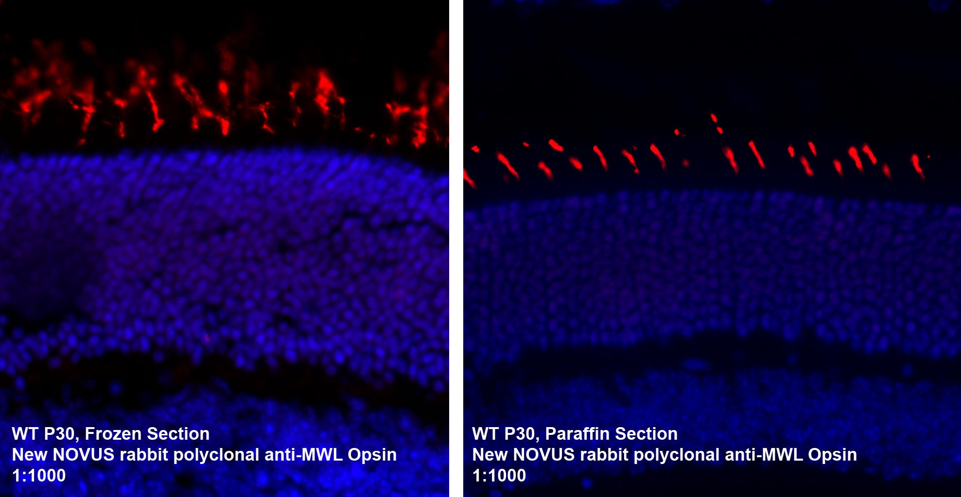
![Immunohistochemistry-Paraffin: Opsin 1 (Medium Wave) Antibody [NB110-74730]](https://resources.rndsystems.com/images/products/Opsin-1-Medium-Wave-Antibody-Immunohistochemistry-Paraffin-NB110-74730-img0001.jpg "Immunohistochemistry-Paraffin: Opsin 1 (Medium Wave) Antibody [NB110-74730]")
Immunohistochemistry-Paraffin: Opsin 1 (Medium Wave) Antibody [NB110-74730]
Immunohistochemistry-Paraffin: Opsin 1 (Medium Wave) Antibody [NB110-74730] - IF analysis of Opsin 1 in paraffin embedded and frozen mouse retina tissues. Image courtesy of product review submitted by Linda Vuong.![Immunohistochemistry: Opsin 1 (Medium Wave) Antibody [NB110-74730]](https://resources.rndsystems.com/images/products/Opsin-1-Medium-Wave-Antibody-Immunohistochemistry-NB110-74730-img0004.jpg "Immunohistochemistry: Opsin 1 (Medium Wave) Antibody [NB110-74730]")
Immunohistochemistry: Opsin 1 (Medium Wave) Antibody [NB110-74730]
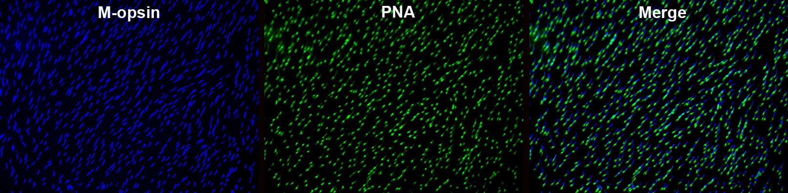
Immunohistochemistry: Opsin 1 (Medium Wave) Antibody [NB110-74730] - Retinal whole mount of BALB/c mouse stained against anti M-opsin secondary (AF647) antibody and peanut agglutinin (Fluorescein). This image was submitted via customer Review. Antibody [NB110-74730] -")
Opsin 1 (Medium Wave) Antibody [NB110-74730] -
HC-P on paraffin sections of mouse eye. The animal was perfused using Autoperfuser at a pressure of 130 mmHg with 300 ml 4% FA before being processed for paraffin embedding. HIER: Tris-EDTA, pH 9 for 20 min using Thermo PT Module. Blocking: 0.2% LFDM in TBST filtered thru 0.2 µm. Detection was done using Novolink HRP polymer from Leica following manufacturers instructions; DAB chromogen: Candela DAB chromogen from Osenses. Primary antibody: dilution 1: 500, incubated 30 min at RT using Autostainer. Sections were counterstained with Harris Hematoxylin. Antibody [NB110-74730] -")
Opsin 1 (Medium Wave) Antibody [NB110-74730] -
IHC-P on paraffin sections of mouse eye. The animal was perfused using Autoperfuser at a pressure of 130 mmHg with 300 ml 4% FA before being processed for paraffin embedding. HIER: Tris-EDTA, pH 9 for 20 min using Thermo PT Module. Blocking: 0.2% LFDM in TBST filtered thru 0.2 µm. Detection was done using Novolink HRP polymer from Leica following manufacturers instructions; DAB chromogen: Candela DAB chromogen from Osenses. Primary antibody: dilution 1: 500, incubated 30 min at RT using Autostainer. Sections were counterstained with Harris Hematoxylin.Applications for Opsin 1 (Medium Wave) Antibody
Immunohistochemistry
Immunohistochemistry-Frozen
Immunohistochemistry-Paraffin
Western Blot
Reviewed Applications
Read 3 reviews rated 4.7 using NB110-74730 in the following applications:
Formulation, Preparation, and Storage
Purification
Reconstitution
Formulation
Preservative
Concentration
Shipping
Stability & Storage
Calculators
Background: Opsin 1 (Medium Wave)
Alternate Names
Gene Symbol
UniProt
Additional Opsin 1 (Medium Wave) Products
Product Documents for Opsin 1 (Medium Wave) Antibody
Certificate of Analysis
To download a Certificate of Analysis, please enter a lot or batch number in the search box below.
Product Specific Notices for Opsin 1 (Medium Wave) Antibody
This product is for research use only and is not approved for use in humans or in clinical diagnosis. Primary Antibodies are guaranteed for 1 year from date of receipt.
Citations for Opsin 1 (Medium Wave) Antibody
 Powered by Bioz
Customer Reviews for Opsin 1 (Medium Wave) Antibody (3)
Have you used Opsin 1 (Medium Wave) Antibody?
Submit a review and receive an Amazon gift card!
$25/€18/£15/$25CAN/¥2500 Yen for a review with an image
$10/€7/£6/$10CAN/¥1110 Yen for a review without an image
Submit a review
Customer Images
-
Application: Immunohistochemistry of intact specimenSample Tested: mouse retinaSpecies: MouseVerified Customer | Posted 07/18/2017Retinal whole mount of BALB/c mouse stained against anti M-opsin (Novus Biologicals NB110-74730; + AlexaFluor 647 nm secondary AB) antibody and peanut agglutinin (+ Fluorescein).Anti-rabbit AlexaFluor 647 nm secondary antibody

-
Application: Immunohistochemistry-ParaffinSample Tested: Mouse retina embedded in paraffinSpecies: MouseVerified Customer | Posted 06/21/2016
-
Application: ImmunohistochemistrySample Tested: Mouse retina embedded in paraffin and frozen mediumSpecies: MouseVerified Customer | Posted 08/14/2012
There are no reviews that match your criteria.
Protocols
Find general support by application which include: protocols, troubleshooting, illustrated assays, videos and webinars.
- Antigen Retrieval Protocol (PIER)
- Antigen Retrieval for Frozen Sections Protocol
- Appropriate Fixation of IHC/ICC Samples
- Cellular Response to Hypoxia Protocols
- Chromogenic IHC Staining of Formalin-Fixed Paraffin-Embedded (FFPE) Tissue Protocol
- Chromogenic Immunohistochemistry Staining of Frozen Tissue
- ClariTSA™ Fluorophore Kits
- Detection & Visualization of Antibody Binding
- Fluorescent IHC Staining of Frozen Tissue Protocol
- Graphic Protocol for Heat-induced Epitope Retrieval
- Graphic Protocol for the Preparation and Fluorescent IHC Staining of Frozen Tissue Sections
- Graphic Protocol for the Preparation and Fluorescent IHC Staining of Paraffin-embedded Tissue Sections
- Graphic Protocol for the Preparation of Gelatin-coated Slides for Histological Tissue Sections
- ICC Cell Smear Protocol for Suspension Cells
- ICC Immunocytochemistry Protocol Videos
- ICC for Adherent Cells
- IHC Sample Preparation (Frozen sections vs Paraffin)
- Immunocytochemistry (ICC) Protocol
- Immunocytochemistry Troubleshooting
- Immunofluorescence of Organoids Embedded in Cultrex Basement Membrane Extract
- Immunofluorescent IHC Staining of Formalin-Fixed Paraffin-Embedded (FFPE) Tissue Protocol
- Immunohistochemistry (IHC) and Immunocytochemistry (ICC) Protocols
- Immunohistochemistry Frozen Troubleshooting
- Immunohistochemistry Paraffin Troubleshooting
- Preparing Samples for IHC/ICC Experiments
- Preventing Non-Specific Staining (Non-Specific Binding)
- Primary Antibody Selection & Optimization
- Protocol for Heat-Induced Epitope Retrieval (HIER)
- Protocol for Making a 4% Formaldehyde Solution in PBS
- Protocol for VisUCyte™ HRP Polymer Detection Reagent
- Protocol for the Fluorescent ICC Staining of Cell Smears - Graphic
- Protocol for the Fluorescent ICC Staining of Cultured Cells on Coverslips - Graphic
- Protocol for the Preparation & Fixation of Cells on Coverslips
- Protocol for the Preparation and Chromogenic IHC Staining of Frozen Tissue Sections
- Protocol for the Preparation and Chromogenic IHC Staining of Frozen Tissue Sections - Graphic
- Protocol for the Preparation and Chromogenic IHC Staining of Paraffin-embedded Tissue Sections
- Protocol for the Preparation and Chromogenic IHC Staining of Paraffin-embedded Tissue Sections - Graphic
- Protocol for the Preparation and Fluorescent ICC Staining of Cells on Coverslips
- Protocol for the Preparation and Fluorescent ICC Staining of Non-adherent Cells
- Protocol for the Preparation and Fluorescent ICC Staining of Stem Cells on Coverslips
- Protocol for the Preparation and Fluorescent IHC Staining of Frozen Tissue Sections
- Protocol for the Preparation and Fluorescent IHC Staining of Paraffin-embedded Tissue Sections
- Protocol for the Preparation of Gelatin-coated Slides for Histological Tissue Sections
- Protocol for the Preparation of a Cell Smear for Non-adherent Cell ICC - Graphic
- R&D Systems Quality Control Western Blot Protocol
- TUNEL and Active Caspase-3 Detection by IHC/ICC Protocol
- The Importance of IHC/ICC Controls
- Troubleshooting Guide: Immunohistochemistry
- Troubleshooting Guide: Western Blot Figures
- Western Blot Conditions
- Western Blot Protocol
- Western Blot Protocol for Cell Lysates
- Western Blot Troubleshooting
- Western Blot Troubleshooting Guide
- View all Protocols, Troubleshooting, Illustrated assays and Webinars
FAQs for Opsin 1 (Medium Wave) Antibody
-
Q: I would to know if the synthetic peptide used as immunogen to generate the anti-opsin1 antibody (NB110-74730) belongs to the N-terminus or C-terminus of opsin1?
A: The antigen for our Opsin 1 (Medium Wave) antibody [NB110-74730] is located between aa region 2-50, so it is N-terminal.