P-Selectin/CD62P Antibody (Psel.KO.2.7) [DyLight 488]
Novus Biologicals | Catalog # NB100-65392G
Key Product Details
Validated by
Species Reactivity
Validated:
Cited:
Applications
Validated:
Cited:
Label
Antibody Source
Product Specifications
Immunogen
Reactivity Notes
Localization
Specificity
Clonality
Host
Isotype
Description
Scientific Data Images for P-Selectin/CD62P Antibody (Psel.KO.2.7) [DyLight 488]

![P-Selectin/CD62P Antibody (Psel.KO.2.7) [DyLight 488]](https://resources.rndsystems.com/images/products/nb100-65392g_mouse-monoclonal-p-selectin-cd62p-antibody-psel-ko-2-7-dylight-488-31020241682190.jpg "Flow Cytometry: P-Selectin/CD62P Antibody (Psel.KO.2.7) [DyLight 488] [NB100-65392G] -")
Flow Cytometry: P-Selectin/CD62P Antibody (Psel.KO.2.7) [DyLight 488] [NB100-65392G] -
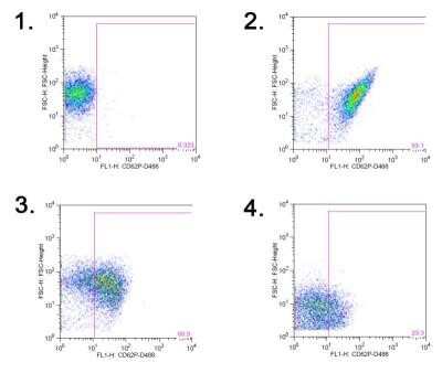
Flow Cytometry: P-Selectin/CD62P Antibody (Psel.KO.2.7) [DyLight 488] [NB100-65392G] - Flow cytometry gating strategy for quantification of platelet-derived microparticles in equine platelet samples.A: Platelet events in citrate-anticoagulated platelet-rich plasma were identified & gated as CD41-positive cells (R1 region) in a CD41 fluorescence versus forward scatter (FSC) dotplot. The R1 region or gate was established on an isotype control for the CD41 antibody. Representative image from platelets exposed to the RacL11 strain of EHV-1 at 1 plaque forming unit (PFU)/cell. B: Platelet-derived microparticles (PDMPs) were defined as small events (<101 log FSC units) positive for Annexin V & CD41. The PDMP percentage was obtained from the lower right quadrant of an Annexin V fluorescence versus FSC dotplot of the R1 gate (CD41-positive events), with the quadrants being defined on a negative sample in which 1 mM EDTA was added to the buffer with Annexin V. The PDMP percentage was 0.1% in this representative image of platelets exposed to rabbit kidney 13 (RK) cell lysate at an equivalent volume to 1 PFU/cell (mock-infected negative control). The events in the upper left & right quadrants are platelets that are negative (94.0%) & positive for Annexin V (0.9%), respectively. C: Representative image of PDMP quantification in platelets exposed to RacL11 at 1 PFU/cell. In this sample, there are 12.1% PDMP (lower right quadrant) & 22.1% of platelets are weakly positive for Annexin V (upper right quadrant). Image collected & cropped by CiteAb from the following publication (https://pubmed.ncbi.nlm.nih.gov/25905776), licensed under a CC-BY license. Not internally tested by Novus Biologicals.![P-Selectin/CD62P Antibody (Psel.KO.2.7) [DyLight 488]](https://resources.rndsystems.com/images/products/nb100-65392g_mouse-monoclonal-p-selectin-cd62p-antibody-psel-ko-2-7-dylight-488-31020241682170.jpg "Flow Cytometry: P-Selectin/CD62P Antibody (Psel.KO.2.7) [DyLight 488] [NB100-65392G] -")
Flow Cytometry: P-Selectin/CD62P Antibody (Psel.KO.2.7) [DyLight 488] [NB100-65392G] -
Flow Cytometry: P-Selectin/CD62P Antibody (Psel.KO.2.7) [DyLight 488] [NB100-65392G] - Flow cytometry gating strategy for quantification of platelet-derived microparticles in equine platelet samples.A: Platelet events in citrate-anticoagulated platelet-rich plasma were identified & gated as CD41-positive cells (R1 region) in a CD41 fluorescence versus forward scatter (FSC) dotplot. The R1 region or gate was established on an isotype control for the CD41 antibody. Representative image from platelets exposed to the RacL11 strain of EHV-1 at 1 plaque forming unit (PFU)/cell. B: Platelet-derived microparticles (PDMPs) were defined as small events (<101 log FSC units) positive for Annexin V & CD41. The PDMP percentage was obtained from the lower right quadrant of an Annexin V fluorescence versus FSC dotplot of the R1 gate (CD41-positive events), with the quadrants being defined on a negative sample in which 1 mM EDTA was added to the buffer with Annexin V. The PDMP percentage was 0.1% in this representative image of platelets exposed to rabbit kidney 13 (RK) cell lysate at an equivalent volume to 1 PFU/cell (mock-infected negative control). The events in the upper left & right quadrants are platelets that are negative (94.0%) & positive for Annexin V (0.9%), respectively. C: Representative image of PDMP quantification in platelets exposed to RacL11 at 1 PFU/cell. In this sample, there are 12.1% PDMP (lower right quadrant) & 22.1% of platelets are weakly positive for Annexin V (upper right quadrant). Image collected & cropped by CiteAb from the following publication (https://pubmed.ncbi.nlm.nih.gov/25905776), licensed under a CC-BY license. Not internally tested by Novus Biologicals.![P-Selectin/CD62P Antibody (Psel.KO.2.7) [DyLight 488]](https://resources.rndsystems.com/images/products/nb100-65392g_mouse-monoclonal-p-selectin-cd62p-antibody-psel-ko-2-7-dylight-488-310202415535190.jpg "Flow Cytometry: P-Selectin/CD62P Antibody (Psel.KO.2.7) [DyLight 488] [NB100-65392G] -")
Flow Cytometry: P-Selectin/CD62P Antibody (Psel.KO.2.7) [DyLight 488] [NB100-65392G] -
Flow Cytometry: P-Selectin/CD62P Antibody (Psel.KO.2.7) [DyLight 488] [NB100-65392G] - Flow cytometry gating strategy for quantification of platelet-derived microparticles in equine platelet samples.A: Platelet events in citrate-anticoagulated platelet-rich plasma were identified & gated as CD41-positive cells (R1 region) in a CD41 fluorescence versus forward scatter (FSC) dotplot. The R1 region or gate was established on an isotype control for the CD41 antibody. Representative image from platelets exposed to the RacL11 strain of EHV-1 at 1 plaque forming unit (PFU)/cell. B: Platelet-derived microparticles (PDMPs) were defined as small events (<101 log FSC units) positive for Annexin V & CD41. The PDMP percentage was obtained from the lower right quadrant of an Annexin V fluorescence versus FSC dotplot of the R1 gate (CD41-positive events), with the quadrants being defined on a negative sample in which 1 mM EDTA was added to the buffer with Annexin V. The PDMP percentage was 0.1% in this representative image of platelets exposed to rabbit kidney 13 (RK) cell lysate at an equivalent volume to 1 PFU/cell (mock-infected negative control). The events in the upper left & right quadrants are platelets that are negative (94.0%) & positive for Annexin V (0.9%), respectively. C: Representative image of PDMP quantification in platelets exposed to RacL11 at 1 PFU/cell. In this sample, there are 12.1% PDMP (lower right quadrant) & 22.1% of platelets are weakly positive for Annexin V (upper right quadrant). Image collected & cropped by CiteAb from the following publication (https://pubmed.ncbi.nlm.nih.gov/25905776), licensed under a CC-BY license. Not internally tested by Novus Biologicals.Applications for P-Selectin/CD62P Antibody (Psel.KO.2.7) [DyLight 488]
Flow Cytometry
Immunocytochemistry/ Immunofluorescence
Immunohistochemistry
Immunohistochemistry-Frozen
Immunohistochemistry-Paraffin
Immunoprecipitation
Reviewed Applications
Read 1 review rated 4 using NB100-65392G in the following applications:
Spectra Viewer
Plan Your Experiments
Use our spectra viewer to interactively plan your experiments, assessing multiplexing options. View the excitation and emission spectra for our fluorescent dye range and other commonly used dyes.
Spectra Viewer
Flow Cytometry Panel Builder
Bio-Techne Knows Flow Cytometry
Save time and reduce costly mistakes by quickly finding compatible reagents using the Panel Builder Tool.
Advanced Features
- Spectra Viewer - Custom analysis of spectra from multiple fluorochromes
- Spillover Popups - Visualize the spectra of individual fluorochromes
- Antigen Density Selector - Match fluorochrome brightness with antigen density

Formulation, Preparation, and Storage
Purification
Formulation
Preservative
Concentration
Shipping
Stability & Storage
Background: P-Selectin/CD62P
Alternate Names
Gene Symbol
Additional P-Selectin/CD62P Products
Product Documents for P-Selectin/CD62P Antibody (Psel.KO.2.7) [DyLight 488]
Certificate of Analysis
To download a Certificate of Analysis, please enter a lot or batch number in the search box below.
Product Specific Notices for P-Selectin/CD62P Antibody (Psel.KO.2.7) [DyLight 488]
DyLight (R) is a trademark of Thermo Fisher Scientific Inc. and its subsidiaries.
This product is for research use only and is not approved for use in humans or in clinical diagnosis. Primary Antibodies are guaranteed for 1 year from date of receipt.
Citations for P-Selectin/CD62P Antibody (Psel.KO.2.7) [DyLight 488]
 Powered by Bioz
Customer Reviews for P-Selectin/CD62P Antibody (Psel.KO.2.7) [DyLight 488] (1)
Have you used P-Selectin/CD62P Antibody (Psel.KO.2.7) [DyLight 488]?
Submit a review and receive an Amazon gift card!
$25/€18/£15/$25CAN/¥2500 Yen for a review with an image
$10/€7/£6/$10CAN/¥1110 Yen for a review without an image
Submit a review
-
Application: Flow CytometrySample Tested:Species: OtherVerified Customer | Posted 11/01/2013
There are no reviews that match your criteria.
Protocols
Find general support by application which include: protocols, troubleshooting, illustrated assays, videos and webinars.
- 7-Amino Actinomycin D (7-AAD) Cell Viability Flow Cytometry Protocol
- Antigen Retrieval Protocol (PIER)
- Antigen Retrieval for Frozen Sections Protocol
- Appropriate Fixation of IHC/ICC Samples
- Cellular Response to Hypoxia Protocols
- Chromogenic IHC Staining of Formalin-Fixed Paraffin-Embedded (FFPE) Tissue Protocol
- Chromogenic Immunohistochemistry Staining of Frozen Tissue
- ClariTSA™ Fluorophore Kits
- Detection & Visualization of Antibody Binding
- Extracellular Membrane Flow Cytometry Protocol
- Flow Cytometry Protocol for Cell Surface Markers
- Flow Cytometry Protocol for Staining Membrane Associated Proteins
- Flow Cytometry Staining Protocols
- Flow Cytometry Troubleshooting Guide
- Fluorescent IHC Staining of Frozen Tissue Protocol
- Graphic Protocol for Heat-induced Epitope Retrieval
- Graphic Protocol for the Preparation and Fluorescent IHC Staining of Frozen Tissue Sections
- Graphic Protocol for the Preparation and Fluorescent IHC Staining of Paraffin-embedded Tissue Sections
- Graphic Protocol for the Preparation of Gelatin-coated Slides for Histological Tissue Sections
- ICC Cell Smear Protocol for Suspension Cells
- ICC Immunocytochemistry Protocol Videos
- ICC for Adherent Cells
- IHC Sample Preparation (Frozen sections vs Paraffin)
- Immunocytochemistry (ICC) Protocol
- Immunocytochemistry Troubleshooting
- Immunofluorescence of Organoids Embedded in Cultrex Basement Membrane Extract
- Immunofluorescent IHC Staining of Formalin-Fixed Paraffin-Embedded (FFPE) Tissue Protocol
- Immunohistochemistry (IHC) and Immunocytochemistry (ICC) Protocols
- Immunohistochemistry Frozen Troubleshooting
- Immunohistochemistry Paraffin Troubleshooting
- Immunoprecipitation Protocol
- Intracellular Flow Cytometry Protocol Using Alcohol (Methanol)
- Intracellular Flow Cytometry Protocol Using Detergents
- Intracellular Nuclear Staining Flow Cytometry Protocol Using Detergents
- Intracellular Staining Flow Cytometry Protocol Using Alcohol Permeabilization
- Intracellular Staining Flow Cytometry Protocol Using Detergents to Permeabilize Cells
- Preparing Samples for IHC/ICC Experiments
- Preventing Non-Specific Staining (Non-Specific Binding)
- Primary Antibody Selection & Optimization
- Propidium Iodide Cell Viability Flow Cytometry Protocol
- Protocol for Heat-Induced Epitope Retrieval (HIER)
- Protocol for Liperfluo
- Protocol for Making a 4% Formaldehyde Solution in PBS
- Protocol for VisUCyte™ HRP Polymer Detection Reagent
- Protocol for the Characterization of Human Th22 Cells
- Protocol for the Characterization of Human Th9 Cells
- Protocol for the Fluorescent ICC Staining of Cell Smears - Graphic
- Protocol for the Fluorescent ICC Staining of Cultured Cells on Coverslips - Graphic
- Protocol for the Preparation & Fixation of Cells on Coverslips
- Protocol for the Preparation and Chromogenic IHC Staining of Frozen Tissue Sections
- Protocol for the Preparation and Chromogenic IHC Staining of Frozen Tissue Sections - Graphic
- Protocol for the Preparation and Chromogenic IHC Staining of Paraffin-embedded Tissue Sections
- Protocol for the Preparation and Chromogenic IHC Staining of Paraffin-embedded Tissue Sections - Graphic
- Protocol for the Preparation and Fluorescent ICC Staining of Cells on Coverslips
- Protocol for the Preparation and Fluorescent ICC Staining of Non-adherent Cells
- Protocol for the Preparation and Fluorescent ICC Staining of Stem Cells on Coverslips
- Protocol for the Preparation and Fluorescent IHC Staining of Frozen Tissue Sections
- Protocol for the Preparation and Fluorescent IHC Staining of Paraffin-embedded Tissue Sections
- Protocol for the Preparation of Gelatin-coated Slides for Histological Tissue Sections
- Protocol for the Preparation of a Cell Smear for Non-adherent Cell ICC - Graphic
- Protocol: Annexin V and PI Staining by Flow Cytometry
- Protocol: Annexin V and PI Staining for Apoptosis by Flow Cytometry
- TUNEL and Active Caspase-3 Detection by IHC/ICC Protocol
- The Importance of IHC/ICC Controls
- Troubleshooting Guide: Fluorokine Flow Cytometry Kits
- Troubleshooting Guide: Immunohistochemistry
- View all Protocols, Troubleshooting, Illustrated assays and Webinars