p14ARF/CDKN2A Antibody - BSA Free
Novus Biologicals | Catalog # NB200-111
![Knockdown Validated: p14ARF/CDKN2A Antibody - BSA Free [NB200-111]](https://resources.rndsystems.com/images/products/p14ARF-CDKN2A-Antibody-Western-Blot-NB200-111-img0012.jpg "Western Blot: p14ARF/CDKN2A Antibody - BSA Free [NB200-111]")
Key Product Details
Validated by
Species Reactivity
Validated:
Cited:
Applications
Validated:
Cited:
Label
Antibody Source
Format
Product Specifications
Immunogen
Reactivity Notes
Localization
Clonality
Host
Isotype
Scientific Data Images for p14ARF/CDKN2A Antibody - BSA Free
![Immunohistochemistry: p14ARF/CDKN2A Antibody - BSA Free [NB200-111]](https://resources.rndsystems.com/images/products/p14ARF-CDKN2A-Antibody-Immunocytochemistry-Immunofluorescence-NB200-111-img0011.jpg "Immunohistochemistry: p14ARF/CDKN2A Antibody - BSA Free [NB200-111]")
![Immunocytochemistry/ Immunofluorescence: p14ARF/CDKN2A Antibody - BSA Free [NB200-111]](https://resources.rndsystems.com/images/products/p14ARF-CDKN2A-Antibody-Immunocytochemistry-Immunofluorescence-NB200-111-img0006.jpg "Immunocytochemistry/ Immunofluorescence: p14ARF/CDKN2A Antibody - BSA Free [NB200-111]")
Immunocytochemistry/ Immunofluorescence: p14ARF/CDKN2A Antibody - BSA Free [NB200-111]
Immunocytochemistry/Immunofluorescence: p14ARF/CDKN2A Antibody [NB200-111] - p14ARF antibody was tested in HeLa cells with Dylight 488 (green). Nuclei were counterstained with DAPI (blue). Tubulin was stained with alpha tubulin (red).![Western Blot: p14ARF/CDKN2A AntibodyBSA Free [NB200-111]](https://resources.rndsystems.com/images/products/p14ARF-CDKN2A-Antibody-Western-Blot-NB200-111-img0009.jpg "Western Blot: p14ARF/CDKN2A AntibodyBSA Free [NB200-111]")
![Flow Cytometry: p14ARF/CDKN2A Antibody - BSA Free [NB200-111]](https://resources.rndsystems.com/images/products/p14ARF-CDKN2A-Antibody-Flow-Cytometry-NB200-111-img0007.jpg "Flow Cytometry: p14ARF/CDKN2A Antibody - BSA Free [NB200-111]")
Flow Cytometry: p14ARF/CDKN2A Antibody - BSA Free [NB200-111]
Flow Cytometry: p14ARF/CDKN2A Antibody [NB200-111] - p14ARF antibody was tested at 1:400 in HeLa cells using an Alexa Fluor 488 secondary (shown in purple). M1 is defined by unstained cells.![Western Blot: p14ARF/CDKN2A AntibodyBSA Free [NB200-111]](https://resources.rndsystems.com/images/products/p14ARF-CDKN2A-Antibody-Western-Blot-NB200-111-img0008.jpg "Western Blot: p14ARF/CDKN2A AntibodyBSA Free [NB200-111]")
Western Blot: p14ARF/CDKN2A AntibodyBSA Free [NB200-111]
Western Blot: p14ARF/CDKN2A Antibody [NB200-111] - Analysis of HeLa Whole Cell Lysate (NB800-PC1) using p14ARF antibody (lot C) employing ECL detection method.![Immunohistochemistry-Frozen: p14ARF/CDKN2A Antibody - BSA Free [NB200-111]](https://resources.rndsystems.com/images/products/p14ARF-CDKN2A-Antibody-Immunohistochemistry-Frozen-NB200-111-img0010.jpg "Immunohistochemistry-Frozen: p14ARF/CDKN2A Antibody - BSA Free [NB200-111]")
Immunohistochemistry-Frozen: p14ARF/CDKN2A Antibody - BSA Free [NB200-111]
Immunohistochemistry-Frozen: p14ARF/CDKN2A Antibody [NB200-111] - Mouse brain section, hippocampal (10 um thickness). 3% PFA perfused. 1:1000 dilultion. Image from verified customer review. Image using the Biotin form of this antibody.
Western Blot: p14ARF/CDKN2A Antibody - BSA Free [NB200-111] -
Western Blot: p14ARF/CDKN2A Antibody - BSA Free [NB200-111] - Inhibition of AKT decreases p53mut stability(A) T24 cells & PSN1 cells were treated with MK-2206 (5 μM) or DMSO for 24 hrs. Cells were fixed & stained with DAPI & anti-mutant p53 (OP29 clone). (B) T24 cells were pre-treated with MK-2206 (5 μM) or DMSO for 24 hrs before the addition of fresh media containing cyclohexamide (100 μM) (CHX) in combination with MK-2206 (5 μM) or DMSO for the times indicated. Nuclear extracts were prepared from treated cells & blotted with the indicated antibodies. Quantification is relative to initiation of CHX treatment for both conditions *, MK-2206 is 40% of DMSO control but taken as 1.0 for relative assessment. (C) H1299 cells were transfected with HA-tagged-ubiquitin & mutant p53 (R175H or R248W) as indicated. Transfected cells were treated with DMSO, MK-2206 (5 μM) or Nutlin3A (5 μM) for 16hrs as indicated. p53 immunoprecipitates & whole cell lysates were probed with the indicated antibodies. (D) T24 cells were transfected with non-targeting control or p14ARF siRNA & treated with DMSO, MK-2206 (5 μM) or the NPM oligomerisation inhibitor NCS348884 (4 μM) (Qi et al., 2008) as indicated. (E) T24 cells were transfected with non-targeting control, AKT1, or p14ARF siRNA. Cells were treated with NCS348884 (4 μM), Nutlin3A (5 μM) or DMSO as indicated. Whole cell lysates were probed with the indicated antibodies. (F) KPC mice derived KRASG12D p53 Floxed (p53Fl), KRASG12D p53R172H ARF+/+ & KRASG12D p53R172H ARF−/− pancreatic tumor cells were treated with MK-2206 (1μM), Nutlin3A (5μM) or DMSO as indicated. Whole cell lysates were probed with the indicated antibodies. Image collected & cropped by CiteAb from the following publication (https://pubmed.ncbi.nlm.nih.gov/25071014), licensed under a CC-BY license. Not internally tested by Novus Biologicals.
Immunocytochemistry/ Immunofluorescence: p14ARF/CDKN2A Antibody - BSA Free [NB200-111] -
Immunocytochemistry/ Immunofluorescence: p14ARF/CDKN2A Antibody - BSA Free [NB200-111] - Inhibition of AKT modulates p53 stability in-vivo & synergizes with ionizing radiation to inhibit tumor growth(A) PSN1 xenografts (PSN1 cells co-injected with LTC-14 stellate cells) established in the flank of athymic nude mice were treated with MK-2206 (60 mg/kg-320 mg/kg) as indicated or beta -cyclo-dextrin (1.5 mg/ml) carrier. Xenograft tumors were lysed & lysates probed by western blot with the indicated antibodies. (B-D) Sections of PSN1 xenografts treated with three consecutive doses of MK-2206 (60 mg/kg). (B) Sections of PSN1 xenografts & in-vitro PSN1 cells fixed & stained with anti-NPM (red) & anti-p14ARF (green). (C) PSN1 xenografts treated with MK-2206 or carrier were stained with DAPI, anti-p53 (DO1) or p53mut (OP29 clone) (D) PSN1 xenografts treated with MK-2206 (60 mg/kg) or carrier were stained by immunohistochemical methods with anti-pS473-AKT, anti-phospho-S48-NPM (pS48-NPM) or p53. (E) PSN1 xenografts established in the flank of athymic nude mice were injected subcutaneously with two alternate day doses of MK-2206 (60 mg/kg) or carrier. Mice were subsequently treated with a single dose of IR (6 Gy) & tumor volumes measured regularly with callipers. Dash lines indicate tumor growth differential at 250 mm3. Image collected & cropped by CiteAb from the following publication (https://pubmed.ncbi.nlm.nih.gov/25071014), licensed under a CC-BY license. Not internally tested by Novus Biologicals.
Western Blot: p14ARF/CDKN2A Antibody - BSA Free [NB200-111] -
Western Blot: p14ARF/CDKN2A Antibody - BSA Free [NB200-111] - Inhibition of AKT decreases p53mut stability(A) T24 cells & PSN1 cells were treated with MK-2206 (5 μM) or DMSO for 24 hrs. Cells were fixed & stained with DAPI & anti-mutant p53 (OP29 clone). (B) T24 cells were pre-treated with MK-2206 (5 μM) or DMSO for 24 hrs before the addition of fresh media containing cyclohexamide (100 μM) (CHX) in combination with MK-2206 (5 μM) or DMSO for the times indicated. Nuclear extracts were prepared from treated cells & blotted with the indicated antibodies. Quantification is relative to initiation of CHX treatment for both conditions *, MK-2206 is 40% of DMSO control but taken as 1.0 for relative assessment. (C) H1299 cells were transfected with HA-tagged-ubiquitin & mutant p53 (R175H or R248W) as indicated. Transfected cells were treated with DMSO, MK-2206 (5 μM) or Nutlin3A (5 μM) for 16hrs as indicated. p53 immunoprecipitates & whole cell lysates were probed with the indicated antibodies. (D) T24 cells were transfected with non-targeting control or p14ARF siRNA & treated with DMSO, MK-2206 (5 μM) or the NPM oligomerisation inhibitor NCS348884 (4 μM) (Qi et al., 2008) as indicated. (E) T24 cells were transfected with non-targeting control, AKT1, or p14ARF siRNA. Cells were treated with NCS348884 (4 μM), Nutlin3A (5 μM) or DMSO as indicated. Whole cell lysates were probed with the indicated antibodies. (F) KPC mice derived KRASG12D p53 Floxed (p53Fl), KRASG12D p53R172H ARF+/+ & KRASG12D p53R172H ARF−/− pancreatic tumor cells were treated with MK-2206 (1μM), Nutlin3A (5μM) or DMSO as indicated. Whole cell lysates were probed with the indicated antibodies. Image collected & cropped by CiteAb from the following publication (https://pubmed.ncbi.nlm.nih.gov/25071014), licensed under a CC-BY license. Not internally tested by Novus Biologicals.
Western Blot: p14ARF/CDKN2A Antibody - BSA Free [NB200-111] -
Western Blot: p14ARF/CDKN2A Antibody - BSA Free [NB200-111] - Inhibition of AKT promotes enhanced MDM2 activity via the increased association between NPM & p14ARF(A) Npm−/−, p53−/−double null MEF were infected with pBABE retrovirus empty vector & pBABE expressing FLAG-tagged-NPM-WT, NPM-S48A or S48E as indicated. Immunopurification of NPM was done by pulling down with the Flag tag (middle panel) followed by elution of complexes by the Flag peptide & subsequent immunopurification of endogenous MDM2 (lower panel). (B) Nuclear immunoprecipitates of MDM2 from T24 cells treated with MK-2206 (5 μM, 24 hrs). Immunoprecipitates & lysates were blotted with the indicated antibodies. (C) T24 cells were treated with MK-2206 (5 μM) as indicated. p14ARF was immunoprecipitated from whole cell lysates & nuclear extracts & the association with NPM & MDM2 determined by western blot. Immunoprecipitates & lysates were blotted with the indicated antibodies. (D) MDM2 & (E) p53 ubiquinitation assay in H1299 cells transfected with wild type p53, HA-tagged ubiquitin & treated for 16 hrs with DMSO, MK-2206 (5 μM) or Nutlin3A (5 μM) as indicated. Immunoprecipitates & whole cell lysates were probed with the indicated antibodies. Image collected & cropped by CiteAb from the following publication (https://pubmed.ncbi.nlm.nih.gov/25071014), licensed under a CC-BY license. Not internally tested by Novus Biologicals.
Western Blot: p14ARF/CDKN2A Antibody - BSA Free [NB200-111] -
Western Blot: p14ARF/CDKN2A Antibody - BSA Free [NB200-111] - Inhibition of AKT promotes enhanced MDM2 activity via the increased association between NPM & p14ARF(A) Npm−/−, p53−/−double null MEF were infected with pBABE retrovirus empty vector & pBABE expressing FLAG-tagged-NPM-WT, NPM-S48A or S48E as indicated. Immunopurification of NPM was done by pulling down with the Flag tag (middle panel) followed by elution of complexes by the Flag peptide & subsequent immunopurification of endogenous MDM2 (lower panel). (B) Nuclear immunoprecipitates of MDM2 from T24 cells treated with MK-2206 (5 μM, 24 hrs). Immunoprecipitates & lysates were blotted with the indicated antibodies. (C) T24 cells were treated with MK-2206 (5 μM) as indicated. p14ARF was immunoprecipitated from whole cell lysates & nuclear extracts & the association with NPM & MDM2 determined by western blot. Immunoprecipitates & lysates were blotted with the indicated antibodies. (D) MDM2 & (E) p53 ubiquinitation assay in H1299 cells transfected with wild type p53, HA-tagged ubiquitin & treated for 16 hrs with DMSO, MK-2206 (5 μM) or Nutlin3A (5 μM) as indicated. Immunoprecipitates & whole cell lysates were probed with the indicated antibodies. Image collected & cropped by CiteAb from the following publication (https://pubmed.ncbi.nlm.nih.gov/25071014), licensed under a CC-BY license. Not internally tested by Novus Biologicals.Applications for p14ARF/CDKN2A Antibody - BSA Free
Flow Cytometry
Immunocytochemistry/ Immunofluorescence
Immunohistochemistry
Immunohistochemistry-Frozen
Immunohistochemistry-Paraffin
Immunoprecipitation
Western Blot
Reviewed Applications
Read 2 reviews rated 4 using NB200-111 in the following applications:
Flow Cytometry Panel Builder
Bio-Techne Knows Flow Cytometry
Save time and reduce costly mistakes by quickly finding compatible reagents using the Panel Builder Tool.
Advanced Features
- Spectra Viewer - Custom analysis of spectra from multiple fluorochromes
- Spillover Popups - Visualize the spectra of individual fluorochromes
- Antigen Density Selector - Match fluorochrome brightness with antigen density

Formulation, Preparation, and Storage
Purification
Formulation
Format
Preservative
Concentration
Shipping
Stability & Storage
Background: p14ARF/CDKN2A
Alternate Names
Entrez Gene IDs
Gene Symbol
UniProt
Additional p14ARF/CDKN2A Products
Product Documents for p14ARF/CDKN2A Antibody - BSA Free
Certificate of Analysis
To download a Certificate of Analysis, please enter a lot or batch number in the search box below.
Product Specific Notices for p14ARF/CDKN2A Antibody - BSA Free
This product is for research use only and is not approved for use in humans or in clinical diagnosis. Primary Antibodies are guaranteed for 1 year from date of receipt.
Citations for p14ARF/CDKN2A Antibody - BSA Free
 Powered by Bioz
Customer Reviews for p14ARF/CDKN2A Antibody - BSA Free (2)
Have you used p14ARF/CDKN2A Antibody - BSA Free?
Submit a review and receive an Amazon gift card!
$25/€18/£15/$25CAN/¥2500 Yen for a review with an image
$10/€7/£6/$10CAN/¥1110 Yen for a review without an image
Submit a review
Customer Images
-(01-ml)_NB200-111_7876.jpg)
-
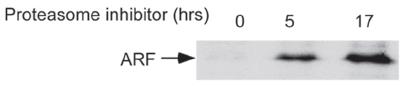
Application: Western BlotSample Tested: OCI/AML3 cells and human AML NPM cytoplasmic mutant cell lineSpecies: HumanVerified Customer | Posted 09/01/2017ARF expression levels in OCI/AML3 cells after proteasome treatment

-
Application: Western BlotSample Tested: Young and senescent cellsSpecies: HumanVerified Customer | Posted 05/29/2014p14/ARF levels in yourng and senescent cells
There are no reviews that match your criteria.
Protocols
View specific protocols for p14ARF/CDKN2A Antibody - BSA Free (NB200-111):
Western Blot Procedure
1) Lyse HeLa or BT549 cells in Laemli buffer (2% SDS, 62.5 mM Tris pH 6.8, 10% glycerol, 5% 2-mercaptoethanol).
2) Boil the lysate for 5 minutes.
3) Load 25 ug of total cell extract, per lane, in a 15% SDS-PAGE minigel.
4) Transfer protein to PVDF membrane in 40 mM Tris base, 20 mM NaAcetate, 2 mM EDTA pH 7.4, 0.05% SDS, 20% methanol for 1 hour at 4 degrees C.
5) Rinse membrane in PBS-T and then block membrane in 5% nonfat dry milk/PBS-T (0.01M phosphate, 0.0027M KCl, 0.137M NaCl pH 7.4, 0.1% Tween-20) for 1 hour at room temperature or overnight at 4 degrees C.
6) Incubate membrane overnight at 4 degrees C with properly diluted NB200-111 (see data sheet) in PBS, 0.2% Tween-20, 5% nonfat dry milk.
7) Rinse the membrane 2X with 40 ml PBS-T. Wash membrane at room temperature, with 40 ml of PBS-T, 1X for 15 minutes and 2X for 5 minutes, each.
8) Incubate membrane with HRP conjugated anti-rabbit, diluted in PBS-T with 5% nonfat dry milk, for 1 hour at room temperature.
9) Wash membrane at room temperature, with 40 ml of PBS-T, 1X for 15 minutes and 4X for 5 minutes, each.
10) Develop with ECL reagents (Amersham) and autoradiography. Expose for 1+ minutes. For BT549 cells, 1 minute exposure is needed. For HeLa cells, additional exposure time may be required. In addition, a stronger ECL (Pierce) may be necessary.
NOTE: HeLa whole cell extracts (NB800-PC1) were used as a positive control for this antibody.
Find general support by application which include: protocols, troubleshooting, illustrated assays, videos and webinars.
- 7-Amino Actinomycin D (7-AAD) Cell Viability Flow Cytometry Protocol
- Antigen Retrieval Protocol (PIER)
- Antigen Retrieval for Frozen Sections Protocol
- Appropriate Fixation of IHC/ICC Samples
- Cellular Response to Hypoxia Protocols
- Chromogenic IHC Staining of Formalin-Fixed Paraffin-Embedded (FFPE) Tissue Protocol
- Chromogenic Immunohistochemistry Staining of Frozen Tissue
- ClariTSA™ Fluorophore Kits
- Detection & Visualization of Antibody Binding
- Extracellular Membrane Flow Cytometry Protocol
- Flow Cytometry Protocol for Cell Surface Markers
- Flow Cytometry Protocol for Staining Membrane Associated Proteins
- Flow Cytometry Staining Protocols
- Flow Cytometry Troubleshooting Guide
- Fluorescent IHC Staining of Frozen Tissue Protocol
- Graphic Protocol for Heat-induced Epitope Retrieval
- Graphic Protocol for the Preparation and Fluorescent IHC Staining of Frozen Tissue Sections
- Graphic Protocol for the Preparation and Fluorescent IHC Staining of Paraffin-embedded Tissue Sections
- Graphic Protocol for the Preparation of Gelatin-coated Slides for Histological Tissue Sections
- ICC Cell Smear Protocol for Suspension Cells
- ICC Immunocytochemistry Protocol Videos
- ICC for Adherent Cells
- IHC Sample Preparation (Frozen sections vs Paraffin)
- Immunocytochemistry (ICC) Protocol
- Immunocytochemistry Troubleshooting
- Immunofluorescence of Organoids Embedded in Cultrex Basement Membrane Extract
- Immunofluorescent IHC Staining of Formalin-Fixed Paraffin-Embedded (FFPE) Tissue Protocol
- Immunohistochemistry (IHC) and Immunocytochemistry (ICC) Protocols
- Immunohistochemistry Frozen Troubleshooting
- Immunohistochemistry Paraffin Troubleshooting
- Immunoprecipitation Protocol
- Intracellular Flow Cytometry Protocol Using Alcohol (Methanol)
- Intracellular Flow Cytometry Protocol Using Detergents
- Intracellular Nuclear Staining Flow Cytometry Protocol Using Detergents
- Intracellular Staining Flow Cytometry Protocol Using Alcohol Permeabilization
- Intracellular Staining Flow Cytometry Protocol Using Detergents to Permeabilize Cells
- Preparing Samples for IHC/ICC Experiments
- Preventing Non-Specific Staining (Non-Specific Binding)
- Primary Antibody Selection & Optimization
- Propidium Iodide Cell Viability Flow Cytometry Protocol
- Protocol for Heat-Induced Epitope Retrieval (HIER)
- Protocol for Liperfluo
- Protocol for Making a 4% Formaldehyde Solution in PBS
- Protocol for VisUCyte™ HRP Polymer Detection Reagent
- Protocol for the Characterization of Human Th22 Cells
- Protocol for the Characterization of Human Th9 Cells
- Protocol for the Fluorescent ICC Staining of Cell Smears - Graphic
- Protocol for the Fluorescent ICC Staining of Cultured Cells on Coverslips - Graphic
- Protocol for the Preparation & Fixation of Cells on Coverslips
- Protocol for the Preparation and Chromogenic IHC Staining of Frozen Tissue Sections
- Protocol for the Preparation and Chromogenic IHC Staining of Frozen Tissue Sections - Graphic
- Protocol for the Preparation and Chromogenic IHC Staining of Paraffin-embedded Tissue Sections
- Protocol for the Preparation and Chromogenic IHC Staining of Paraffin-embedded Tissue Sections - Graphic
- Protocol for the Preparation and Fluorescent ICC Staining of Cells on Coverslips
- Protocol for the Preparation and Fluorescent ICC Staining of Non-adherent Cells
- Protocol for the Preparation and Fluorescent ICC Staining of Stem Cells on Coverslips
- Protocol for the Preparation and Fluorescent IHC Staining of Frozen Tissue Sections
- Protocol for the Preparation and Fluorescent IHC Staining of Paraffin-embedded Tissue Sections
- Protocol for the Preparation of Gelatin-coated Slides for Histological Tissue Sections
- Protocol for the Preparation of a Cell Smear for Non-adherent Cell ICC - Graphic
- Protocol: Annexin V and PI Staining by Flow Cytometry
- Protocol: Annexin V and PI Staining for Apoptosis by Flow Cytometry
- R&D Systems Quality Control Western Blot Protocol
- TUNEL and Active Caspase-3 Detection by IHC/ICC Protocol
- The Importance of IHC/ICC Controls
- Troubleshooting Guide: Fluorokine Flow Cytometry Kits
- Troubleshooting Guide: Immunohistochemistry
- Troubleshooting Guide: Western Blot Figures
- Western Blot Conditions
- Western Blot Protocol
- Western Blot Protocol for Cell Lysates
- Western Blot Troubleshooting
- Western Blot Troubleshooting Guide
- View all Protocols, Troubleshooting, Illustrated assays and Webinars
FAQs for p14ARF/CDKN2A Antibody - BSA Free
-
Q: Do you measure the isotype purity of NB200-111? I realize this is antigen affinity purified, and will be almost completely IgG, but do you measure for IgA/M amount by chance?
A: We unfortunately do not measure isotype purity for our products.
-
Q:
While looking for antibodies targeting human p14ARF, I found a question: Based on the information on UniProt, human p14ARF has the length of only 132 a.a. However, the immunogen of NB200-159 is described as... between residue 125 and the C-terminus (residue 173)...,...between residues 100-173... for NB110-59085, and...between residues 50-150... for NB200-111.
A: Since these antibodies were produced, UniProt has since truncated the protein sequence. These antibodies are still directed towards p14ARF and the immunogens will be updated on our website to the updated amino acid ranges. Since you won't be able to see this until this weekend or early next week, the immunogen for NB200-111 falls between amino acids 50 and 132. The immunogen for NB200-159 falls between amino acids 90 and 132 and the immunogen for NB110-59085 falls between amino acids 50 and 132.
-
Q: Do you measure the isotype purity of NB200-111? I realize this is antigen affinity purified, and will be almost completely IgG, but do you measure for IgA/M amount by chance?
A: We unfortunately do not measure isotype purity for our products.
-
Q:
While looking for antibodies targeting human p14ARF, I found a question: Based on the information on UniProt, human p14ARF has the length of only 132 a.a. However, the immunogen of NB200-159 is described as... between residue 125 and the C-terminus (residue 173)...,...between residues 100-173... for NB110-59085, and...between residues 50-150... for NB200-111.
A: Since these antibodies were produced, UniProt has since truncated the protein sequence. These antibodies are still directed towards p14ARF and the immunogens will be updated on our website to the updated amino acid ranges. Since you won't be able to see this until this weekend or early next week, the immunogen for NB200-111 falls between amino acids 50 and 132. The immunogen for NB200-159 falls between amino acids 90 and 132 and the immunogen for NB110-59085 falls between amino acids 50 and 132.