p19ARF/CDKN2A Antibody - BSA Free
Novus Biologicals | Catalog # NB200-106
![Western Blot: p19ARF/CDKN2A AntibodyBSA Free [NB200-106]](https://resources.rndsystems.com/images/products/p19ARF-CDKN2A-Antibody-Western-Blot-NB200-106-img0010.jpg "Western Blot: p19ARF/CDKN2A AntibodyBSA Free [NB200-106]")
Key Product Details
Species Reactivity
Validated:
Cited:
Applications
Validated:
Cited:
Label
Antibody Source
Format
Product Specifications
Immunogen
Reactivity Notes
Localization
Clonality
Host
Isotype
Scientific Data Images for p19ARF/CDKN2A Antibody - BSA Free
Western Blot: p19ARF/CDKN2A AntibodyBSA Free [NB200-106]
Western Blot: p19ARF/CDKN2A Antibody [NB200-106] - Analysis of p19ARF/CDKN2A in (A) NIH/3T3 and (B) MEF cells.![Immunocytochemistry/ Immunofluorescence: p19ARF/CDKN2A Antibody - BSA Free [NB200-106]](https://resources.rndsystems.com/images/products/p19ARF-Antibody-Immunocytochemistry-Immunofluorescence-NB200-106-img0004.jpg "Immunocytochemistry/ Immunofluorescence: p19ARF/CDKN2A Antibody - BSA Free [NB200-106]")
Immunocytochemistry/ Immunofluorescence: p19ARF/CDKN2A Antibody - BSA Free [NB200-106]
Immunocytochemistry/Immunofluorescence: p19ARF/CDKN2A Antibody [NB200-106] - Antibody was tested in MEF cells with DyLight 488 (green). Nuclei and alpha-tubulin were counterstained with DAPI (blue) and DyLight 550 (red).![Immunohistochemistry: p19ARF/CDKN2A Antibody - BSA Free [NB200-106]](https://resources.rndsystems.com/images/products/p19ARF-Antibody-Immunocytochemistry-Immunofluorescence-NB200-106-img0006.jpg "Immunohistochemistry: p19ARF/CDKN2A Antibody - BSA Free [NB200-106]")
Immunohistochemistry: p19ARF/CDKN2A Antibody - BSA Free [NB200-106]
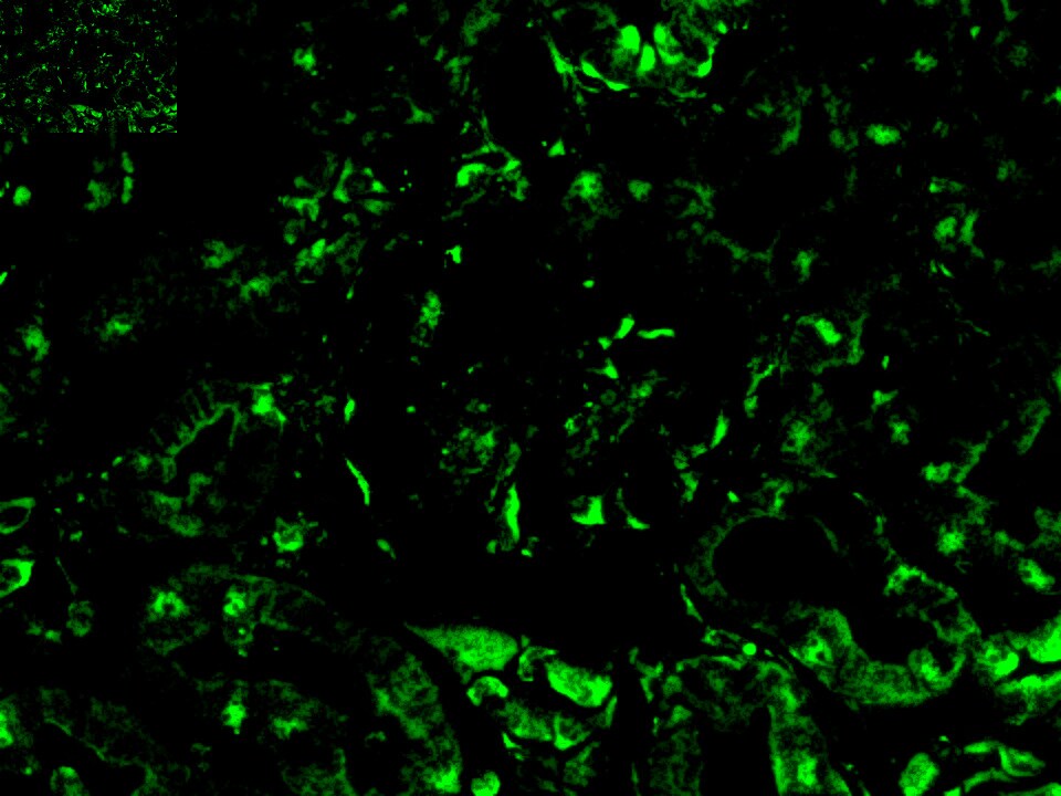
Immunohistochemistry: p19ARF/CDKN2A Antibody [NB200-106] - Adult mouse kidney (glomerulus). Image from verified customer review.![Immunohistochemistry-Paraffin: p19ARF/CDKN2A Antibody - BSA Free [NB200-106]](https://resources.rndsystems.com/images/products/p19ARF-Antibody-Immunohistochemistry-Paraffin-NB200-106-img0005.jpg "Immunohistochemistry-Paraffin: p19ARF/CDKN2A Antibody - BSA Free [NB200-106]")
Immunohistochemistry-Paraffin: p19ARF/CDKN2A Antibody - BSA Free [NB200-106]
Immunohistochemistry-Paraffin: p19ARF/CDKN2A Antibody [NB200-106] - Analysis of FFPE mouse spleen using p19ARF/CDKN2A antibody at 10 ug/mL on a Bond Rx autostainer (Leica Biosystems). The assay involved 20 minutes of heat induced antigen retrieval (HIER) using 10mM sodium citrate buffer (pH 6.0) and endogenous peroxidase quenching with peroxide block. The sections were incubated with primary antibody for 30 minutes and Bond Polymer Refine Detection (Leica Biosystems) with DAB was used for signal development followed by counterstaining with hematoxylin. Whole slide scanning and capturing of representative images was performed using Aperio AT2 (Leica Biosystems). Nuclear staining in the germinal centers of spleen was observed. Staining was performed by Histowiz.
Western Blot: p19ARF/CDKN2A Antibody - BSA Free [NB200-106] -
Senescence marker p21 protein is elevated in CypB‐deficient cells and tissues. (A) Western blot showing levels of p21, p53, p19, and CypB in wild‐type and CypB‐null primary multipotent stromal cells, with quantitative data of p21 from two independent experiments on two individual lines generated from each mouse strain. *p < 0.01, unpaired two‐tailed t test. (B) Western blot showing levels of p21 and CypB in wild‐type and CypB null primary osteoblasts with quantitative data of p21 from five PpiB+/+ lines and three PpiB−/− lines. *p < 0.05, unpaired two‐tailed t test. (C) Western blot showing levels of p21 and CypB in IAT collected from CypB wild‐type (n = 3) and null mice (n = 4) and the quantitative data of p21 abundance in IAT. *p < 0.05, unpaired two‐tailed t test. Image collected and cropped by CiteAb from the following open publication (https://pubmed.ncbi.nlm.nih.gov/36248275), licensed under a CC-BY license. Not internally tested by Novus Biologicals.Applications for p19ARF/CDKN2A Antibody - BSA Free
ELISA
Immunocytochemistry/ Immunofluorescence
Immunohistochemistry
Immunohistochemistry-Frozen
Immunohistochemistry-Paraffin
Western Blot
Reviewed Applications
Read 1 review rated 3 using NB200-106 in the following applications:
Formulation, Preparation, and Storage
Purification
Formulation
Format
Preservative
Concentration
Shipping
Stability & Storage
Background: p19ARF/CDKN2A
Alternate Names
Gene Symbol
Additional p19ARF/CDKN2A Products
Product Documents for p19ARF/CDKN2A Antibody - BSA Free
Certificate of Analysis
To download a Certificate of Analysis, please enter a lot or batch number in the search box below.
Product Specific Notices for p19ARF/CDKN2A Antibody - BSA Free
This product is for research use only and is not approved for use in humans or in clinical diagnosis. Primary Antibodies are guaranteed for 1 year from date of receipt.
Citations for p19ARF/CDKN2A Antibody - BSA Free
 Powered by Bioz
Customer Reviews for p19ARF/CDKN2A Antibody - BSA Free (1)
Have you used p19ARF/CDKN2A Antibody - BSA Free?
Submit a review and receive an Amazon gift card!
$25/€18/£15/$25CAN/¥2500 Yen for a review with an image
$10/€7/£6/$10CAN/¥1110 Yen for a review without an image
Submit a review
Customer Images
-
Application: ImmunofluorescenceSample Tested: Adult kidneySpecies: MouseVerified Customer | Posted 05/07/2018mouse glomerulus

There are no reviews that match your criteria.
Protocols
View specific protocols for p19ARF/CDKN2A Antibody - BSA Free (NB200-106):
Immunocytochemistry Protocol
Culture cells to appropriate density in 35 mm culture dishes or 6-well plates.
1. Remove culture medium and wash the cells briefly in PBS. Add 10% formalin to the dish and fix at room temperature for 10 minutes.
2. Remove the formalin and wash the cells in PBS.
3. Permeablize the cells with 0.5% Triton X100 or other suitable detergent for 10 min.
4. Remove the permeablization buffer and wash three times for 10 minutes each in PBS with 0.1% Tween 20. Be sure to not let the specimen dry out.
5. To block nonspecific antibody binding, incubate in 10% normal goat serum from 1 hour to overnight at room temperature.
6. Add primary antibody at appropriate dilution and incubate overnight at 4C.
7. Remove primary antibody and replace with PBS. Wash three times for 10 minutes each.
8. Add secondary antibody at appropriate dilution. Incubate for 1 hour at room temperature.
9. Remove secondary antibody and replace with PBS. Wash three times for 10 minutes each.
10. Counter stain DNA with DAPi if required.
*The above information is only intended as a guide. The researcher should determine what protocol best meets their needs. Please follow proper laboratory procedures for the disposal of formalin.
Antigen Unmasking:
Bring slides to a boil in 10 mM sodium citrate buffer (pH 6.0) then maintain at a sub-boiling temperature for 10 minutes. Cool slides on bench-top for 30 minutes (keep slides in the sodium citrate buffer all the time).
Staining:
1. Wash sections in deionized water three times for 5 minutes each.
2. Wash sections in PBS for 5 minutes.
3. Block each section with 100-400 ul blocking solution (1% BSA in PBS) for 1 hour at room temperature.
4. Remove blocking solution and add 100-400 ul diluted primary antibody. Incubate overnight at 4 C.
5. Remove antibody solution and wash sections in wash buffer three times for 5 minutes each.
6. Add 100-400 ul HRP polymer conjugated secondary antibody. Incubate 30 minutes at room temperature.
7. Wash sections three times in wash buffer for 5 minutes each.
8. Add 100-400 ul DAB substrate to each section and monitor staining closely.
9. As soon as the sections develop, immerse slides in deionized water.
10. Counterstain sections in hematoxylin.
11. Wash sections in deionized water two times for 5 minutes each.
12. Dehydrate sections.
13. Mount coverslips.
Western Blot Protocol
1. Perform SDS-PAGE on samples to be analyzed, loading 10-25 ug of total protein per lane.
2. Transfer proteins to PVDF membrane according to the instructions provided by the manufacturer of the membrane and transfer apparatus.
3. Stain the membrane with Ponceau S (or similar product) to assess transfer success, and mark molecular weight standards where appropriate.
4. Rinse the blot TBS -0.05% Tween 20 (TBST).
5. Block the membrane in 5% Non-fat milk in TBST (blocking buffer) for at least 1 hour.
6. Wash the membrane in TBST three times for 10 minutes each.
7. Dilute primary antibody in blocking buffer and incubate overnight at 4C with gentle rocking.
8. Wash the membrane in TBST three times for 10 minutes each.
9. Incubate the membrane in diluted HRP conjugated secondary antibody in blocking buffer (as per manufacturer's instructions) for 1 hour at room temperature.
10. Wash the blot in TBST three times for 10 minutes each (this step can be repeated as required to reduce background).
11. Apply the detection reagent of choice in accordance with the manufacturers instructions
Find general support by application which include: protocols, troubleshooting, illustrated assays, videos and webinars.
- Antigen Retrieval Protocol (PIER)
- Antigen Retrieval for Frozen Sections Protocol
- Appropriate Fixation of IHC/ICC Samples
- Cellular Response to Hypoxia Protocols
- Chromogenic IHC Staining of Formalin-Fixed Paraffin-Embedded (FFPE) Tissue Protocol
- Chromogenic Immunohistochemistry Staining of Frozen Tissue
- ClariTSA™ Fluorophore Kits
- Detection & Visualization of Antibody Binding
- ELISA Sample Preparation & Collection Guide
- ELISA Troubleshooting Guide
- Fluorescent IHC Staining of Frozen Tissue Protocol
- Graphic Protocol for Heat-induced Epitope Retrieval
- Graphic Protocol for the Preparation and Fluorescent IHC Staining of Frozen Tissue Sections
- Graphic Protocol for the Preparation and Fluorescent IHC Staining of Paraffin-embedded Tissue Sections
- Graphic Protocol for the Preparation of Gelatin-coated Slides for Histological Tissue Sections
- How to Run an R&D Systems DuoSet ELISA
- How to Run an R&D Systems Quantikine ELISA
- How to Run an R&D Systems Quantikine™ QuicKit™ ELISA
- ICC Cell Smear Protocol for Suspension Cells
- ICC Immunocytochemistry Protocol Videos
- ICC for Adherent Cells
- IHC Sample Preparation (Frozen sections vs Paraffin)
- Immunocytochemistry (ICC) Protocol
- Immunocytochemistry Troubleshooting
- Immunofluorescence of Organoids Embedded in Cultrex Basement Membrane Extract
- Immunofluorescent IHC Staining of Formalin-Fixed Paraffin-Embedded (FFPE) Tissue Protocol
- Immunohistochemistry (IHC) and Immunocytochemistry (ICC) Protocols
- Immunohistochemistry Frozen Troubleshooting
- Immunohistochemistry Paraffin Troubleshooting
- Preparing Samples for IHC/ICC Experiments
- Preventing Non-Specific Staining (Non-Specific Binding)
- Primary Antibody Selection & Optimization
- Protocol for Heat-Induced Epitope Retrieval (HIER)
- Protocol for Making a 4% Formaldehyde Solution in PBS
- Protocol for VisUCyte™ HRP Polymer Detection Reagent
- Protocol for the Fluorescent ICC Staining of Cell Smears - Graphic
- Protocol for the Fluorescent ICC Staining of Cultured Cells on Coverslips - Graphic
- Protocol for the Preparation & Fixation of Cells on Coverslips
- Protocol for the Preparation and Chromogenic IHC Staining of Frozen Tissue Sections
- Protocol for the Preparation and Chromogenic IHC Staining of Frozen Tissue Sections - Graphic
- Protocol for the Preparation and Chromogenic IHC Staining of Paraffin-embedded Tissue Sections
- Protocol for the Preparation and Chromogenic IHC Staining of Paraffin-embedded Tissue Sections - Graphic
- Protocol for the Preparation and Fluorescent ICC Staining of Cells on Coverslips
- Protocol for the Preparation and Fluorescent ICC Staining of Non-adherent Cells
- Protocol for the Preparation and Fluorescent ICC Staining of Stem Cells on Coverslips
- Protocol for the Preparation and Fluorescent IHC Staining of Frozen Tissue Sections
- Protocol for the Preparation and Fluorescent IHC Staining of Paraffin-embedded Tissue Sections
- Protocol for the Preparation of Gelatin-coated Slides for Histological Tissue Sections
- Protocol for the Preparation of a Cell Smear for Non-adherent Cell ICC - Graphic
- Quantikine HS ELISA Kit Assay Principle, Alkaline Phosphatase
- Quantikine HS ELISA Kit Principle, Streptavidin-HRP Polymer
- R&D Systems Quality Control Western Blot Protocol
- Sandwich ELISA (Colorimetric) – Biotin/Streptavidin Detection Protocol
- Sandwich ELISA (Colorimetric) – Direct Detection Protocol
- TUNEL and Active Caspase-3 Detection by IHC/ICC Protocol
- The Importance of IHC/ICC Controls
- Troubleshooting Guide: ELISA
- Troubleshooting Guide: Immunohistochemistry
- Troubleshooting Guide: Western Blot Figures
- Western Blot Conditions
- Western Blot Protocol
- Western Blot Protocol for Cell Lysates
- Western Blot Troubleshooting
- Western Blot Troubleshooting Guide
- View all Protocols, Troubleshooting, Illustrated assays and Webinars
FAQs for p19ARF/CDKN2A Antibody - BSA Free
-
Q: Is there a recommended antigen retrieval method for this product for use in IHC-P?
A: For NB200-106 I would recommend our standard HIER Citrate Buffer pH 6.0.