p27/Kip1 Antibody (SX53G8) - Azide and BSA Free
Novus Biologicals | Catalog # NBP2-34718
Key Product Details
Species Reactivity
Human, Mouse, Rat, Monkey
Applications
Immunohistochemistry, Immunohistochemistry-Paraffin, Western Blot, Flow Cytometry, Immunocytochemistry/ Immunofluorescence, CyTOF-ready
Label
Unconjugated
Antibody Source
Monoclonal Mouse IgG1 kappa Clone # SX53G8
Format
Azide and BSA Free
Loading...
Product Specifications
Immunogen
Purified GST-p27 fusion protein of human origin (Uniprot: P46527)
Localization
Nuclear
Specificity
This monoclonal antibody recognizes a 27kDa protein, identified as the p27Kip1, a cell cycle regulatory mitotic inhibitor. It is highly specific and shows no cross-reaction with other related mitotic inhibitors. In Western blotting of cell lysates from 7 human breast cancer cell lines (ZR75-1, ZR75-30, MCF-7, MDAMB453, T47D, CAL51, 734B), the antibody labels a single band corresponding to p27Kip1. It functions as a negative regulator of G1 progression and has been proposed to function as a possible mediator of TGF- induced G1 arrest. p27Kip1 is a candidate tumor suppressor gene. Reportedly, low p27 expression has been associated with unfavorable prognosis in renal cell carcinoma, colon carcinoma, breast carcinomas, non-small-cell lung carcinoma, hepatocellular carcinoma, multiple myeloma, and lymph node metastases in papillary carcinoma of the thyroid, as well as a more aggressive phenotype in carcinoma of the cervix.
Clonality
Monoclonal
Host
Mouse
Isotype
IgG1 kappa
Description
1.0 mg/ml of antibody purified from Bioreactor Concentrate by Protein A/G. Prepared in 10mM PBS WITHOUT BSA & azide. Also available at 200 ug/ml WITH BSA & azide (NBP2-32889).
Antibody with azide - store at 2 to 8C. Antibody without azide - store at -20 to -80C.
Antibody with azide - store at 2 to 8C. Antibody without azide - store at -20 to -80C.
Scientific Data Images for p27/Kip1 Antibody (SX53G8) - Azide and BSA Free
![Immunocytochemistry/ Immunofluorescence: p27/Kip1 Antibody (SX53G8) - Azide and BSA Free [NBP2-34718]](https://resources.rndsystems.com/images/products/p27-Kip1-Antibody-SX53G8-Azide-and-BSA-Free-Immunocytochemistry-Immunofluorescence-NBP2-34718-img0001.jpg "Immunocytochemistry/ Immunofluorescence: p27/Kip1 Antibody (SX53G8) - Azide and BSA Free [NBP2-34718]")
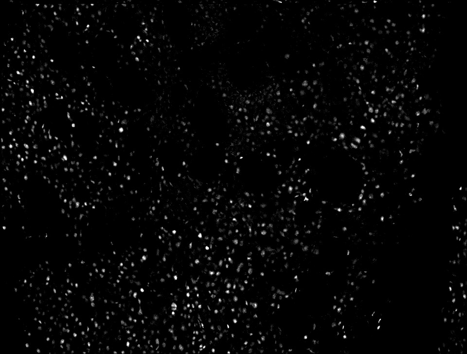
Immunocytochemistry/ Immunofluorescence: p27/Kip1 Antibody (SX53G8) - Azide and BSA Free [NBP2-34718]
Immunocytochemistry/Immunofluorescence: p27/Kip1 Antibody (SX53G8) - Azide and BSA Free [NBP2-34718] - Analysis of PFA-fixed MCF-7 cells labeling p27 with p27/Kip1 Antibody (SX53G8) followed by Goat anti-Mouse IgG-CF488 (Green). Membrane is stained with Phalloidin-CF640.![Immunohistochemistry-Paraffin: p27/Kip1 Antibody (SX53G8) - Azide and BSA Free [NBP2-34718]](https://resources.rndsystems.com/images/products/p27-Kip1-Antibody-SX53G8-Azide-and-BSA-Free-Immunohistochemistry-Paraffin-NBP2-34718-img0007.jpg "Immunohistochemistry-Paraffin: p27/Kip1 Antibody (SX53G8) - Azide and BSA Free [NBP2-34718]")
Immunohistochemistry-Paraffin: p27/Kip1 Antibody (SX53G8) - Azide and BSA Free [NBP2-34718]
Immunohistochemistry-Paraffin: p27/Kip1 Antibody (SX53G8) - Azide and BSA Free [NBP2-34718] - p27/Kip1 antibody in CODEX stain of human bone marrow tissue. Image from verified customer review.![Flow Cytometry: p27/Kip1 Antibody (SX53G8) - Azide and BSA Free [NBP2-34718]](https://resources.rndsystems.com/images/products/p27-Kip1-Antibody-SX53G8-Azide-and-BSA-Free-Flow-Cytometry-NBP2-34718-img0006.jpg "Flow Cytometry: p27/Kip1 Antibody (SX53G8) - Azide and BSA Free [NBP2-34718]")
Flow Cytometry: p27/Kip1 Antibody (SX53G8) - Azide and BSA Free [NBP2-34718]
Flow Cytometry: p27/Kip1 Antibody (SX53G8) - Azide and BSA Free [NBP2-34718] - Flow Cytometry of human p27/Kip1 on HeLa cells. Black: cells alone; Grey: Isotype Control; Green: CF488-labeled p27/Kip1 Monoclonal Antibody (SX53G8).![Immunohistochemistry-Paraffin: p27/Kip1 Antibody (SX53G8) - Azide and BSA Free [NBP2-34718]](https://resources.rndsystems.com/images/products/p27-Kip1-Antibody-SX53G8-Azide-and-BSA-Free-Immunohistochemistry-Paraffin-NBP2-34718-img0004.jpg "Immunohistochemistry-Paraffin: p27/Kip1 Antibody (SX53G8) - Azide and BSA Free [NBP2-34718]")
Immunohistochemistry-Paraffin: p27/Kip1 Antibody (SX53G8) - Azide and BSA Free [NBP2-34718]
Immunohistochemistry-Paraffin: p27/Kip1 Antibody (SX53G8) - Azide and BSA Free [NBP2-34718] - Formalin-paraffin human colon stained with p27 MAb (SX53G8)![Immunohistochemistry-Paraffin: p27/Kip1 Antibody (SX53G8) - Azide and BSA Free [NBP2-34718]](https://resources.rndsystems.com/images/products/p27-Kip1-Antibody-SX53G8-Azide-and-BSA-Free-Immunohistochemistry-Paraffin-NBP2-34718-img0005.jpg "Immunohistochemistry-Paraffin: p27/Kip1 Antibody (SX53G8) - Azide and BSA Free [NBP2-34718]")
Immunohistochemistry-Paraffin: p27/Kip1 Antibody (SX53G8) - Azide and BSA Free [NBP2-34718]
Immunohistochemistry-Paraffin: p27/Kip1 Antibody (SX53G8) - Azide and BSA Free [NBP2-34718] - Formalin-fixed, paraffin-embedded human prostate Carcinoma stained with p27 Monoclonal Antibody (sx53G8)![Flow Cytometry: p27/Kip1 Antibody (SX53G8) - Azide and BSA Free [NBP2-34718]](https://resources.rndsystems.com/images/products/p27-Kip1-Antibody-SX53G8-Azide-and-BSA-Free-Flow-Cytometry-NBP2-34718-img0003.jpg "Flow Cytometry: p27/Kip1 Antibody (SX53G8) - Azide and BSA Free [NBP2-34718]")
Flow Cytometry: p27/Kip1 Antibody (SX53G8) - Azide and BSA Free [NBP2-34718]
Flow Cytometry: p27/Kip1 Antibody (SX53G8) - Azide and BSA Free [NBP2-34718] - Analysis of PFA-fixed MCF-7 cells using p27/Kip1 Antibody (SX53G8) followed by Goat anti- Mouse- IgG-CF488 (Blue); Isotype Control (Red).![p27/Kip1 Antibody (SX53G8) - Azide and BSA Free Western Blot: p27/Kip1 Antibody (SX53G8) - Azide and BSA Free [NBP2-34718] -](https://resources.rndsystems.com/images/products/nbp2-34718_mouse-monoclonal-p27-kip1-antibody-sx53g8-azide-and-bsa-free-2620251694723.jpeg "Western Blot: p27/Kip1 Antibody (SX53G8) - Azide and BSA Free [NBP2-34718] -")
Western Blot: p27/Kip1 Antibody (SX53G8) - Azide and BSA Free [NBP2-34718] -
Western blot analysis of Raw lysate using p27/Kip1 Antibody (SX53G8) - Azide and BSA Free.![p27/Kip1 Antibody (SX53G8) - Azide and BSA Free Western Blot: p27/Kip1 Antibody (SX53G8) - Azide and BSA Free [NBP2-34718] -](https://resources.rndsystems.com/images/products/nbp2-34718_mouse-monoclonal-p27-kip1-antibody-sx53g8-azide-and-bsa-free-2620251694761.jpeg "Western Blot: p27/Kip1 Antibody (SX53G8) - Azide and BSA Free [NBP2-34718] -")
Western Blot: p27/Kip1 Antibody (SX53G8) - Azide and BSA Free [NBP2-34718] -
Western blot analysis of 3T3 lysate using p27/Kip1 Antibody (SX53G8) - Azide and BSA Free.Applications for p27/Kip1 Antibody (SX53G8) - Azide and BSA Free
Application
Recommended Usage
Flow Cytometry
0.5-1ug/million cells
Immunocytochemistry/ Immunofluorescence
0.5-1ug/ml
Immunohistochemistry-Paraffin
0.5-1ug/ml
Western Blot
0.5-1ug/ml
Application Notes
Immunohistochemistry (Formalin-fixed): 0.25-0.5ug/ml for 30 minutes at RT. Staining of formalin-fixed tissues requires heating tissue sections in 10mM Tris with 1mM EDTA, pH 9.0, for 45 min at 95C followed by cooling at RT for 20 minutes.
Optimal dilution for a specific application should be determined.
Optimal dilution for a specific application should be determined.
Reviewed Applications
Read 1 review rated 5 using NBP2-34718 in the following applications:
Flow Cytometry Panel Builder
Bio-Techne Knows Flow Cytometry
Save time and reduce costly mistakes by quickly finding compatible reagents using the Panel Builder Tool.
Advanced Features
- Spectra Viewer - Custom analysis of spectra from multiple fluorochromes
- Spillover Popups - Visualize the spectra of individual fluorochromes
- Antigen Density Selector - Match fluorochrome brightness with antigen density

Formulation, Preparation, and Storage
Purification
Protein A or G purified
Formulation
10 mM PBS
Format
Azide and BSA Free
Preservative
No Preservative
Concentration
1.0 mg/ml
Shipping
The product is shipped with polar packs. Upon receipt, store it immediately at the temperature recommended below.
Stability & Storage
Store at -20 to -80C. Avoid freeze-thaw cycles.
Background: p27/Kip1
Long Name
p27 Cyclin Dependent Kinase 4 Inhibitor 1B
Alternate Names
CDKN1B, Kip1
Gene Symbol
CDKN1B
Additional p27/Kip1 Products
Product Documents for p27/Kip1 Antibody (SX53G8) - Azide and BSA Free
Certificate of Analysis
To download a Certificate of Analysis, please enter a lot or batch number in the search box below.
Product Specific Notices for p27/Kip1 Antibody (SX53G8) - Azide and BSA Free
This product is for research use only and is not approved for use in humans or in clinical diagnosis. Primary Antibodies are guaranteed for 1 year from date of receipt.
Related Research Areas
Customer Reviews for p27/Kip1 Antibody (SX53G8) - Azide and BSA Free (1)
5 out of 5
1 Customer Rating
Have you used p27/Kip1 Antibody (SX53G8) - Azide and BSA Free?
Submit a review and receive an Amazon gift card!
$25/€18/£15/$25CAN/¥2500 Yen for a review with an image
$10/€7/£6/$10CAN/¥1110 Yen for a review without an image
Submit a review
Customer Images
Showing
1
-
1 of
1 review
Showing All
Filter By:
-
Application: Immunohistochemistry-ParaffinSample Tested: bone marrowSpecies: HumanVerified Customer | Posted 10/25/2022p27 antibody in CODEX stain of human bone marrow tissue

There are no reviews that match your criteria.
Protocols
Find general support by application which include: protocols, troubleshooting, illustrated assays, videos and webinars.
- 7-Amino Actinomycin D (7-AAD) Cell Viability Flow Cytometry Protocol
- Antigen Retrieval Protocol (PIER)
- Antigen Retrieval for Frozen Sections Protocol
- Appropriate Fixation of IHC/ICC Samples
- Cellular Response to Hypoxia Protocols
- Chromogenic IHC Staining of Formalin-Fixed Paraffin-Embedded (FFPE) Tissue Protocol
- Chromogenic Immunohistochemistry Staining of Frozen Tissue
- ClariTSA™ Fluorophore Kits
- Detection & Visualization of Antibody Binding
- Extracellular Membrane Flow Cytometry Protocol
- Flow Cytometry Protocol for Cell Surface Markers
- Flow Cytometry Protocol for Staining Membrane Associated Proteins
- Flow Cytometry Staining Protocols
- Flow Cytometry Troubleshooting Guide
- Fluorescent IHC Staining of Frozen Tissue Protocol
- Graphic Protocol for Heat-induced Epitope Retrieval
- Graphic Protocol for the Preparation and Fluorescent IHC Staining of Frozen Tissue Sections
- Graphic Protocol for the Preparation and Fluorescent IHC Staining of Paraffin-embedded Tissue Sections
- Graphic Protocol for the Preparation of Gelatin-coated Slides for Histological Tissue Sections
- ICC Cell Smear Protocol for Suspension Cells
- ICC Immunocytochemistry Protocol Videos
- ICC for Adherent Cells
- IHC Sample Preparation (Frozen sections vs Paraffin)
- Immunocytochemistry (ICC) Protocol
- Immunocytochemistry Troubleshooting
- Immunofluorescence of Organoids Embedded in Cultrex Basement Membrane Extract
- Immunofluorescent IHC Staining of Formalin-Fixed Paraffin-Embedded (FFPE) Tissue Protocol
- Immunohistochemistry (IHC) and Immunocytochemistry (ICC) Protocols
- Immunohistochemistry Frozen Troubleshooting
- Immunohistochemistry Paraffin Troubleshooting
- Intracellular Flow Cytometry Protocol Using Alcohol (Methanol)
- Intracellular Flow Cytometry Protocol Using Detergents
- Intracellular Nuclear Staining Flow Cytometry Protocol Using Detergents
- Intracellular Staining Flow Cytometry Protocol Using Alcohol Permeabilization
- Intracellular Staining Flow Cytometry Protocol Using Detergents to Permeabilize Cells
- Preparing Samples for IHC/ICC Experiments
- Preventing Non-Specific Staining (Non-Specific Binding)
- Primary Antibody Selection & Optimization
- Propidium Iodide Cell Viability Flow Cytometry Protocol
- Protocol for Heat-Induced Epitope Retrieval (HIER)
- Protocol for Liperfluo
- Protocol for Making a 4% Formaldehyde Solution in PBS
- Protocol for VisUCyte™ HRP Polymer Detection Reagent
- Protocol for the Characterization of Human Th22 Cells
- Protocol for the Characterization of Human Th9 Cells
- Protocol for the Fluorescent ICC Staining of Cell Smears - Graphic
- Protocol for the Fluorescent ICC Staining of Cultured Cells on Coverslips - Graphic
- Protocol for the Preparation & Fixation of Cells on Coverslips
- Protocol for the Preparation and Chromogenic IHC Staining of Frozen Tissue Sections
- Protocol for the Preparation and Chromogenic IHC Staining of Frozen Tissue Sections - Graphic
- Protocol for the Preparation and Chromogenic IHC Staining of Paraffin-embedded Tissue Sections
- Protocol for the Preparation and Chromogenic IHC Staining of Paraffin-embedded Tissue Sections - Graphic
- Protocol for the Preparation and Fluorescent ICC Staining of Cells on Coverslips
- Protocol for the Preparation and Fluorescent ICC Staining of Non-adherent Cells
- Protocol for the Preparation and Fluorescent ICC Staining of Stem Cells on Coverslips
- Protocol for the Preparation and Fluorescent IHC Staining of Frozen Tissue Sections
- Protocol for the Preparation and Fluorescent IHC Staining of Paraffin-embedded Tissue Sections
- Protocol for the Preparation of Gelatin-coated Slides for Histological Tissue Sections
- Protocol for the Preparation of a Cell Smear for Non-adherent Cell ICC - Graphic
- Protocol: Annexin V and PI Staining by Flow Cytometry
- Protocol: Annexin V and PI Staining for Apoptosis by Flow Cytometry
- R&D Systems Quality Control Western Blot Protocol
- TUNEL and Active Caspase-3 Detection by IHC/ICC Protocol
- The Importance of IHC/ICC Controls
- Troubleshooting Guide: Fluorokine Flow Cytometry Kits
- Troubleshooting Guide: Immunohistochemistry
- Troubleshooting Guide: Western Blot Figures
- Western Blot Conditions
- Western Blot Protocol
- Western Blot Protocol for Cell Lysates
- Western Blot Troubleshooting
- Western Blot Troubleshooting Guide
- View all Protocols, Troubleshooting, Illustrated assays and Webinars
Loading...
Associated Pathways
