![Immunohistochemistry-Paraffin: pan Cadherin Antibody [NB200-592]](https://resources.rndsystems.com/images/products/N-Cadherin-Antibody-Immunohistochemistry-Paraffin-NB200-592-img0003.jpg "Immunohistochemistry-Paraffin: pan Cadherin Antibody [NB200-592]")
Loading...
Key Product Details
Validated by
Biological Validation
Species Reactivity
Validated:
Human, Mouse, Rat, Amphibian, Avian, Bovine, Canine, Chicken, Fish, Rabbit, Xenopus
Cited:
Human, Mouse
Applications
Validated:
Immunohistochemistry, Immunohistochemistry-Paraffin, Immunohistochemistry-Frozen, Western Blot, Immunocytochemistry/ Immunofluorescence
Cited:
Immunohistochemistry-Paraffin, Western Blot, Immunocytochemistry/ Immunofluorescence, IF/IHC
Label
Unconjugated
Antibody Source
Polyclonal Rabbit IgG
Loading...
Product Specifications
Immunogen
Synthetic peptide: DYDYLNDWGPRFKKLADMYGGGDD conjugated to KLH by a Glutaraldehyde linker. Sequence corrresponds to the C-terminal amino acids of N-Cadherin (Chicken), with an extra N-terminal lysine residue (24 amino acids).
Localization
Cell membrane
Marker
Mesenchymal Cells Marker
Specificity
Recognizes all cadherin members. Rabbit Anti-Pan Cadherin shows specific reactivity with a distinct 135 kDa band on chicken or rabbit heart extract blots using indirect immunoblotting.
Clonality
Polyclonal
Host
Rabbit
Isotype
IgG
Scientific Data Images for pan Cadherin Antibody
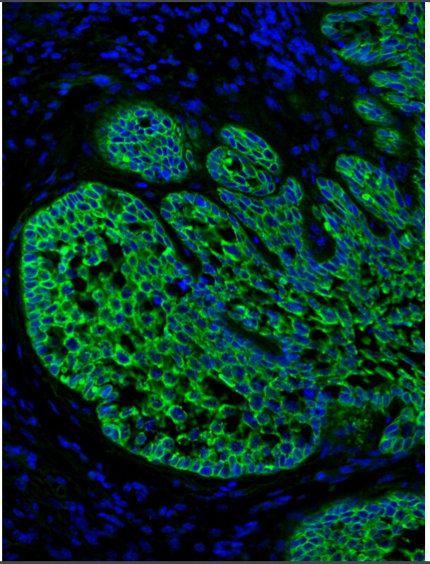
Immunohistochemistry-Paraffin: pan Cadherin Antibody [NB200-592]
Immunohistochemistry-Paraffin: pan Cadherin Antibody [NB200-592] - Mouse cervical cancer tissue stained with N-Cadherin antibody (green). Nuclei were counterstained with DAPI (Blue). Antigen retrieval was done with pepsin treatment. Image from verified customer review.![Western Blot: pan Cadherin Antibody [NB200-592]](https://resources.rndsystems.com/images/products/pan-Cadherin-Antibody-Western-Blot-NB200-592-img0005.jpg "Western Blot: pan Cadherin Antibody [NB200-592]")
Western Blot: pan Cadherin Antibody [NB200-592]
Western Blot: pan Cadherin Antibody [NB200-592] - Whole extract of Chicken Cardiac Muscle were separated on SDS-PAGE and probed with Anti-Pan Cadherin antibody produced in Rabbit. The antibody was developed using 1:1000 Anti-Rabbit IgG (whole molecule)-Peroxidase antibody produced in Goat.Lanes:1. 1:200 antibody2. 1:400 antibody3. Negative Control![Immunohistochemistry: pan Cadherin Antibody [NB200-592]](https://resources.rndsystems.com/images/products/pan-Cadherin-Antibody-Immunohistochemistry-NB200-592-img0004.jpg "Immunohistochemistry: pan Cadherin Antibody [NB200-592]")
Immunohistochemistry: pan Cadherin Antibody [NB200-592]
Immunohistochemistry: pan Cadherin Antibody [NB200-592] - Immunoperoxidase staining of a formalin-fixed, paraffin-embedded section of human heart using Rabbit Anti-Pan Cadherin.Applications for pan Cadherin Antibody
Application
Recommended Usage
Immunocytochemistry/ Immunofluorescence
1:100
Immunohistochemistry
1:10-1:500
Immunohistochemistry-Frozen
1:10-1:500
Immunohistochemistry-Paraffin
1:1000
Western Blot
1:200
Application Notes
This antibody is useful for: Western Blot. IHC-P: Perform enzymatic antigen retrieval before commencing with IHC staining protocol. WB: On chicken and rabbit heart extract this antibody detects a single band at 135 kD.
Reviewed Applications
Read 1 review rated 5 using NB200-592 in the following applications:
Formulation, Preparation, and Storage
Purification
Unpurified
Formulation
Whole antisera
Preservative
0.09% Sodium Azide
Concentration
This product is unpurified. The exact concentration of antibody is not quantifiable.
Shipping
The product is shipped with polar packs. Upon receipt, store it immediately at the temperature recommended below.
Stability & Storage
Store at -20C. Avoid freeze-thaw cycles.
Background: Cadherin
Long Name
Cadherin Pan Specific
Alternate Names
Arc-1, cadherin 1, E-cadherin (epithelial), cadherin 1, type 1, E-cadherin (epithelial), cadherin-1, calcium-dependent adhesion protein, epithelial, CAM 120/80, CD324, CD324 antigen, CDHE, cell-CAM 120/80, ECAD, E-cadherin, Epithelial cadherin, LCAM, UVOE-Cadherin, uvomorulin
Gene Symbol
CDH1
Additional Cadherin Products
Product Documents for pan Cadherin Antibody
Certificate of Analysis
To download a Certificate of Analysis, please enter a lot or batch number in the search box below.
Product Specific Notices for pan Cadherin Antibody
This product is for research use only and is not approved for use in humans or in clinical diagnosis. Primary Antibodies are guaranteed for 1 year from date of receipt.
Related Research Areas
Citations for pan Cadherin Antibody
 Powered by Bioz
Customer Reviews for pan Cadherin Antibody (1)
5 out of 5
1 Customer Rating
Have you used pan Cadherin Antibody?
Submit a review and receive an Amazon gift card!
$25/€18/£15/$25CAN/¥2500 Yen for a review with an image
$10/€7/£6/$10CAN/¥1110 Yen for a review without an image
Submit a review
Customer Images
Showing
1
-
1 of
1 review
Showing All
Filter By:
-
Application: Immunohistochemistry-ParaffinSample Tested: Mouse cervixSpecies: MouseVerified Customer | Posted 12/20/2016cervical cancer in the mouse was stained for N-cadherin (green). Nuclei are shown in blue.antigen retrieval with pepsin treatment

There are no reviews that match your criteria.
Protocols
Find general support by application which include: protocols, troubleshooting, illustrated assays, videos and webinars.
- Antigen Retrieval Protocol (PIER)
- Antigen Retrieval for Frozen Sections Protocol
- Appropriate Fixation of IHC/ICC Samples
- Cellular Response to Hypoxia Protocols
- Chromogenic IHC Staining of Formalin-Fixed Paraffin-Embedded (FFPE) Tissue Protocol
- Chromogenic Immunohistochemistry Staining of Frozen Tissue
- ClariTSA™ Fluorophore Kits
- Detection & Visualization of Antibody Binding
- Fluorescent IHC Staining of Frozen Tissue Protocol
- Graphic Protocol for Heat-induced Epitope Retrieval
- Graphic Protocol for the Preparation and Fluorescent IHC Staining of Frozen Tissue Sections
- Graphic Protocol for the Preparation and Fluorescent IHC Staining of Paraffin-embedded Tissue Sections
- Graphic Protocol for the Preparation of Gelatin-coated Slides for Histological Tissue Sections
- ICC Cell Smear Protocol for Suspension Cells
- ICC Immunocytochemistry Protocol Videos
- ICC for Adherent Cells
- IHC Sample Preparation (Frozen sections vs Paraffin)
- Immunocytochemistry (ICC) Protocol
- Immunocytochemistry Troubleshooting
- Immunofluorescence of Organoids Embedded in Cultrex Basement Membrane Extract
- Immunofluorescent IHC Staining of Formalin-Fixed Paraffin-Embedded (FFPE) Tissue Protocol
- Immunohistochemistry (IHC) and Immunocytochemistry (ICC) Protocols
- Immunohistochemistry Frozen Troubleshooting
- Immunohistochemistry Paraffin Troubleshooting
- Preparing Samples for IHC/ICC Experiments
- Preventing Non-Specific Staining (Non-Specific Binding)
- Primary Antibody Selection & Optimization
- Protocol for Heat-Induced Epitope Retrieval (HIER)
- Protocol for Making a 4% Formaldehyde Solution in PBS
- Protocol for VisUCyte™ HRP Polymer Detection Reagent
- Protocol for the Fluorescent ICC Staining of Cell Smears - Graphic
- Protocol for the Fluorescent ICC Staining of Cultured Cells on Coverslips - Graphic
- Protocol for the Preparation & Fixation of Cells on Coverslips
- Protocol for the Preparation and Chromogenic IHC Staining of Frozen Tissue Sections
- Protocol for the Preparation and Chromogenic IHC Staining of Frozen Tissue Sections - Graphic
- Protocol for the Preparation and Chromogenic IHC Staining of Paraffin-embedded Tissue Sections
- Protocol for the Preparation and Chromogenic IHC Staining of Paraffin-embedded Tissue Sections - Graphic
- Protocol for the Preparation and Fluorescent ICC Staining of Cells on Coverslips
- Protocol for the Preparation and Fluorescent ICC Staining of Non-adherent Cells
- Protocol for the Preparation and Fluorescent ICC Staining of Stem Cells on Coverslips
- Protocol for the Preparation and Fluorescent IHC Staining of Frozen Tissue Sections
- Protocol for the Preparation and Fluorescent IHC Staining of Paraffin-embedded Tissue Sections
- Protocol for the Preparation of Gelatin-coated Slides for Histological Tissue Sections
- Protocol for the Preparation of a Cell Smear for Non-adherent Cell ICC - Graphic
- R&D Systems Quality Control Western Blot Protocol
- TUNEL and Active Caspase-3 Detection by IHC/ICC Protocol
- The Importance of IHC/ICC Controls
- Troubleshooting Guide: Immunohistochemistry
- Troubleshooting Guide: Western Blot Figures
- Western Blot Conditions
- Western Blot Protocol
- Western Blot Protocol for Cell Lysates
- Western Blot Troubleshooting
- Western Blot Troubleshooting Guide
- View all Protocols, Troubleshooting, Illustrated assays and Webinars
Loading...