PARD3/Par3 Antibody - BSA Free
Novus Biologicals | Catalog # NBP1-88861
![Western Blot: PARD3/Par3 Antibody [NBP1-88861]](https://resources.rndsystems.com/images/products/PARD3-Par3-Antibody-Western-Blot-NBP1-88861-img0017.jpg "Western Blot: PARD3/Par3 Antibody [NBP1-88861]")
Loading...
Key Product Details
Validated by
Orthogonal Validation
Species Reactivity
Validated:
Human
Cited:
Human, Mouse
Applications
Validated:
Immunohistochemistry, Immunohistochemistry-Paraffin, Western Blot, Immunocytochemistry/ Immunofluorescence, Simple Western, Immunoprecipitation
Cited:
Immunohistochemistry, Immunohistochemistry-Paraffin, Immunohistochemistry-Frozen, Immunocytochemistry/ Immunofluorescence, Immunoprecipitation, IF/IHC
Label
Unconjugated
Antibody Source
Polyclonal Rabbit IgG
Format
BSA Free
Loading...
Product Specifications
Immunogen
This antibody was developed against Recombinant Protein corresponding to amino acids: LKGLGDMFRIQAKTREFRERQARERDYAEIQDFHRTFGCDDELMYGGVSSYEGSMALNARPQSPREGHMMDALYAQVKKPRNSKPSPVDSNR
Reactivity Notes
Mouse reactivity reported from a verified customer review.
Clonality
Polyclonal
Host
Rabbit
Isotype
IgG
Scientific Data Images for PARD3/Par3 Antibody - BSA Free
![Simple Western: PARD3/Par3 Antibody [NBP1-88861]](https://resources.rndsystems.com/images/products/PARD3-Par3-Antibody-Simple-Western-NBP1-88861-img0009.jpg "Simple Western: PARD3/Par3 Antibody [NBP1-88861]")
Simple Western: PARD3/Par3 Antibody [NBP1-88861]
Simple Western: PARD3/Par3 Antibody [NBP1-88861] - Simple Western lane view shows a specific band for PARD3/Par3 in 0.2 mg/ml of RT-4 lysate. This experiment was performed under reducing conditions using the 66-440 kDa separation system.![Immunocytochemistry/ Immunofluorescence: PARD3/Par3 Antibody [NBP1-88861]](https://resources.rndsystems.com/images/products/PARD3-Par3-Antibody-Immunocytochemistry-Immunofluorescence-NBP1-88861-img0016.jpg "Immunocytochemistry/ Immunofluorescence: PARD3/Par3 Antibody [NBP1-88861]")
Immunocytochemistry/ Immunofluorescence: PARD3/Par3 Antibody [NBP1-88861]
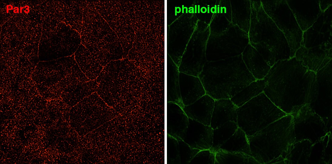
Immunocytochemistry/Immunofluorescence: PARD3/Par3 Antibody [NBP1-88861] - Staining of human cell line U-2 OS shows localization to cell junctions. Antibody staining is shown in green.![Immunocytochemistry/ Immunofluorescence: PARD3/Par3 Antibody [NBP1-88861]](https://resources.rndsystems.com/images/products/PARD3-Par3-Antibody-Immunocytochemistry-Immunofluorescence-NBP1-88861-img0012.jpg "Immunocytochemistry/ Immunofluorescence: PARD3/Par3 Antibody [NBP1-88861]")
Immunocytochemistry/ Immunofluorescence: PARD3/Par3 Antibody [NBP1-88861]
Immunocytochemistry/Immunofluorescence: PARD3/Par3 Antibody [NBP1-88861] - Staining PARD3 in mouse trophoblast stem cells. Verified customer review from 1DegreeBio.![Immunohistochemistry-Paraffin: PARD3/Par3 Antibody [NBP1-88861]](https://resources.rndsystems.com/images/products/PARD3-Par3-Antibody-Immunohistochemistry-Paraffin-NBP1-88861-img0014.jpg "Immunohistochemistry-Paraffin: PARD3/Par3 Antibody [NBP1-88861]")
Immunohistochemistry-Paraffin: PARD3/Par3 Antibody [NBP1-88861]
Immunohistochemistry-Paraffin: PARD3/Par3 Antibody [NBP1-88861] - Staining of human colon shows distinct luminal membranous positivity in glandular cells.![Immunocytochemistry/ Immunofluorescence: PARD3/Par3 Antibody [NBP1-88861]](https://resources.rndsystems.com/images/products/PARD3-Par3-Antibody-Immunocytochemistry-Immunofluorescence-NBP1-88861-img0023.jpg "Immunocytochemistry/ Immunofluorescence: PARD3/Par3 Antibody [NBP1-88861]")
Immunocytochemistry/ Immunofluorescence: PARD3/Par3 Antibody [NBP1-88861]
PARD3-Par3-Antibody-Immunocytochemistry-Immunofluorescence-NBP1-88861-img0023.jpg![Immunohistochemistry-Paraffin: PARD3/Par3 Antibody [NBP1-88861]](https://resources.rndsystems.com/images/products/PARD3-Par3-Antibody-Immunohistochemistry-Paraffin-NBP1-88861-img0022.jpg "Immunohistochemistry-Paraffin: PARD3/Par3 Antibody [NBP1-88861]")
Immunohistochemistry-Paraffin: PARD3/Par3 Antibody [NBP1-88861]
Immunohistochemistry-Paraffin: PARD3/Par3 Antibody [NBP1-88861] - Staining of human skin shows weak to moderate cytoplasmic positivity in epidermal cells.![Immunocytochemistry/ Immunofluorescence: PARD3/Par3 Antibody [NBP1-88861]](https://resources.rndsystems.com/images/products/PARD3-Par3-Antibody-Immunocytochemistry-Immunofluorescence-NBP1-88861-img0013.jpg "Immunocytochemistry/ Immunofluorescence: PARD3/Par3 Antibody [NBP1-88861]")
Immunocytochemistry/ Immunofluorescence: PARD3/Par3 Antibody [NBP1-88861]
Immunocytochemistry/Immunofluorescence: PARD3/Par3 Antibody [NBP1-88861] - Staining of human cell line 8505C cells (anaplastic thyroid cancer cell line). Image from verified customer review.![Immunohistochemistry-Paraffin: PARD3/Par3 Antibody [NBP1-88861]](https://resources.rndsystems.com/images/products/PARD3-Par3-Antibody-Immunohistochemistry-Paraffin-NBP1-88861-img0019.jpg "Immunohistochemistry-Paraffin: PARD3/Par3 Antibody [NBP1-88861]")
Immunohistochemistry-Paraffin: PARD3/Par3 Antibody [NBP1-88861]
Immunohistochemistry-Paraffin: PARD3/Par3 Antibody [NBP1-88861] - Staining of human kidney shows weak to moderate membranous positivity in cells in tubules.![Immunohistochemistry-Paraffin: PARD3/Par3 Antibody [NBP1-88861]](https://resources.rndsystems.com/images/products/PARD3-Par3-Antibody-Immunohistochemistry-Paraffin-NBP1-88861-img0020.jpg "Immunohistochemistry-Paraffin: PARD3/Par3 Antibody [NBP1-88861]")
Immunohistochemistry-Paraffin: PARD3/Par3 Antibody [NBP1-88861]
Immunohistochemistry-Paraffin: PARD3/Par3 Antibody [NBP1-88861] - Staining of human cerebellum shows moderate positivity in neuropil.![Immunohistochemistry-Paraffin: PARD3/Par3 Antibody [NBP1-88861]](https://resources.rndsystems.com/images/products/PARD3-Par3-Antibody-Immunohistochemistry-Paraffin-NBP1-88861-img0021.jpg "Immunohistochemistry-Paraffin: PARD3/Par3 Antibody [NBP1-88861]")
Immunohistochemistry-Paraffin: PARD3/Par3 Antibody [NBP1-88861]
Immunohistochemistry-Paraffin: PARD3/Par3 Antibody [NBP1-88861] - Staining of human fallopian tube shows weak to moderate positivity in luminal membrane in glandular cells.![Simple Western: PARD3/Par3 Antibody [NBP1-88861]](https://resources.rndsystems.com/images/products/PARD3-Par3-Antibody-Simple-Western-NBP1-88861-img0015.jpg "Simple Western: PARD3/Par3 Antibody [NBP1-88861]")
Simple Western: PARD3/Par3 Antibody [NBP1-88861]
Simple Western: PARD3/Par3 Antibody [NBP1-88861] - Electropherogram image(s) of corresponding Simple Western lane view. PARD3/Par3 antibody was used at 1:100 dilution on RT-4 lysate(s).
Immunocytochemistry/ Immunofluorescence: PARD3/Par3 Antibody [NBP1-88861] -
Immunocytochemistry/ Immunofluorescence: PARD3/Par3 Antibody [NBP1-88861] - MCAM is required to establish cell autonomous polarity. (A) In elongating myotubes (10T1/2 cells treated with testosterone for 7 days) VANGL2 is localized asymmetrically at the tip of the cell. (B) The VANGL2 enriched tip of the cell is marked by MSN. (C) In MCAM knockout C164 cells myotube elongation fails, MSN labels the whole plasma membrane & VANGL2 is spread across the cytoplasm. (D) Highly polarized localization of MCAM & SCRIB at the distal end of growing wild-type myotube. Separate channels of the boxed area are shown on the right. (E) In MCAM knockout cells SCRIB levels remain low & it is spread evenly in the cell. (F) In wild-type myotubes PAR3 remains cytoplasmic, whereas (G) in MCAM knockout C164 cells it can be detected at the cell cortex. (H) RT-qPCR demonstrates reduced expression of Scrib in MCAM mutant cell lines. Cells were treated for 7 days with BMP2 or testosterone (n=3; **P<0.01; ***P<0.001; two-tailed t-test; mean±s.e.m.). (I) Deletion of MCAM endocytosis motif leads to similar polarity defects as complete MCAM elimination. VANGL2 is evenly spread in U125 cells & PAR3 accumulates in cell cortex. (J) In chondrogenic differentiation VANGL2 was observed asymmetrically in limited number of cells. In MCAM mutant cell lines (C149, C164, U125) VANGL2 accumulated around the nucleus. (K) Initiation of myogenic (4-day culture with testosterone) & chondrogenic differentiation (4-day culture with BMP2) led to downregulation of ERK1/2 phosphorylation (p-ERK1/2). Instead in MCAM mutant cell lines ERK1/2 phosphorylation increased. Scale bars: 25 µm. Image collected & cropped by CiteAb from the following publication (https://journals.biologists.com/bio/article/doi/10.1242/bio.027771/2567…), licensed under a CC-BY license. Not internally tested by Novus Biologicals.
Immunocytochemistry/ Immunofluorescence: PARD3/Par3 Antibody [NBP1-88861] -
Immunocytochemistry/ Immunofluorescence: PARD3/Par3 Antibody [NBP1-88861] - MCAM is required to establish cell autonomous polarity. (A) In elongating myotubes (10T1/2 cells treated with testosterone for 7 days) VANGL2 is localized asymmetrically at the tip of the cell. (B) The VANGL2 enriched tip of the cell is marked by MSN. (C) In MCAM knockout C164 cells myotube elongation fails, MSN labels the whole plasma membrane & VANGL2 is spread across the cytoplasm. (D) Highly polarized localization of MCAM & SCRIB at the distal end of growing wild-type myotube. Separate channels of the boxed area are shown on the right. (E) In MCAM knockout cells SCRIB levels remain low & it is spread evenly in the cell. (F) In wild-type myotubes PAR3 remains cytoplasmic, whereas (G) in MCAM knockout C164 cells it can be detected at the cell cortex. (H) RT-qPCR demonstrates reduced expression of Scrib in MCAM mutant cell lines. Cells were treated for 7 days with BMP2 or testosterone (n=3; **P<0.01; ***P<0.001; two-tailed t-test; mean±s.e.m.). (I) Deletion of MCAM endocytosis motif leads to similar polarity defects as complete MCAM elimination. VANGL2 is evenly spread in U125 cells & PAR3 accumulates in cell cortex. (J) In chondrogenic differentiation VANGL2 was observed asymmetrically in limited number of cells. In MCAM mutant cell lines (C149, C164, U125) VANGL2 accumulated around the nucleus. (K) Initiation of myogenic (4-day culture with testosterone) & chondrogenic differentiation (4-day culture with BMP2) led to downregulation of ERK1/2 phosphorylation (p-ERK1/2). Instead in MCAM mutant cell lines ERK1/2 phosphorylation increased. Scale bars: 25 µm. Image collected & cropped by CiteAb from the following publication (https://journals.biologists.com/bio/article/doi/10.1242/bio.027771/2567…), licensed under a CC-BY license. Not internally tested by Novus Biologicals.Applications for PARD3/Par3 Antibody - BSA Free
Application
Recommended Usage
Immunocytochemistry/ Immunofluorescence
0.25-2 ug/ml
Immunohistochemistry
1:200 - 1:500
Immunohistochemistry-Paraffin
1:200-1:500
Immunoprecipitation
Reported in scientific literature (PMID:34158849).
Simple Western
1:100
Western Blot
0.04-0.4 ug/ml
Application Notes
ICC/IF Fixation Permeabilization: Use PFA/Triton X-100. IHC-Paraffin HIER pH6 retrieval is recommended. In Simple Western only 10 - 15 uL of the recommended dilution is used per data point.
See Simple Western Antibody Database for Simple Western validation: Tested in RT-4, separated by Size, antibody dilution of 1:100, apparent MW was 190 kDa
See Simple Western Antibody Database for Simple Western validation: Tested in RT-4, separated by Size, antibody dilution of 1:100, apparent MW was 190 kDa
Reviewed Applications
Read 2 reviews rated 4 using NBP1-88861 in the following applications:
Formulation, Preparation, and Storage
Purification
Affinity purified
Formulation
PBS (pH 7.2) and 40% Glycerol
Format
BSA Free
Preservative
0.02% Sodium Azide
Concentration
Concentrations vary lot to lot. See vial label for concentration. If unlisted please contact technical services.
Shipping
The product is shipped with polar packs. Upon receipt, store it immediately at the temperature recommended below.
Stability & Storage
Store at 4C short term. Aliquot and store at -20C long term. Avoid freeze-thaw cycles.
Background: PARD3/Par3
Long Name
Par-3 Partitioning Defective 3 Homolog
Alternate Names
ASIP, Baz, Bazooka, PAR3, PAR3A, SE2-5L16, SE2-5LT1, SE2-5T2
Gene Symbol
PARD3
Additional PARD3/Par3 Products
Product Documents for PARD3/Par3 Antibody - BSA Free
Certificate of Analysis
To download a Certificate of Analysis, please enter a lot or batch number in the search box below.
Product Specific Notices for PARD3/Par3 Antibody - BSA Free
This product is for research use only and is not approved for use in humans or in clinical diagnosis. Primary Antibodies are guaranteed for 1 year from date of receipt.
Related Research Areas
Citations for PARD3/Par3 Antibody - BSA Free
 Powered by Bioz
Customer Reviews for PARD3/Par3 Antibody - BSA Free (2)
4 out of 5
2 Customer Ratings
Have you used PARD3/Par3 Antibody - BSA Free?
Submit a review and receive an Amazon gift card!
$25/€18/£15/$25CAN/¥2500 Yen for a review with an image
$10/€7/£6/$10CAN/¥1110 Yen for a review without an image
Submit a review
Customer Images
-(01-ml)_NBP1-88861_7161.Jpg)
Showing
1
-
2 of
2 reviews
Showing All
Filter By:
-
Application: ImmunofluorescenceSample Tested: cell line derived from anaplastic thyroic cancerSpecies: HumanVerified Customer | Posted 04/29/20148505C cells (anaplastic thyroid cancer cell line)

-
Application: ImmunocytochemistrySample Tested: Mouse trophoblast stem cellsSpecies: MouseVerified Customer | Posted 11/07/2013
There are no reviews that match your criteria.
Protocols
Find general support by application which include: protocols, troubleshooting, illustrated assays, videos and webinars.
- Antigen Retrieval Protocol (PIER)
- Antigen Retrieval for Frozen Sections Protocol
- Appropriate Fixation of IHC/ICC Samples
- Cellular Response to Hypoxia Protocols
- Chromogenic IHC Staining of Formalin-Fixed Paraffin-Embedded (FFPE) Tissue Protocol
- Chromogenic Immunohistochemistry Staining of Frozen Tissue
- ClariTSA™ Fluorophore Kits
- Detection & Visualization of Antibody Binding
- Fluorescent IHC Staining of Frozen Tissue Protocol
- Graphic Protocol for Heat-induced Epitope Retrieval
- Graphic Protocol for the Preparation and Fluorescent IHC Staining of Frozen Tissue Sections
- Graphic Protocol for the Preparation and Fluorescent IHC Staining of Paraffin-embedded Tissue Sections
- Graphic Protocol for the Preparation of Gelatin-coated Slides for Histological Tissue Sections
- ICC Cell Smear Protocol for Suspension Cells
- ICC Immunocytochemistry Protocol Videos
- ICC for Adherent Cells
- IHC Sample Preparation (Frozen sections vs Paraffin)
- Immunocytochemistry (ICC) Protocol
- Immunocytochemistry Troubleshooting
- Immunofluorescence of Organoids Embedded in Cultrex Basement Membrane Extract
- Immunofluorescent IHC Staining of Formalin-Fixed Paraffin-Embedded (FFPE) Tissue Protocol
- Immunohistochemistry (IHC) and Immunocytochemistry (ICC) Protocols
- Immunohistochemistry Frozen Troubleshooting
- Immunohistochemistry Paraffin Troubleshooting
- Immunoprecipitation Protocol
- Preparing Samples for IHC/ICC Experiments
- Preventing Non-Specific Staining (Non-Specific Binding)
- Primary Antibody Selection & Optimization
- Protocol for Heat-Induced Epitope Retrieval (HIER)
- Protocol for Making a 4% Formaldehyde Solution in PBS
- Protocol for VisUCyte™ HRP Polymer Detection Reagent
- Protocol for the Fluorescent ICC Staining of Cell Smears - Graphic
- Protocol for the Fluorescent ICC Staining of Cultured Cells on Coverslips - Graphic
- Protocol for the Preparation & Fixation of Cells on Coverslips
- Protocol for the Preparation and Chromogenic IHC Staining of Frozen Tissue Sections
- Protocol for the Preparation and Chromogenic IHC Staining of Frozen Tissue Sections - Graphic
- Protocol for the Preparation and Chromogenic IHC Staining of Paraffin-embedded Tissue Sections
- Protocol for the Preparation and Chromogenic IHC Staining of Paraffin-embedded Tissue Sections - Graphic
- Protocol for the Preparation and Fluorescent ICC Staining of Cells on Coverslips
- Protocol for the Preparation and Fluorescent ICC Staining of Non-adherent Cells
- Protocol for the Preparation and Fluorescent ICC Staining of Stem Cells on Coverslips
- Protocol for the Preparation and Fluorescent IHC Staining of Frozen Tissue Sections
- Protocol for the Preparation and Fluorescent IHC Staining of Paraffin-embedded Tissue Sections
- Protocol for the Preparation of Gelatin-coated Slides for Histological Tissue Sections
- Protocol for the Preparation of a Cell Smear for Non-adherent Cell ICC - Graphic
- R&D Systems Quality Control Western Blot Protocol
- TUNEL and Active Caspase-3 Detection by IHC/ICC Protocol
- The Importance of IHC/ICC Controls
- Troubleshooting Guide: Immunohistochemistry
- Troubleshooting Guide: Western Blot Figures
- Western Blot Conditions
- Western Blot Protocol
- Western Blot Protocol for Cell Lysates
- Western Blot Troubleshooting
- Western Blot Troubleshooting Guide
- View all Protocols, Troubleshooting, Illustrated assays and Webinars
Loading...