PDGFR alpha Antibody (APA5) - BSA Free
Novus Biologicals | Catalog # NBP1-43350
![Flow Cytometry: PDGFR alpha Antibody (APA5) - BSA Free [NBP1-43350]](https://resources.rndsystems.com/images/products/PDGF-R-alpha-Antibody-APA5-Flow-Cytometry-NBP1-43350-img0003.jpg "Flow Cytometry: PDGFR alpha Antibody (APA5) - BSA Free [NBP1-43350]")
Key Product Details
Species Reactivity
Human, Mouse
Applications
Immunohistochemistry, Immunohistochemistry-Frozen, Western Blot, Flow Cytometry
Label
Unconjugated
Antibody Source
Monoclonal Rat IgG2a Kappa Clone # APA5
Format
BSA Free
Loading...
Product Specifications
Immunogen
The immunogen for this antibody was CD140a.
Reactivity Notes
Human reactivity reported from a verified customer review.
Clonality
Monoclonal
Host
Rat
Isotype
IgG2a Kappa
Scientific Data Images for PDGFR alpha Antibody (APA5) - BSA Free
Flow Cytometry: PDGFR alpha Antibody (APA5) - BSA Free [NBP1-43350]
Flow Cytometry: PDGF R alpha Antibody (APA5) [NBP1-43350] - Analysis using the Biotin conjugate of NBP1-43350. Staining of NIH/3T3 cell line with 0.5 ug of Anti-Mouse CD140a (PDGF Receptor a) Biotin followed by Streptavidin PE. Appropriate isotype controls were used (open histogram).![Flow Cytometry: PDGFR alpha Antibody (APA5) - BSA Free [NBP1-43350]](https://resources.rndsystems.com/images/products/PDGF-R-alpha-Antibody-APA5-Flow-Cytometry-NBP1-43350-img0001.jpg "Flow Cytometry: PDGFR alpha Antibody (APA5) - BSA Free [NBP1-43350]")
Flow Cytometry: PDGFR alpha Antibody (APA5) - BSA Free [NBP1-43350]
Flow Cytometry: PDGF R alpha Antibody (APA5) [NBP1-43350] - Staining of NIH/3T3 cell line with 0.5 ug of Rat IgG2a kappa Isotype Control Purified (open histogram) or 0.5 ug of Anti-Mouse CD140a (PDGF Receptor a) Purified (filled histogram) followed by Anti-Rat IgG Biotin and Streptavidin PE. Total viable cells were used for analysisApplications for PDGFR alpha Antibody (APA5) - BSA Free
Application
Recommended Usage
Flow Cytometry
1:10 - 1:1000
Immunohistochemistry
1:10 - 1:500
Immunohistochemistry-Frozen
1:10 - 1:500
Western Blot
1:100 - 1:2000
Application Notes
The APA5 antibody has been tested by flow cytometric analysis of the NIH-3T3 cell line. This can be used at less than or equal to 1 ug per test. Cell number should be determined empirically but can range from 10^5 to 10^8cells/test.
In flow cytometry, we recommend using a cell surface staining protocol and fixation and permeabilization is not required to get staining. If fixation/permeabilization is needed for other markers in the same experiment, we recommend doing all surface staining prior to the fix/perm procedure. This antibody may not bind to a fixed epitope.
In flow cytometry, we recommend using a cell surface staining protocol and fixation and permeabilization is not required to get staining. If fixation/permeabilization is needed for other markers in the same experiment, we recommend doing all surface staining prior to the fix/perm procedure. This antibody may not bind to a fixed epitope.
Reviewed Applications
Read 1 review rated 5 using NBP1-43350 in the following applications:
Flow Cytometry Panel Builder
Bio-Techne Knows Flow Cytometry
Save time and reduce costly mistakes by quickly finding compatible reagents using the Panel Builder Tool.
Advanced Features
- Spectra Viewer - Custom analysis of spectra from multiple fluorochromes
- Spillover Popups - Visualize the spectra of individual fluorochromes
- Antigen Density Selector - Match fluorochrome brightness with antigen density

Formulation, Preparation, and Storage
Purification
Protein A or G purified
Formulation
PBS (pH 7.2)
Format
BSA Free
Preservative
0.09% Sodium Azide
Concentration
0.5 mg/ml
Shipping
The product is shipped with polar packs. Upon receipt, store it immediately at the temperature recommended below.
Stability & Storage
Store at 4C. Do not freeze.
Background: PDGF R alpha
Long Name
Platelet-derived Growth Factor Receptor alpha
Alternate Names
CD140a, PDGFR alpha, PDGFRA
Entrez Gene IDs
18595 (Mouse)
Gene Symbol
PDGFRA
UniProt
Additional PDGF R alpha Products
Product Documents for PDGFR alpha Antibody (APA5) - BSA Free
Certificate of Analysis
To download a Certificate of Analysis, please enter a lot or batch number in the search box below.
Product Specific Notices for PDGFR alpha Antibody (APA5) - BSA Free
This product is for research use only and is not approved for use in humans or in clinical diagnosis. Primary Antibodies are guaranteed for 1 year from date of receipt.
Customer Reviews for PDGFR alpha Antibody (APA5) - BSA Free (1)
5 out of 5
1 Customer Rating
Have you used PDGFR alpha Antibody (APA5) - BSA Free?
Submit a review and receive an Amazon gift card!
$25/€18/£15/$25CAN/¥2500 Yen for a review with an image
$10/€7/£6/$10CAN/¥1110 Yen for a review without an image
Submit a review
Customer Images
Showing
1
-
1 of
1 review
Showing All
Filter By:
-
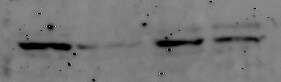
Application: Western BlotSample Tested: A549 human lung carcinoma cell lineSpecies: HumanVerified Customer | Posted 03/07/202030ug protein was loaded each lane and antibody dilution was 1:1000.

There are no reviews that match your criteria.
Protocols
Find general support by application which include: protocols, troubleshooting, illustrated assays, videos and webinars.
- 7-Amino Actinomycin D (7-AAD) Cell Viability Flow Cytometry Protocol
- Antigen Retrieval Protocol (PIER)
- Antigen Retrieval for Frozen Sections Protocol
- Appropriate Fixation of IHC/ICC Samples
- Cellular Response to Hypoxia Protocols
- Chromogenic IHC Staining of Formalin-Fixed Paraffin-Embedded (FFPE) Tissue Protocol
- Chromogenic Immunohistochemistry Staining of Frozen Tissue
- ClariTSA™ Fluorophore Kits
- Detection & Visualization of Antibody Binding
- Extracellular Membrane Flow Cytometry Protocol
- Flow Cytometry Protocol for Cell Surface Markers
- Flow Cytometry Protocol for Staining Membrane Associated Proteins
- Flow Cytometry Staining Protocols
- Flow Cytometry Troubleshooting Guide
- Fluorescent IHC Staining of Frozen Tissue Protocol
- Graphic Protocol for Heat-induced Epitope Retrieval
- Graphic Protocol for the Preparation and Fluorescent IHC Staining of Frozen Tissue Sections
- Graphic Protocol for the Preparation and Fluorescent IHC Staining of Paraffin-embedded Tissue Sections
- Graphic Protocol for the Preparation of Gelatin-coated Slides for Histological Tissue Sections
- IHC Sample Preparation (Frozen sections vs Paraffin)
- Immunofluorescent IHC Staining of Formalin-Fixed Paraffin-Embedded (FFPE) Tissue Protocol
- Immunohistochemistry (IHC) and Immunocytochemistry (ICC) Protocols
- Immunohistochemistry Frozen Troubleshooting
- Immunohistochemistry Paraffin Troubleshooting
- Intracellular Flow Cytometry Protocol Using Alcohol (Methanol)
- Intracellular Flow Cytometry Protocol Using Detergents
- Intracellular Nuclear Staining Flow Cytometry Protocol Using Detergents
- Intracellular Staining Flow Cytometry Protocol Using Alcohol Permeabilization
- Intracellular Staining Flow Cytometry Protocol Using Detergents to Permeabilize Cells
- Preparing Samples for IHC/ICC Experiments
- Preventing Non-Specific Staining (Non-Specific Binding)
- Primary Antibody Selection & Optimization
- Propidium Iodide Cell Viability Flow Cytometry Protocol
- Protocol for Heat-Induced Epitope Retrieval (HIER)
- Protocol for Liperfluo
- Protocol for Making a 4% Formaldehyde Solution in PBS
- Protocol for VisUCyte™ HRP Polymer Detection Reagent
- Protocol for the Characterization of Human Th22 Cells
- Protocol for the Characterization of Human Th9 Cells
- Protocol for the Preparation & Fixation of Cells on Coverslips
- Protocol for the Preparation and Chromogenic IHC Staining of Frozen Tissue Sections
- Protocol for the Preparation and Chromogenic IHC Staining of Frozen Tissue Sections - Graphic
- Protocol for the Preparation and Chromogenic IHC Staining of Paraffin-embedded Tissue Sections
- Protocol for the Preparation and Chromogenic IHC Staining of Paraffin-embedded Tissue Sections - Graphic
- Protocol for the Preparation and Fluorescent IHC Staining of Frozen Tissue Sections
- Protocol for the Preparation and Fluorescent IHC Staining of Paraffin-embedded Tissue Sections
- Protocol for the Preparation of Gelatin-coated Slides for Histological Tissue Sections
- Protocol: Annexin V and PI Staining by Flow Cytometry
- Protocol: Annexin V and PI Staining for Apoptosis by Flow Cytometry
- R&D Systems Quality Control Western Blot Protocol
- TUNEL and Active Caspase-3 Detection by IHC/ICC Protocol
- The Importance of IHC/ICC Controls
- Troubleshooting Guide: Fluorokine Flow Cytometry Kits
- Troubleshooting Guide: Immunohistochemistry
- Troubleshooting Guide: Western Blot Figures
- Western Blot Conditions
- Western Blot Protocol
- Western Blot Protocol for Cell Lysates
- Western Blot Troubleshooting
- Western Blot Troubleshooting Guide
- View all Protocols, Troubleshooting, Illustrated assays and Webinars


