
Perilipin-4 Antibody - BSA Free
Novus Biologicals | Catalog # NBP2-13776

![Immunohistochemistry-Paraffin: Perilipin-4 Antibody [NBP2-13776]](https://resources.rndsystems.com/images/products/Perilipin-4-Antibody-Immunohistochemistry-Paraffin-NBP2-13776-img0023.jpg "Immunohistochemistry-Paraffin: Perilipin-4 Antibody [NBP2-13776]")
Loading...
Key Product Details
Validated by
Independent Antibodies
Species Reactivity
Validated:
Human
Cited:
Mouse
Applications
Validated:
Immunohistochemistry, Immunohistochemistry-Paraffin, Western Blot, Immunocytochemistry/ Immunofluorescence
Cited:
Immunohistochemistry-Frozen, Western Blot, Immunocytochemistry/ Immunofluorescence, IF/IHC
Label
Unconjugated
Antibody Source
Polyclonal Rabbit IgG
Format
BSA Free
Loading...
Product Specifications
Immunogen
This antibody was developed against a recombinant protein corresponding to the amino acids: GVDTSKTVLTGTKDTVCSGVTGAMNVAKGTIQTGVDTTKTVLTGTKNTVCSGVTGAVNLAKEAIQGGLDTTKSMVMGTKDTMSTGLTGAANVAKGAMQTGLNTTQNIATGT
Clonality
Polyclonal
Host
Rabbit
Isotype
IgG
Scientific Data Images for Perilipin-4 Antibody - BSA Free
Immunohistochemistry-Paraffin: Perilipin-4 Antibody [NBP2-13776]
Immunohistochemistry-Paraffin: Perilipin-4 Antibody [NBP2-13776] - Staining of human adipose tissue, breast, colon and liver using Anti-Perilipin-4 antibody NBP2-13776 (A) shows similar protein distribution across tissues to independent antibody NBP2-38731 (B).![Western Blot: Perilipin-4 Antibody [NBP2-13776]](https://resources.rndsystems.com/images/products/Perilipin-4-Antibody-Western-Blot-NBP2-13776-img0024.jpg "Western Blot: Perilipin-4 Antibody [NBP2-13776]")
Western Blot: Perilipin-4 Antibody [NBP2-13776]
Western Blot: Perilipin-4 Antibody [NBP2-13776] - Analysis in human adipose tissue.![Immunocytochemistry/ Immunofluorescence: Perilipin-4 Antibody [NBP2-13776]](https://resources.rndsystems.com/images/products/Perilipin-4-Antibody-Immunocytochemistry-Immunofluorescence-NBP2-13776-img0007.jpg "Immunocytochemistry/ Immunofluorescence: Perilipin-4 Antibody [NBP2-13776]")
Immunocytochemistry/ Immunofluorescence: Perilipin-4 Antibody [NBP2-13776]
Immunocytochemistry/Immunofluorescence: Perilipin-4 Antibody [NBP2-13776] - Staining of human cell line RT4 shows localization to plasma membrane, cytosol & lipid droplets. Antibody staining is shown in green.![Immunohistochemistry-Paraffin: Perilipin-4 Antibody [NBP2-13776]](https://resources.rndsystems.com/images/products/Perilipin-4-Antibody-Immunohistochemistry-Paraffin-NBP2-13776-img0009.jpg "Immunohistochemistry-Paraffin: Perilipin-4 Antibody [NBP2-13776]")
Immunohistochemistry-Paraffin: Perilipin-4 Antibody [NBP2-13776]
Immunohistochemistry-Paraffin: Perilipin-4 Antibody [NBP2-13776] - Staining of human liver shows no positivity in hepatocytes as expected.![Immunohistochemistry-Paraffin: Perilipin-4 Antibody [NBP2-13776]](https://resources.rndsystems.com/images/products/Perilipin-4-Antibody-Immunohistochemistry-Paraffin-NBP2-13776-img0010.jpg "Immunohistochemistry-Paraffin: Perilipin-4 Antibody [NBP2-13776]")
Immunohistochemistry-Paraffin: Perilipin-4 Antibody [NBP2-13776]
Immunohistochemistry-Paraffin: Perilipin-4 Antibody [NBP2-13776] - Staining of human breast shows moderate membranous positivity in adipocytes.![Immunohistochemistry-Paraffin: Perilipin-4 Antibody [NBP2-13776]](https://resources.rndsystems.com/images/products/Perilipin-4-Antibody-Immunohistochemistry-Paraffin-NBP2-13776-img0011.jpg "Immunohistochemistry-Paraffin: Perilipin-4 Antibody [NBP2-13776]")
Immunohistochemistry-Paraffin: Perilipin-4 Antibody [NBP2-13776]
Immunohistochemistry-Paraffin: Perilipin-4 Antibody [NBP2-13776] - Staining of human adipose tissue shows moderate membranous positivity in adipocytes.![Immunohistochemistry-Paraffin: Perilipin-4 Antibody [NBP2-13776]](https://resources.rndsystems.com/images/products/Perilipin-4-Antibody-Immunohistochemistry-Paraffin-NBP2-13776-img0022.jpg "Immunohistochemistry-Paraffin: Perilipin-4 Antibody [NBP2-13776]")
Immunohistochemistry-Paraffin: Perilipin-4 Antibody [NBP2-13776]
Immunohistochemistry-Paraffin: Perilipin-4 Antibody [NBP2-13776] - Staining of human colon shows no positivity in glandular cells as expected.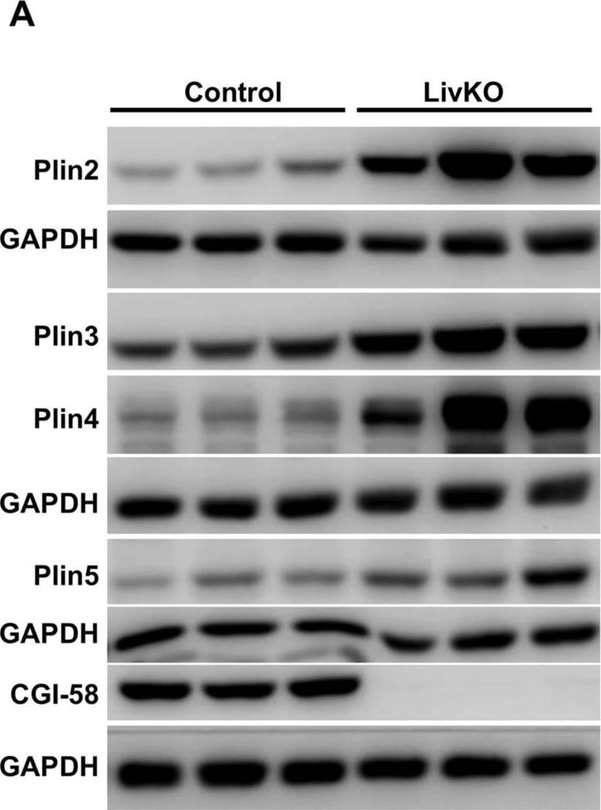
Western Blot: Perilipin-4 Antibody - BSA Free [NBP2-13776] -
Relative levels of hepatic proteins and mRNAs of PAT family genes in the male mice fed the Western diet for 3 weeks starting at 6 weeks of age. (A) Immunoblots of perilipins and control proteins. (B) Densitometry of the immunoblots in A (n = 5–6). (C) Relative levels of hepatic mRNAs for perilipins in the mice at necropsy (n = 5–6). *P < 0.05; **P < 0.01. Image collected and cropped by CiteAb from the following open publication (https://pubmed.ncbi.nlm.nih.gov/32170127), licensed under a CC-BY license. Not internally tested by Novus Biologicals.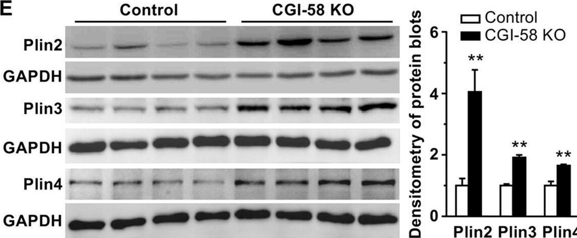
Western Blot: Perilipin-4 Antibody - BSA Free [NBP2-13776] -
Deletion of CGI-58 in Huh7 human hepatoma cell line increases cellular lipids and perilipin expression. (A) The sgRNA sequence targeting Exon 3 of human CGI-58 gene and the sequencing result of the DNA from CGI-58 knockout (KO) Huh7 cells. (B) Immunoblots of the KO and control Huh7 cell lysates. (C) Fluorescence microscopic images of LDs stained with Bodipy (green) and nuclei stained with DAPI (blue) in the KO and control cells. (D) Triglyceride (TG) content in the KO and control cells (n = 5). (E) Western blots of perilipins in the KO and control cell lysates. The blots were quantified by densitometry. (F) Relative levels of mRNAs for perilipins in the KO and control cells (n = 4). GAPDH was used as an internal invariant control. *P < 0.05; **P < 0.01. Image collected and cropped by CiteAb from the following open publication (https://pubmed.ncbi.nlm.nih.gov/32170127), licensed under a CC-BY license. Not internally tested by Novus Biologicals.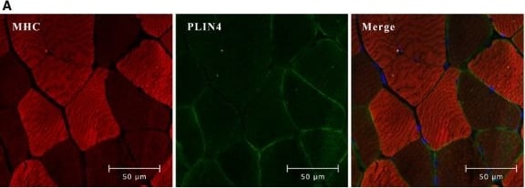
Immunohistochemistry-Paraffin: Perilipin-4 Antibody - BSA Free [NBP2-13776] -
Perilipins (PLIN)4 is mostly expressed in slow muscle fibers. (A) Representative images (400X) from confocal fluorescence microscopy of sections of paraffin-embedded biopsies from m. vastus lateralis (n = 10), stained with antibodies against myosin heavy chain fast (red, mouse anti-myosin heavy chain; skeletal, fast) and PLIN4 (green, guinea pig anti-PLIN4). Fast muscle fibers are stained in bright red color, whereas slow fibers appear as dark. (B) M. soleus and m. gastrocnemius were dissected from male C57BL/6N mice (n = 5) and Plin4 mRNA expression was determined by quantitative RT-PCR. Plin4 mRNA was normalized to Tbp mRNA. Results are presented as means +/- SEM, *P < 0.05 (two-tailed Student’s t-test). Image collected and cropped by CiteAb from the following open publication (https://pubmed.ncbi.nlm.nih.gov/26265748), licensed under a CC-BY license. Not internally tested by Novus Biologicals.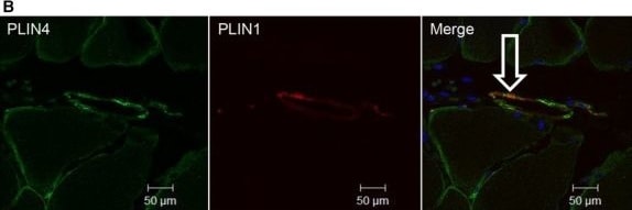
Immunohistochemistry-Paraffin: Perilipin-4 Antibody - BSA Free [NBP2-13776] -
Intramuscular adipocytes. (A) Sections of paraffin-embedded biopsies from m. vastus lateralis (n = 8) were stained with PLIN1 antibody (red, mouse anti-PLIN1) to detect adipocytes (A). Representative images (200X) were obtained by wide-field fluorescence microscopy. High magnification (1000X) of adipocytes is shown in the insert. (B) The sections were stained for PLIN4 (green, rabbit anti-PLIN4) and PLIN1 (red, mouse anti-PLIN1), showing partly co-localization in intramuscular adipocytes (n = 4). The merged image (200X) shows co-localization of the proteins (white arrow). The image was obtained by confocal fluorescence microscopy. Image collected and cropped by CiteAb from the following open publication (https://pubmed.ncbi.nlm.nih.gov/26265748), licensed under a CC-BY license. Not internally tested by Novus Biologicals.Applications for Perilipin-4 Antibody - BSA Free
Application
Recommended Usage
Immunocytochemistry/ Immunofluorescence
0.25-2 ug/ml
Immunohistochemistry
1:5000 - 1:10000
Immunohistochemistry-Paraffin
1:5000 - 1:10000
Western Blot
0.04 - 0.4 ug/ml
Application Notes
IHC-Paraffin, HIER pH 6 retrieval is recommended. ICC/IF, Fixation Permeabilization: Use PFA/Triton X-100.
Reviewed Applications
Read 1 review rated 4 using NBP2-13776 in the following applications:
Formulation, Preparation, and Storage
Purification
Affinity purified
Formulation
PBS (pH 7.2) and 40% Glycerol
Format
BSA Free
Preservative
0.02% Sodium Azide
Concentration
Concentrations vary lot to lot. See vial label for concentration. If unlisted please contact technical services.
Shipping
The product is shipped with polar packs. Upon receipt, store it immediately at the temperature recommended below.
Stability & Storage
Store at 4C short term. Aliquot and store at -20C long term. Avoid freeze-thaw cycles.
Background: Perilipin-4
Alternate Names
KIAA1881, MDRV, MRUPAV, NIPA2, S3-12
Gene Symbol
PLIN4
Additional Perilipin-4 Products
Product Documents for Perilipin-4 Antibody - BSA Free
Certificate of Analysis
To download a Certificate of Analysis, please enter a lot or batch number in the search box below.
Product Specific Notices for Perilipin-4 Antibody - BSA Free
This product is for research use only and is not approved for use in humans or in clinical diagnosis. Primary Antibodies are guaranteed for 1 year from date of receipt.
Citations for Perilipin-4 Antibody - BSA Free
 Powered by Bioz
Customer Reviews for Perilipin-4 Antibody - BSA Free (1)
4 out of 5
1 Customer Rating
Have you used Perilipin-4 Antibody - BSA Free?
Submit a review and receive an Amazon gift card!
$25/€18/£15/$25CAN/¥2500 for a review with an image
$10/€7/£6/$10CAN/¥1110 for a review without an image
Submit a review
Customer Images
Showing
1
-
1 of
1 review
Showing All
Filter By:
-
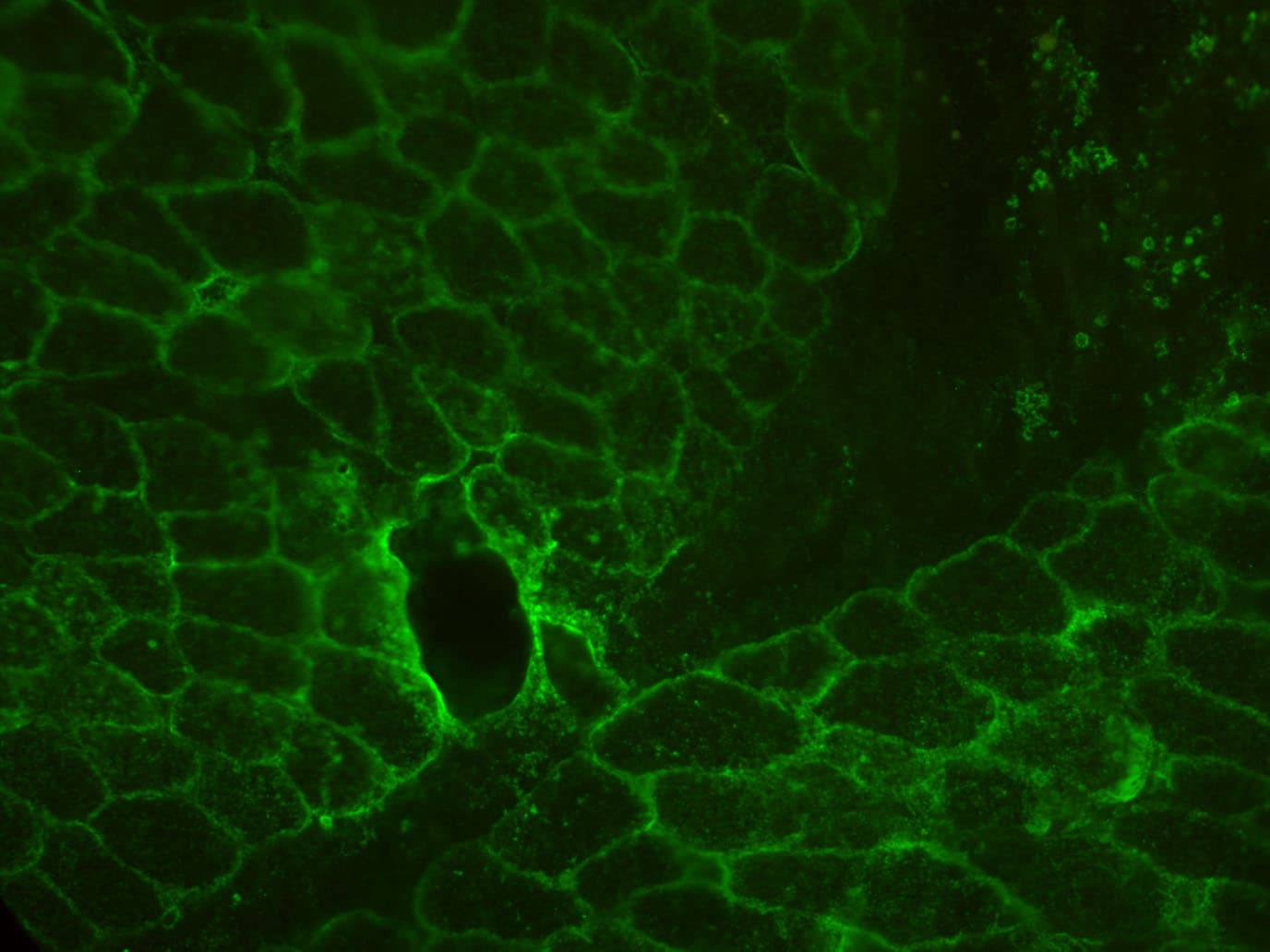
Application: Immunohistochemistry-FrozenSample Tested: Skeletal muscleSpecies: PigVerified Customer | Posted 09/15/2020Detection of PLIN4 in pig muscle cross section dilution 1:200 in PBS with 2% NGS secondary antibody 1:1000 Alexa Fluor 488 goat anti-rabbit IgGno fluorescence was detected when blocking the antibody binding in advance with the blocking peptide

There are no reviews that match your criteria.
Protocols
Find general support by application which include: protocols, troubleshooting, illustrated assays, videos and webinars.
- Antigen Retrieval Protocol (PIER)
- Antigen Retrieval for Frozen Sections Protocol
- Appropriate Fixation of IHC/ICC Samples
- Cellular Response to Hypoxia Protocols
- Chromogenic IHC Staining of Formalin-Fixed Paraffin-Embedded (FFPE) Tissue Protocol
- Chromogenic Immunohistochemistry Staining of Frozen Tissue
- ClariTSA™ Fluorophore Kits
- Detection & Visualization of Antibody Binding
- Fluorescent IHC Staining of Frozen Tissue Protocol
- Graphic Protocol for Heat-induced Epitope Retrieval
- Graphic Protocol for the Preparation and Fluorescent IHC Staining of Frozen Tissue Sections
- Graphic Protocol for the Preparation and Fluorescent IHC Staining of Paraffin-embedded Tissue Sections
- Graphic Protocol for the Preparation of Gelatin-coated Slides for Histological Tissue Sections
- ICC Cell Smear Protocol for Suspension Cells
- ICC Immunocytochemistry Protocol Videos
- ICC for Adherent Cells
- IHC Sample Preparation (Frozen sections vs Paraffin)
- Immunocytochemistry (ICC) Protocol
- Immunocytochemistry Troubleshooting
- Immunofluorescence of Organoids Embedded in Cultrex Basement Membrane Extract
- Immunofluorescent IHC Staining of Formalin-Fixed Paraffin-Embedded (FFPE) Tissue Protocol
- Immunohistochemistry (IHC) and Immunocytochemistry (ICC) Protocols
- Immunohistochemistry Frozen Troubleshooting
- Immunohistochemistry Paraffin Troubleshooting
- Preparing Samples for IHC/ICC Experiments
- Preventing Non-Specific Staining (Non-Specific Binding)
- Primary Antibody Selection & Optimization
- Protocol for Heat-Induced Epitope Retrieval (HIER)
- Protocol for Making a 4% Formaldehyde Solution in PBS
- Protocol for VisUCyte™ HRP Polymer Detection Reagent
- Protocol for the Fluorescent ICC Staining of Cell Smears - Graphic
- Protocol for the Fluorescent ICC Staining of Cultured Cells on Coverslips - Graphic
- Protocol for the Preparation & Fixation of Cells on Coverslips
- Protocol for the Preparation and Chromogenic IHC Staining of Frozen Tissue Sections
- Protocol for the Preparation and Chromogenic IHC Staining of Frozen Tissue Sections - Graphic
- Protocol for the Preparation and Chromogenic IHC Staining of Paraffin-embedded Tissue Sections
- Protocol for the Preparation and Chromogenic IHC Staining of Paraffin-embedded Tissue Sections - Graphic
- Protocol for the Preparation and Fluorescent ICC Staining of Cells on Coverslips
- Protocol for the Preparation and Fluorescent ICC Staining of Non-adherent Cells
- Protocol for the Preparation and Fluorescent ICC Staining of Stem Cells on Coverslips
- Protocol for the Preparation and Fluorescent IHC Staining of Frozen Tissue Sections
- Protocol for the Preparation and Fluorescent IHC Staining of Paraffin-embedded Tissue Sections
- Protocol for the Preparation of Gelatin-coated Slides for Histological Tissue Sections
- Protocol for the Preparation of a Cell Smear for Non-adherent Cell ICC - Graphic
- R&D Systems Quality Control Western Blot Protocol
- TUNEL and Active Caspase-3 Detection by IHC/ICC Protocol
- The Importance of IHC/ICC Controls
- Troubleshooting Guide: Immunohistochemistry
- Troubleshooting Guide: Western Blot Figures
- Western Blot Conditions
- Western Blot Protocol
- Western Blot Protocol for Cell Lysates
- Western Blot Troubleshooting
- Western Blot Troubleshooting Guide
- View all Protocols, Troubleshooting, Illustrated assays and Webinars
FAQs for Perilipin-4 Antibody - BSA Free
Showing
1
-
1 of
1 FAQ
Showing All
-
Q: Can you please provide me the concentration of the lot A96877?
A: I can confirm that lot number A96877 is supplied at 0.2mg/ml.
Loading...