PGAM1 Antibody - BSA Free
Novus Biologicals | Catalog # NBP1-49532
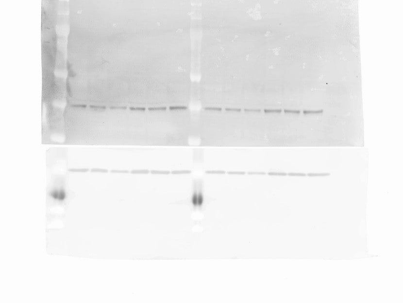
![Western Blot: PGAM1 AntibodyBSA Free [NBP1-49532]](https://resources.rndsystems.com/images/products/PGAM1-Antibody-Western-Blot-NBP1-49532-img0008.jpg "Western Blot: PGAM1 AntibodyBSA Free [NBP1-49532]")
Key Product Details
Species Reactivity
Validated:
Cited:
Applications
Validated:
Cited:
Label
Antibody Source
Format
Product Specifications
Immunogen
Clonality
Host
Isotype
Scientific Data Images for PGAM1 Antibody - BSA Free
Western Blot: PGAM1 AntibodyBSA Free [NBP1-49532]
Western Blot: PGAM1 Antibody [NBP1-49532] - Analysis of PGAM1 in Jurkat whole cell extracts![Immunocytochemistry/ Immunofluorescence: PGAM1 Antibody - BSA Free [NBP1-49532]](https://resources.rndsystems.com/images/products/PGAM1-Antibody-Immunocytochemistry-Immunofluorescence-NBP1-49532-img0005.jpg "Immunocytochemistry/ Immunofluorescence: PGAM1 Antibody - BSA Free [NBP1-49532]")
Immunocytochemistry/ Immunofluorescence: PGAM1 Antibody - BSA Free [NBP1-49532]
Immunocytochemistry/Immunofluorescence: PGAM1 Antibody [NBP1-49532] - Analysis of PGAM1 in HeLa cells using NBP1-49532. Nuclei (Blue) are counterstained using Hoechst 33258.![Immunohistochemistry: PGAM1 Antibody - BSA Free [NBP1-49532]](https://resources.rndsystems.com/images/products/PGAM1-Antibody-Immunohistochemistry-NBP1-49532-img0006.jpg "Immunohistochemistry: PGAM1 Antibody - BSA Free [NBP1-49532]")
Immunohistochemistry: PGAM1 Antibody - BSA Free [NBP1-49532]
Immunohistochemistry: PGAM1 Antibody [NBP1-49532] - Analysis of PGAM1 in mouse tongue.![Simple Western: PGAM1 AntibodyBSA Free [NBP1-49532]](https://resources.rndsystems.com/images/products/PGAM1-Antibody-Simple-Western-NBP1-49532-img0009.jpg "Simple Western: PGAM1 AntibodyBSA Free [NBP1-49532]")
Simple Western: PGAM1 AntibodyBSA Free [NBP1-49532]
Simple Western: PGAM1 Antibody [NBP1-49532] - Simple Western lane view shows a specific band for PGAM1 in 0.05 mg/ml of Jurkat lysate. This experiment was performed under reducing conditions using the 12-230 kDa separation system.
Western Blot: PGAM1 Antibody - BSA Free [NBP1-49532] -
Induction of PGAM is essential for myogenic and adipogenic differentiation.A Heatmap of genes related to glycolysis in a time course microarray sampled during differentiation in C2C12 cells. B IB of the indicated proteins in differentiated and undifferentiated C2C12 cells. C mRNA expression levels of the indicated genes in C2C12 cells expressing scramble or Rb shRNA. C2C12 cells were differentiated under low serum conditions for 7 days. Tukey’s HSD test was performed. ****P < 0.0001 and *P < 0.05. D IB of the indicated proteins in C2C12 cells expressing scramble or Pgam2 shRNA those with or without exposure to stimuli to induce myogenic differentiation. E Immunocytochemistry of myosin heavy chain (MHC) in C2C12 cells expressing scramble or Pgam2 shRNA and exposed to stimuli to induce myogenic differentiation for 7 days. F mRNA expression levels of the indicated genes in C2C12 cells expressing scramble or Rb shRNA. Cells were differentiated in the presence or absence of pyruvate for 7 days. Tukey’s HSD test was performed. **P < 0.01, *P < 0.05 and n.s.; not significant. G RNA-seq heatmap of genes related to glycolysis during adipogenic differentiation in 3T3L1 cells. H IB of the indicated proteins in differentiated and undifferentiated 3T3L1 cells. I IB of the indicated proteins in PGAM1-depleted 3T3L1 after exposure to stimuli to induce adipogenic differentiation. J Oil red staining of 3T3L1 cells transduced with the indicated gRNA and treated with vehicle or 4 mM pyruvate under the culture conditions to induce adipogenic differentiation. K IB of the indicated proteins in PGAM1-depleted 3T3L1 cells after exposure to stimuli to induce adipogenic differentiation in the presence or absence of 4 mM pyruvate. All data are shown as the mean +/- SEM. Image collected and cropped by CiteAb from the following open publication (https://www.nature.com/articles/s41419-025-07850-3), licensed under a CC-BY license. Not internally tested by Novus Biologicals.
Western Blot: PGAM1 Antibody - BSA Free [NBP1-49532] -
Induction of PGAM is essential for myogenic and adipogenic differentiation.A Heatmap of genes related to glycolysis in a time course microarray sampled during differentiation in C2C12 cells. B IB of the indicated proteins in differentiated and undifferentiated C2C12 cells. C mRNA expression levels of the indicated genes in C2C12 cells expressing scramble or Rb shRNA. C2C12 cells were differentiated under low serum conditions for 7 days. Tukey’s HSD test was performed. ****P < 0.0001 and *P < 0.05. D IB of the indicated proteins in C2C12 cells expressing scramble or Pgam2 shRNA those with or without exposure to stimuli to induce myogenic differentiation. E Immunocytochemistry of myosin heavy chain (MHC) in C2C12 cells expressing scramble or Pgam2 shRNA and exposed to stimuli to induce myogenic differentiation for 7 days. F mRNA expression levels of the indicated genes in C2C12 cells expressing scramble or Rb shRNA. Cells were differentiated in the presence or absence of pyruvate for 7 days. Tukey’s HSD test was performed. **P < 0.01, *P < 0.05 and n.s.; not significant. G RNA-seq heatmap of genes related to glycolysis during adipogenic differentiation in 3T3L1 cells. H IB of the indicated proteins in differentiated and undifferentiated 3T3L1 cells. I IB of the indicated proteins in PGAM1-depleted 3T3L1 after exposure to stimuli to induce adipogenic differentiation. J Oil red staining of 3T3L1 cells transduced with the indicated gRNA and treated with vehicle or 4 mM pyruvate under the culture conditions to induce adipogenic differentiation. K IB of the indicated proteins in PGAM1-depleted 3T3L1 cells after exposure to stimuli to induce adipogenic differentiation in the presence or absence of 4 mM pyruvate. All data are shown as the mean +/- SEM. Image collected and cropped by CiteAb from the following open publication (https://www.nature.com/articles/s41419-025-07850-3), licensed under a CC-BY license. Not internally tested by Novus Biologicals.Applications for PGAM1 Antibody - BSA Free
Immunocytochemistry/ Immunofluorescence
Immunohistochemistry
Immunohistochemistry-Paraffin
Knockdown Validated
Simple Western
Western Blot
See Simple Western Antibody Database for Simple Western validation: Tested in Jurkat lysate 0.05 mg/mL, separated by Size, antibody dilution of 1:400, apparent MW was 35 kDa. Separated by Size-Wes, Sally Sue/Peggy Sue.
Reviewed Applications
Read 1 review rated 5 using NBP1-49532 in the following applications:
Formulation, Preparation, and Storage
Purification
Formulation
Format
Preservative
Concentration
Shipping
Stability & Storage
Background: PGAM1
Alternate Names
Gene Symbol
UniProt
Additional PGAM1 Products
Product Documents for PGAM1 Antibody - BSA Free
Certificate of Analysis
To download a Certificate of Analysis, please enter a lot or batch number in the search box below.
Product Specific Notices for PGAM1 Antibody - BSA Free
This product is for research use only and is not approved for use in humans or in clinical diagnosis. Primary Antibodies are guaranteed for 1 year from date of receipt.
Citations for PGAM1 Antibody - BSA Free
 Powered by Bioz
Customer Reviews for PGAM1 Antibody - BSA Free (1)
Have you used PGAM1 Antibody - BSA Free?
Submit a review and receive an Amazon gift card!
$25/€18/£15/$25CAN/¥2500 Yen for a review with an image
$10/€7/£6/$10CAN/¥1110 Yen for a review without an image
Submit a review
Customer Images
-
Application: Western BlotSample Tested:Species: OtherVerified Customer | Posted 09/21/2015WB analysis of PGAM1 in Pteropus Alecto

There are no reviews that match your criteria.
Protocols
View specific protocols for PGAM1 Antibody - BSA Free (NBP1-49532):
Immunocytochemistry Protocol
Culture cells to appropriate density in 35mm culture dishes or 6-well plates.
1. Pull off culture medium with and add 10% formalin to the dish. Fix at room temperature for 30 minutes..
2. Take off the formalin and add ice cold methanol (kept in well sealed bottle in -20C). Incubate for 5-10 minutes.
3. Take off methanol and add PBS (You can add 0.1% Tween-20 to PBS used here and all subsequent steps), be sure to not let the specimen dry out. Wash 3 times 10 minutes before proceeding to blocking step.
4. To block nonspecific antibody binding incubate in 10% normal goat serum for a minimum of 1 hr at room temp. Cells can also block overnight at 4C for this step.
5. Add primary antibody at appropriate dilution and incubate at room temp for 2 hrs or overnight at room temp.
6. Remove primary antibody and replace with PBS. Wash 3 x 10 min in PBS.
7. Add secondary antibody at appropriate dilution. Incubate for 1 hr at room temperature
8. Remove antibody and replace with PBS, wash 1 x 10 min in PBS. Add Hoechst 33258 to PBS at 1:25,0000 and incubate for 10 min. Wash a third time with PBS for 10 min (total of 3X10min PBS washes).
9. Cells can be viewed directly after washing. The plates can also be stored in PBS containing Azide and parafilmed. Cells can also be coverslipped using Fluoromount. If storing coverslip be sure to seal the edges with clear nail polish.
*The above information is only intended as a guide. The researcher should determine what protocol best meets their needs. Please follow safe laboratory procedures.
Immunohistochemistry-paraffin embedded sections
Antigen Unmasking:
Bring slides to a boil in 10 mM sodium citrate buffer pH 6.0 then maintain at a sub-boiling temperature for 10 minutes. Cool slides on bench top for 30 minutes.
Staining:
1. Wash sections in dH2O three times for 5 minutes each.
2. Wash section in wash buffer (1X PBS/0.1% Tween-20 (1X PBST)) for 5 minutes.
3. Block each section with 100-400 ul blocking solution (1X PBST, 5% goat serum) for 1 hour at room temperature.
4. Remove blocking solution and add 100-400 ul primary antibody diluted in 1X PBST, 5% goat serum to each section. Incubate overnight at 4C.
5. Remove antibody solution and wash sections in wash buffer three times for 5 minutes each.
6. Add 100-400 ul biotinylated secondary antibody, diluted in 1X PBST, 5% goat serum. Incubate 30 minutes at room temperature.
7. Remove secondary antibody solution and wash sections three times with wash buffer for 5 minutes each.
8. Add 100-400 ul Striptavidin-HRP reagent to each section and incubate for 30 minutes at room temperature.
9. Wash sections three times in wash buffer for 5 minutes each.
10. Add 100-400 ul DAB substrate to each section and monitor staining closely.
11. As soon as the sections develop, immerse slides in dH2O.
12. Counterstain sections in hematoxylin.
13. Wash sections in dH2O two times for 5 minutes each.
14. Dehydrate sections.
15. Mount coverslips.
Western Blot Protocol
1. Perform SDS-PAGE (4-12% MOPS) on samples to be analyzed, loading 40 ug of total protein per lane.
2. Transfer proteins to Nitrocellulose according to the instructions provided by the manufacturer of the transfer apparatus.
3. Rinse membrane with dH2O and then stain the blot using Ponceau S for 1-2 minutes to access the transfer of proteins onto the nitrocellulose membrane. Rinse the blot in water to remove excess stain and mark the lane locations and locations of molecular weight markers using a pencil.
4. Rinse the blot in TBS for approximately 5 minutes.
5. Block the membrane using 5% NFDM + 1% BSA in TBS + Tween, 1 hour at RT.
6. Rinse the membrane in dH2O and then wash the membrane in wash buffer [TBS + 0.1% Tween] 3 times for 10 minutes each.
7. Dilute the rabbit anti-PGAM1 primary antibody (NBP1-49532) in blocking buffer and incubate 1 hour at room temperature.
8. Rinse the membrane in dH2O and then wash the membrane in wash buffer [TBS + 0.1% Tween] 3 times for 10 minutes each.
9. Apply the diluted rabbit-IgG HRP-conjugated secondary antibody in blocking buffer (as per manufacturers instructions) and incubate 1 hour at room temperature.
10. Wash the blot in wash buffer [TBS + 0.1% Tween] 3 times for 10 minutes each (this step can be repeated as required to reduce background).
11. Apply the detection reagent of choice in accordance with the manufacturers instructions (Pierce ECL).
**Note: Tween-20 can be added to the blocking or antibody dilution buffer at a final concentration of 0.05-0.2%, provided it does not interfere with antibody-antigen binding.
Find general support by application which include: protocols, troubleshooting, illustrated assays, videos and webinars.
- Antigen Retrieval Protocol (PIER)
- Antigen Retrieval for Frozen Sections Protocol
- Appropriate Fixation of IHC/ICC Samples
- Cellular Response to Hypoxia Protocols
- Chromogenic IHC Staining of Formalin-Fixed Paraffin-Embedded (FFPE) Tissue Protocol
- Chromogenic Immunohistochemistry Staining of Frozen Tissue
- ClariTSA™ Fluorophore Kits
- Detection & Visualization of Antibody Binding
- Fluorescent IHC Staining of Frozen Tissue Protocol
- Graphic Protocol for Heat-induced Epitope Retrieval
- Graphic Protocol for the Preparation and Fluorescent IHC Staining of Frozen Tissue Sections
- Graphic Protocol for the Preparation and Fluorescent IHC Staining of Paraffin-embedded Tissue Sections
- Graphic Protocol for the Preparation of Gelatin-coated Slides for Histological Tissue Sections
- ICC Cell Smear Protocol for Suspension Cells
- ICC Immunocytochemistry Protocol Videos
- ICC for Adherent Cells
- IHC Sample Preparation (Frozen sections vs Paraffin)
- Immunocytochemistry (ICC) Protocol
- Immunocytochemistry Troubleshooting
- Immunofluorescence of Organoids Embedded in Cultrex Basement Membrane Extract
- Immunofluorescent IHC Staining of Formalin-Fixed Paraffin-Embedded (FFPE) Tissue Protocol
- Immunohistochemistry (IHC) and Immunocytochemistry (ICC) Protocols
- Immunohistochemistry Frozen Troubleshooting
- Immunohistochemistry Paraffin Troubleshooting
- Preparing Samples for IHC/ICC Experiments
- Preventing Non-Specific Staining (Non-Specific Binding)
- Primary Antibody Selection & Optimization
- Protocol for Heat-Induced Epitope Retrieval (HIER)
- Protocol for Making a 4% Formaldehyde Solution in PBS
- Protocol for VisUCyte™ HRP Polymer Detection Reagent
- Protocol for the Fluorescent ICC Staining of Cell Smears - Graphic
- Protocol for the Fluorescent ICC Staining of Cultured Cells on Coverslips - Graphic
- Protocol for the Preparation & Fixation of Cells on Coverslips
- Protocol for the Preparation and Chromogenic IHC Staining of Frozen Tissue Sections
- Protocol for the Preparation and Chromogenic IHC Staining of Frozen Tissue Sections - Graphic
- Protocol for the Preparation and Chromogenic IHC Staining of Paraffin-embedded Tissue Sections
- Protocol for the Preparation and Chromogenic IHC Staining of Paraffin-embedded Tissue Sections - Graphic
- Protocol for the Preparation and Fluorescent ICC Staining of Cells on Coverslips
- Protocol for the Preparation and Fluorescent ICC Staining of Non-adherent Cells
- Protocol for the Preparation and Fluorescent ICC Staining of Stem Cells on Coverslips
- Protocol for the Preparation and Fluorescent IHC Staining of Frozen Tissue Sections
- Protocol for the Preparation and Fluorescent IHC Staining of Paraffin-embedded Tissue Sections
- Protocol for the Preparation of Gelatin-coated Slides for Histological Tissue Sections
- Protocol for the Preparation of a Cell Smear for Non-adherent Cell ICC - Graphic
- R&D Systems Quality Control Western Blot Protocol
- TUNEL and Active Caspase-3 Detection by IHC/ICC Protocol
- The Importance of IHC/ICC Controls
- Troubleshooting Guide: Immunohistochemistry
- Troubleshooting Guide: Western Blot Figures
- Western Blot Conditions
- Western Blot Protocol
- Western Blot Protocol for Cell Lysates
- Western Blot Troubleshooting
- Western Blot Troubleshooting Guide
- View all Protocols, Troubleshooting, Illustrated assays and Webinars
FAQs for PGAM1 Antibody - BSA Free
-
Q: This antibody is reactive with human as noted in the datasheet. Can we use this antibody for dog tissues?
A:
Our antibody NBP1-49532 has not yet been tested with dog PGAM1. However, I have run a UniProt Blast sequence comparison of the immunogen sequence (residues 200-254 of the human protein, UniProt P18669) with canine, and the result was 98% homology with canine PGAM1 (UniProt E2RT65). This strongly suggests that this antibody is suitable for use with dog tissues (although we cannot guarantee success). Note that if you decide to go ahead and test this antibody with a new species, you would be eligible for our Innovator's Reward. The details of this program are shown here: Innovator's Reward program.