PIEZO1 Antibody (2-10)
Novus Biologicals | Catalog # NBP2-75617
![Western Blot: PIEZO1 Antibody (2-10) [NBP2-75617]](https://resources.rndsystems.com/images/products/PIEZO1-Antibody-2-10-Western-Blot-NBP2-75617-img0005.jpg "Western Blot: PIEZO1 Antibody (2-10) [NBP2-75617]")
Loading...
Key Product Details
Validated by
Biological Validation
Species Reactivity
Validated:
Human, Mouse, Rat, Porcine
Cited:
Human
Applications
Validated:
Immunohistochemistry, Immunohistochemistry-Paraffin, Western Blot, Immunocytochemistry/ Immunofluorescence, Simple Western, Electron Microscopy
Cited:
Western Blot, Immunocytochemistry/ Immunofluorescence, Electron Microscopy
Label
Unconjugated
Antibody Source
Monoclonal Mouse IgG2A Clone # 2-10
Loading...
Product Specifications
Immunogen
Recombinant protein within Human PIEZO1 aa 1275-1540 / 2521. (SwissProt: Q92508 Human)
Reactivity Notes
Porcine reactivity reported from a verified customer review. Mouse blocking reagent may be needed for IHC and ICC experiments to reduce high background signal. You can find these reagents under catalog numbers PK-2200-NB and MP-2400-NB. Please contact Technical Support if you have any. Please note that this antibody is reactive to Mouse and derived from the same host, Mouse. Mouse-On-questions.
Localization
Endoplasmic reticulum membrane. Cell membrane.
Clonality
Monoclonal
Host
Mouse
Isotype
IgG2A
Theoretical MW
287 kDa.
Disclaimer note: The observed molecular weight of the protein may vary from the listed predicted molecular weight due to post translational modifications, post translation cleavages, relative charges, and other experimental factors.
Disclaimer note: The observed molecular weight of the protein may vary from the listed predicted molecular weight due to post translational modifications, post translation cleavages, relative charges, and other experimental factors.
Scientific Data Images for PIEZO1 Antibody (2-10)
Western Blot: PIEZO1 Antibody (2-10) [NBP2-75617]
Western Blot: PIEZO1 Antibody (2-10) [NBP2-75617] - Analysis of FAM38A/PIEZO1 on recombinant protein with Mouse anti-FAM38A/PIEZO1 antibody at 1/500 dilution. Lysates/proteins at 50 ng/Lane.Exposure time: 1 minute; 10% SDS-PAGE gel. Proteins were transferred to a PVDF membrane and blocked with 5% NFDM/TBST for 1 hour at room temperature. The primary antibody at 1/500 dilution was used in 5% NFDM/TBST at room temperature for 2 hours. Goat Anti-Mouse IgG - HRP Secondary Antibody at 1:100,000 dilution was used for 1 hour at room temperature.![Immunocytochemistry/ Immunofluorescence: PIEZO1 Antibody (2-10) [NBP2-75617]](https://resources.rndsystems.com/images/products/PIEZO1-Antibody-2-10-Immunocytochemistry-Immunofluorescence-NBP2-75617-img0009.jpg "Immunocytochemistry/ Immunofluorescence: PIEZO1 Antibody (2-10) [NBP2-75617]")
Immunocytochemistry/ Immunofluorescence: PIEZO1 Antibody (2-10) [NBP2-75617]
Immunocytochemistry/Immunofluorescence: PIEZO1 Antibody (2-10) [NBP2-75617] - Analysis of Siha cells labeling FAM38A/PIEZO1 with Mouse anti-PIEZO1 antibody at 1/100 dilution. Cells were fixed in 4% paraformaldehyde for 30 minutes, permeabilized with 0.1% Triton X-100 in PBS for 15 minutes, and then blocked with 2% BSA for 30 minutes at room temperature. Cells were then incubated with Mouse anti-PIEZO1 antibody at 1/100 dilution in 2% BSA overnight at 4. Goat Anti-Mouse IgG H&L (iFluor(TM) 488) was used as the secondary antibody at 1/1,000 dilution. PBS instead of the primary antibody was used as the secondary antibody only control. Nuclear DNA was labelled in blue with DAPI.![Immunohistochemistry-Paraffin: PIEZO1 Antibody (2-10) [NBP2-75617]](https://resources.rndsystems.com/images/products/PIEZO1-Antibody-2-10-Immunohistochemistry-Paraffin-NBP2-75617-img0010.jpg "Immunohistochemistry-Paraffin: PIEZO1 Antibody (2-10) [NBP2-75617]")
Immunohistochemistry-Paraffin: PIEZO1 Antibody (2-10) [NBP2-75617]
Immunohistochemistry-Paraffin: PIEZO1 Antibody (2-10) [NBP2-75617] - Analysis of paraffin-embedded mouse brain tissue with Mouse anti-FAM38A/PIEZO1 antibody at 1/200 dilution. The section was pre-treated using heat mediated antigen retrieval with Tris-EDTA buffer (pH 9.0) for 20 minutes. The tissues were blocked in 1% BSA for 20 minutes at room temperature, washed with ddH2O and PBS, and then probed with the primary antibody at 1/200 dilution for 1 hour at room temperature. The detection was performed using an HRP conjugated compact polymer system. DAB was used as the chromogen. Tissues were counterstained with hematoxylin and mounted with DPX.![Western Blot: PIEZO1 Antibody (2-10) [NBP2-75617]](https://resources.rndsystems.com/images/products/PIEZO1-Antibody-2-10-Western-Blot-NBP2-75617-img0004.jpg "Western Blot: PIEZO1 Antibody (2-10) [NBP2-75617]")
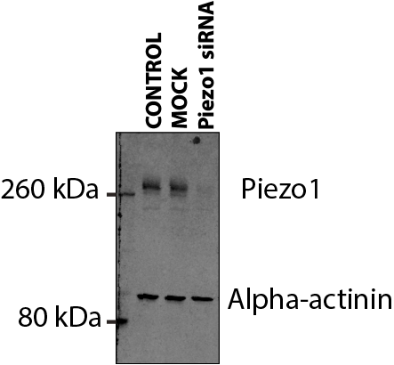
Western Blot: PIEZO1 Antibody (2-10) [NBP2-75617]
Western Blot: PIEZO1 Antibody (2-10) [NBP2-75617] - Porcine endocardial endothelium lysate. Control, mock transfected and Piezo1 siRNA transfected with alpha-actinin as loading control. WB image submitted by a verified customer review.![Immunocytochemistry/ Immunofluorescence: PIEZO1 Antibody (2-10) [NBP2-75617]](https://resources.rndsystems.com/images/products/PIEZO1-Antibody-2-10-Immunocytochemistry-Immunofluorescence-NBP2-75617-img0003.jpg "Immunocytochemistry/ Immunofluorescence: PIEZO1 Antibody (2-10) [NBP2-75617]")
Immunocytochemistry/ Immunofluorescence: PIEZO1 Antibody (2-10) [NBP2-75617]
Immunocytochemistry/Immunofluorescence: PIEZO1 Antibody (2-10) [NBP2-75617] - ICC staining Protein PIEZO (green) in A431 cells. Cells were fixed in paraformaldehyde, permeabilized with 0.25% Triton X-100 in PBS.![Immunocytochemistry/ Immunofluorescence: PIEZO1 Antibody (2-10) [NBP2-75617]](https://resources.rndsystems.com/images/products/PIEZO1-Antibody-2-10-Immunocytochemistry-Immunofluorescence-NBP2-75617-img0008.jpg "Immunocytochemistry/ Immunofluorescence: PIEZO1 Antibody (2-10) [NBP2-75617]")
Immunocytochemistry/ Immunofluorescence: PIEZO1 Antibody (2-10) [NBP2-75617]
Immunocytochemistry/Immunofluorescence: PIEZO1 Antibody (2-10) [NBP2-75617] - Analysis of Hela cells labeling FAM38A/PIEZO1 with Mouse anti-PIEZO1 antibody at 1/100 dilution. Cells were fixed in 4% paraformaldehyde for 30 minutes, permeabilized with 0.1% Triton X-100 in PBS for 15 minutes, and then blocked with 2% BSA for 30 minutes at room temperature. Cells were then incubated with Mouse anti-FAM38A/PIEZO1 antibody at 1/100 dilution in 2% BSA overnight at 4. Goat Anti-Mouse IgG H&L (iFluor(TM) 488) was used as the secondary antibody at 1/1,000 dilution. PBS instead of the primary antibody was used as the secondary antibody only control. Nuclear DNA was labelled in blue with DAPI.![Immunohistochemistry-Paraffin: PIEZO1 Antibody (2-10) [NBP2-75617]](https://resources.rndsystems.com/images/products/PIEZO1-Antibody-2-10-Immunohistochemistry-Paraffin-NBP2-75617-img0001.jpg "Immunohistochemistry-Paraffin: PIEZO1 Antibody (2-10) [NBP2-75617]")
Immunohistochemistry-Paraffin: PIEZO1 Antibody (2-10) [NBP2-75617]
Immunohistochemistry-Paraffin: PIEZO1 Antibody (2-10) [NBP2-75617] - Analysis of paraffin-embedded mouse brain tissue using anti-Protein FAM38A antibody. Counter stained with hematoxylin.![Immunohistochemistry-Paraffin: PIEZO1 Antibody (2-10) [NBP2-75617]](https://resources.rndsystems.com/images/products/PIEZO1-Antibody-2-10-Immunohistochemistry-Paraffin-NBP2-75617-img0006.jpg "Immunohistochemistry-Paraffin: PIEZO1 Antibody (2-10) [NBP2-75617]")
Immunohistochemistry-Paraffin: PIEZO1 Antibody (2-10) [NBP2-75617]
Immunohistochemistry-Paraffin: PIEZO1 Antibody (2-10) [NBP2-75617] - Rat brain tissue with Mouse anti-FAM38A/PIEZO1 antibody at 1/600 dilution.The section was pre-treated using heat mediated antigen retrieval with Tris-EDTA buffer (pH 9.0) for 20 minutes. The tissues were blocked in 1% BSA for 20 minutes at room temperature, washed with ddH2O and PBS, and then probed with the primary antibody at 1/600 dilution for 1 hour at room temperature. The detection was performed using an HRP conjugated compact polymer system. DAB was used as the chromogen. Tissues were counterstained with hematoxylin and mounted with DPX.![Immunohistochemistry-Paraffin: PIEZO1 Antibody (2-10) [NBP2-75617]](https://resources.rndsystems.com/images/products/PIEZO1-Antibody-2-10-Immunohistochemistry-Paraffin-NBP2-75617-img0007.jpg "Immunohistochemistry-Paraffin: PIEZO1 Antibody (2-10) [NBP2-75617]")
Immunohistochemistry-Paraffin: PIEZO1 Antibody (2-10) [NBP2-75617]
Immunohistochemistry-Paraffin: PIEZO1 Antibody (2-10) [NBP2-75617] - Rat hippocampus tissue with Mouse anti-FAM38A/PIEZO1 antibody at 1/600 dilution. The section was pre-treated using heat mediated antigen retrieval with Tris-EDTA buffer (pH 9.0) for 20 minutes. The tissues were blocked in 1% BSA for 20 minutes at room temperature, washed with ddH2O and PBS, and then probed with the primary antibody at 1/600 dilution for 1 hour at room temperature. The detection was performed using an HRP conjugated compact polymer system. DAB was used as the chromogen. Tissues were counterstained with hematoxylin and mounted with DPX.Applications for PIEZO1 Antibody (2-10)
Application
Recommended Usage
Electron Microscopy
Reported in scientific literature (PMID:34489534)
Immunocytochemistry/ Immunofluorescence
1:100
Immunohistochemistry-Paraffin
1:200-1:600
Simple Western
1:50
Western Blot
1:500-1:2000
Application Notes
See Simple Western Antibody Database for Simple Western validation: separated by Size, antibody dilution of 1:50
Reviewed Applications
Read 1 review rated 5 using NBP2-75617 in the following applications:
Formulation, Preparation, and Storage
Purification
Protein A purified
Formulation
PBS (pH7.4), 0.2% BSA, 50% Glycerol
Preservative
0.05% Sodium Azide
Concentration
2 mg/ml
Shipping
The product is shipped with polar packs. Upon receipt, store it immediately at the temperature recommended below.
Stability & Storage
Store at 4C short term. Aliquot and store at -20C long term. Avoid freeze-thaw cycles.
Background: PIEZO1
Long Name
Piezo-type mechanosensitive ion channel component 1
Alternate Names
FAM38A, KIAA0233, Mib, Protein FAM38A
Gene Symbol
PIEZO1
Additional PIEZO1 Products
Product Documents for PIEZO1 Antibody (2-10)
Certificate of Analysis
To download a Certificate of Analysis, please enter a lot or batch number in the search box below.
Product Specific Notices for PIEZO1 Antibody (2-10)
This product is for research use only and is not approved for use in humans or in clinical diagnosis. Primary Antibodies are guaranteed for 1 year from date of receipt.
Citations for PIEZO1 Antibody (2-10)
 Powered by Bioz
Customer Reviews for PIEZO1 Antibody (2-10) (1)
5 out of 5
1 Customer Rating
Have you used PIEZO1 Antibody (2-10)?
Submit a review and receive an Amazon gift card!
$25/€18/£15/$25CAN/¥2500 Yen for a review with an image
$10/€7/£6/$10CAN/¥1110 Yen for a review without an image
Submit a review
Customer Images
Showing
1
-
1 of
1 review
Showing All
Filter By:
-
Application: Western BlotSample Tested: EndothelialSpecies: PigVerified Customer | Posted 07/29/2021Lysate from pig endocardial endothelium. Control mock transfected and Piezo1 siRNA transfected with alpha-actinin as loading control.This was done using 1;1000 NBP2-75617 with secondary anti-mouse IR680 and viewed using the licor odyssey system.

There are no reviews that match your criteria.
Protocols
Find general support by application which include: protocols, troubleshooting, illustrated assays, videos and webinars.
- Antigen Retrieval Protocol (PIER)
- Antigen Retrieval for Frozen Sections Protocol
- Appropriate Fixation of IHC/ICC Samples
- Cellular Response to Hypoxia Protocols
- Chromogenic IHC Staining of Formalin-Fixed Paraffin-Embedded (FFPE) Tissue Protocol
- Chromogenic Immunohistochemistry Staining of Frozen Tissue
- ClariTSA™ Fluorophore Kits
- Detection & Visualization of Antibody Binding
- Fluorescent IHC Staining of Frozen Tissue Protocol
- Graphic Protocol for Heat-induced Epitope Retrieval
- Graphic Protocol for the Preparation and Fluorescent IHC Staining of Frozen Tissue Sections
- Graphic Protocol for the Preparation and Fluorescent IHC Staining of Paraffin-embedded Tissue Sections
- Graphic Protocol for the Preparation of Gelatin-coated Slides for Histological Tissue Sections
- ICC Cell Smear Protocol for Suspension Cells
- ICC Immunocytochemistry Protocol Videos
- ICC for Adherent Cells
- IHC Sample Preparation (Frozen sections vs Paraffin)
- Immunocytochemistry (ICC) Protocol
- Immunocytochemistry Troubleshooting
- Immunofluorescence of Organoids Embedded in Cultrex Basement Membrane Extract
- Immunofluorescent IHC Staining of Formalin-Fixed Paraffin-Embedded (FFPE) Tissue Protocol
- Immunohistochemistry (IHC) and Immunocytochemistry (ICC) Protocols
- Immunohistochemistry Frozen Troubleshooting
- Immunohistochemistry Paraffin Troubleshooting
- Preparing Samples for IHC/ICC Experiments
- Preventing Non-Specific Staining (Non-Specific Binding)
- Primary Antibody Selection & Optimization
- Protocol for Heat-Induced Epitope Retrieval (HIER)
- Protocol for Making a 4% Formaldehyde Solution in PBS
- Protocol for VisUCyte™ HRP Polymer Detection Reagent
- Protocol for the Fluorescent ICC Staining of Cell Smears - Graphic
- Protocol for the Fluorescent ICC Staining of Cultured Cells on Coverslips - Graphic
- Protocol for the Preparation & Fixation of Cells on Coverslips
- Protocol for the Preparation and Chromogenic IHC Staining of Frozen Tissue Sections
- Protocol for the Preparation and Chromogenic IHC Staining of Frozen Tissue Sections - Graphic
- Protocol for the Preparation and Chromogenic IHC Staining of Paraffin-embedded Tissue Sections
- Protocol for the Preparation and Chromogenic IHC Staining of Paraffin-embedded Tissue Sections - Graphic
- Protocol for the Preparation and Fluorescent ICC Staining of Cells on Coverslips
- Protocol for the Preparation and Fluorescent ICC Staining of Non-adherent Cells
- Protocol for the Preparation and Fluorescent ICC Staining of Stem Cells on Coverslips
- Protocol for the Preparation and Fluorescent IHC Staining of Frozen Tissue Sections
- Protocol for the Preparation and Fluorescent IHC Staining of Paraffin-embedded Tissue Sections
- Protocol for the Preparation of Gelatin-coated Slides for Histological Tissue Sections
- Protocol for the Preparation of a Cell Smear for Non-adherent Cell ICC - Graphic
- R&D Systems Quality Control Western Blot Protocol
- TUNEL and Active Caspase-3 Detection by IHC/ICC Protocol
- The Importance of IHC/ICC Controls
- Troubleshooting Guide: Immunohistochemistry
- Troubleshooting Guide: Western Blot Figures
- Western Blot Conditions
- Western Blot Protocol
- Western Blot Protocol for Cell Lysates
- Western Blot Troubleshooting
- Western Blot Troubleshooting Guide
- View all Protocols, Troubleshooting, Illustrated assays and Webinars
Loading...