PKM2 Antibody - BSA Free
Novus Biologicals | Catalog # NBP1-48308
![Western Blot: PKM2 AntibodyBSA Free [NBP1-48308]](https://resources.rndsystems.com/images/products/PKM2-Antibody---BSA-Free-Western-Blot-NBP1-48308-img0018.jpg "Western Blot: PKM2 AntibodyBSA Free [NBP1-48308]")
Key Product Details
Species Reactivity
Validated:
Cited:
Applications
Validated:
Cited:
Label
Antibody Source
Format
Product Specifications
Immunogen
Reactivity Notes
Localization
Clonality
Host
Isotype
Theoretical MW
Disclaimer note: The observed molecular weight of the protein may vary from the listed predicted molecular weight due to post translational modifications, post translation cleavages, relative charges, and other experimental factors.
Scientific Data Images for PKM2 Antibody - BSA Free
Western Blot: PKM2 AntibodyBSA Free [NBP1-48308]
PKM2-Antibody---BSA-Free-Western-Blot-NBP1-48308-img0018.jpg![Immunocytochemistry/ Immunofluorescence: PKM2 Antibody - BSA Free [NBP1-48308]](https://resources.rndsystems.com/images/products/PKM2-Antibody---BSA-Free-Immunocytochemistry-Immunofluorescence-NBP1-48308-img0007.jpg "Immunocytochemistry/ Immunofluorescence: PKM2 Antibody - BSA Free [NBP1-48308]")
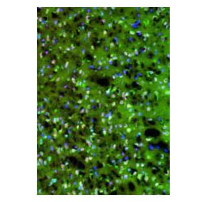
Immunocytochemistry/ Immunofluorescence: PKM2 Antibody - BSA Free [NBP1-48308]
Immunocytochemistry/Immunofluorescence: PKM2 Antibody - BSA Free [NBP1-48308] - Analysis of PKM2 in frozen sections of rat glioblastoma tissue. Image courtsey of anonymous customer review.![Immunohistochemistry: PKM2 Antibody - BSA Free [NBP1-48308]](https://resources.rndsystems.com/images/products/PKM2-Antibody---BSA-Free-Immunohistochemistry-NBP1-48308-img0009.jpg "Immunohistochemistry: PKM2 Antibody - BSA Free [NBP1-48308]")
Immunohistochemistry: PKM2 Antibody - BSA Free [NBP1-48308]
Immunohistochemistry: PKM2 Antibody - BSA Free [NBP1-48308] - Staining of PKM2 in mouse liver tissue.![Flow Cytometry: PKM2 Antibody - BSA Free [NBP1-48308]](https://resources.rndsystems.com/images/products/PKM2-Antibody---BSA-Free-Flow-Cytometry-NBP1-48308-img0019.jpg "Flow Cytometry: PKM2 Antibody - BSA Free [NBP1-48308]")
Flow Cytometry: PKM2 Antibody - BSA Free [NBP1-48308]
Flow Cytometry: PKM2 Antibody - BSA Free [NBP1-48308] - An intracellular stain was performed on Neuro2a cells with PKM2 Antibody NBP1-48308AF488 (blue) and a matched isotype control (orange). Cells were fixed with 4% PFA and then permeabilized with 0.1% saponin. Cells were incubated in an antibody dilution of 10 ug/mL for 30 minutes at room temperature. Both antibodies were conjugated to Alexa Fluor 488.![Western Blot: PKM2 AntibodyBSA Free [NBP1-48308]](https://resources.rndsystems.com/images/products/PKM2-Antibody---BSA-Free-Western-Blot-NBP1-48308-img0010.jpg "Western Blot: PKM2 AntibodyBSA Free [NBP1-48308]")
Western Blot: PKM2 AntibodyBSA Free [NBP1-48308]
Western Blot: PKM2 Antibody - BSA Free [NBP1-48308] - Analysis of HeLa lysates using NBP1-48308. Image courtesy of Gregg Semenza, (PMID: 21620138).![Western Blot: PKM2 AntibodyBSA Free [NBP1-48308]](https://resources.rndsystems.com/images/products/PKM2-Antibody---BSA-Free-Western-Blot-NBP1-48308-img0012.jpg "Western Blot: PKM2 AntibodyBSA Free [NBP1-48308]")
Western Blot: PKM2 AntibodyBSA Free [NBP1-48308]
Western Blot: PKM2 Antibody - BSA Free [NBP1-48308] - Analysis of PKM2 in MCF7 whole cell lysates.![Western Blot: PKM2 AntibodyBSA Free [NBP1-48308]](https://resources.rndsystems.com/images/products/PKM2-Antibody---BSA-Free-Western-Blot-NBP1-48308-img0016.jpg "Western Blot: PKM2 AntibodyBSA Free [NBP1-48308]")
Western Blot: PKM2 AntibodyBSA Free [NBP1-48308]
PKM2-Antibody---BSA-Free-Western-Blot-NBP1-48308-img0016.jpg![Western Blot: PKM2 AntibodyBSA Free [NBP1-48308]](https://resources.rndsystems.com/images/products/PKM2-Antibody---BSA-Free-Western-Blot-NBP1-48308-img0017.jpg "Western Blot: PKM2 AntibodyBSA Free [NBP1-48308]")
Western Blot: PKM2 AntibodyBSA Free [NBP1-48308]
PKM2-Antibody---BSA-Free-Western-Blot-NBP1-48308-img0017.jpg![Immunocytochemistry/ Immunofluorescence: PKM2 Antibody - BSA Free [NBP1-48308]](https://resources.rndsystems.com/images/products/PKM2-Antibody---BSA-Free-Immunocytochemistry-Immunofluorescence-NBP1-48308-img0008.jpg "Immunocytochemistry/ Immunofluorescence: PKM2 Antibody - BSA Free [NBP1-48308]")
Immunocytochemistry/ Immunofluorescence: PKM2 Antibody - BSA Free [NBP1-48308]
Immunocytochemistry/Immunofluorescence: PKM2 Antibody - BSA Free [NBP1-48308] - Analysis of PKM2 in HeLa cells.![Immunohistochemistry: PKM2 Antibody - BSA Free [NBP1-48308]](https://resources.rndsystems.com/images/products/PKM2%20Antibody%20-%20BSA%20Free-Immunohistochemistry-NBP1-48308-img0020.jpg "Immunohistochemistry: PKM2 Antibody - BSA Free [NBP1-48308]")
Immunohistochemistry: PKM2 Antibody - BSA Free [NBP1-48308]
PKM2 Antibody - BSA Free-Immunohistochemistry-NBP1-48308-img0020.jpg![Flow Cytometry: PKM2 Antibody - BSA Free [NBP1-48308]](https://resources.rndsystems.com/images/products/PKM2-Antibody---BSA-Free-Flow-Cytometry-NBP1-48308-img0014.jpg "Flow Cytometry: PKM2 Antibody - BSA Free [NBP1-48308]")
Flow Cytometry: PKM2 Antibody - BSA Free [NBP1-48308]
Flow Cytometry: PKM2 Antibody - BSA Free [NBP1-48308] - An intracellular stain was performed on HeLa cells with PKM2 Antibody NBP1-48308AF488 (blue) and a matched isotype control (orange). Cells were fixed with 4% PFA and then permeabilized with 0.1% saponin. Cells were incubated in an antibody dilution of 5 ug/mL for 30 minutes at room temperature. Both antibodies were conjugated to Alexa Fluor 488.![Simple Western: PKM2 AntibodyBSA Free [NBP1-48308]](https://resources.rndsystems.com/images/products/PKM2-Antibody---BSA-Free-Simple-Western-NBP1-48308-img0013.jpg "Simple Western: PKM2 AntibodyBSA Free [NBP1-48308]")
Simple Western: PKM2 AntibodyBSA Free [NBP1-48308]
Simple Western: PKM2 Antibody - BSA Free [NBP1-48308] - Lane view shows a specific band for PKM2 in 0.1 mg/ml of HeLa lysate. This experiment was performed under reducing conditions using the 12-230kDa separation system. * Non-specific interaction with the 230 kDa Simple Western standard may be seen with this antibody.
Immunohistochemistry-Paraffin: PKM2 Antibody - BSA Free [NBP1-48308] -
Expression of PKM1 and PKM2 in clinical colorectal cancer samples.(a) The protein expression of PKM1 and PKM2 in clinical specimens of cancer tumor (T) and the adjacent normal tissues (N) is shown. PKM1 and PKM2 were detected by Western blotting in under the same experimental conditions at the same time. The full-length blots are presented in Supplementary Figure S3b. (b–d) Immunohistochemical staining of normal colon tissue adjacent to tumor tissue of case 10. Results of H&E staining (b), staining with anti-PKM1 antibody (c), and staining with anti-PKM2 (d) are shown. The boxed regions in “c" and “d" are enlarged in the images below. (e–h) Immunohistochemical staining of clinical colorectal cancer tissue specimen of representative case 3. H&E-stained section with normal tissue (upper right corner) neighboring the tumor area in the section is shown (e), along with the same section stained with anti-PKM2 antibody (f). Enlarged views of boxed areas in “f" show normal colorectal crypt in mucosa (g) and tumor area (h) stained with anti-PKM2 antibody. Image collected and cropped by CiteAb from the following open publication (https://pubmed.ncbi.nlm.nih.gov/25721733), licensed under a CC-BY license. Not internally tested by Novus Biologicals.Applications for PKM2 Antibody - BSA Free
Flow Cytometry
Immunoblotting
Immunocytochemistry/ Immunofluorescence
Immunohistochemistry
Immunohistochemistry-Paraffin
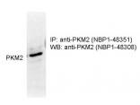
Immunoprecipitation
Simple Western
Western Blot
See Simple Western Antibody Database for Simple Western validation: Tested in HeLa lysate 0.1 mg/mL, separated by Size, antibody dilution of 1:12.5, apparent MW was 74 kDa. Separated by Size-Wes, Sally Sue/Peggy Sue.
Reviewed Applications
Read 3 reviews rated 5 using NBP1-48308 in the following applications:
Flow Cytometry Panel Builder
Bio-Techne Knows Flow Cytometry
Save time and reduce costly mistakes by quickly finding compatible reagents using the Panel Builder Tool.
Advanced Features
- Spectra Viewer - Custom analysis of spectra from multiple fluorochromes
- Spillover Popups - Visualize the spectra of individual fluorochromes
- Antigen Density Selector - Match fluorochrome brightness with antigen density

Formulation, Preparation, and Storage
Purification
Formulation
Format
Preservative
Concentration
Shipping
Stability & Storage
Background: PKM2
Long Name
Alternate Names
Gene Symbol
UniProt
Additional PKM2 Products
Product Documents for PKM2 Antibody - BSA Free
Certificate of Analysis
To download a Certificate of Analysis, please enter a lot or batch number in the search box below.
Product Specific Notices for PKM2 Antibody - BSA Free
This product is for research use only and is not approved for use in humans or in clinical diagnosis. Primary Antibodies are guaranteed for 1 year from date of receipt.
Related Research Areas
Citations for PKM2 Antibody - BSA Free
 Powered by Bioz
Customer Reviews for PKM2 Antibody - BSA Free (3)
Have you used PKM2 Antibody - BSA Free?
Submit a review and receive an Amazon gift card!
$25/€18/£15/$25CAN/¥2500 Yen for a review with an image
$10/€7/£6/$10CAN/¥1110 Yen for a review without an image
Submit a review
Customer Images
-
Application: ImmunocytochemistrySample Tested: Human lung cancer cell linesSpecies: HumanVerified Customer | Posted 06/15/2017
-
Application: Western BlotSample Tested: HeLa cellsSpecies: HumanVerified Customer | Posted 02/17/2011

-
Application: ImmunofluorescenceSample Tested: Rat glioblastoma frozen sectionSpecies: RatVerified Customer | Posted 10/15/2010
There are no reviews that match your criteria.
Protocols
View specific protocols for PKM2 Antibody - BSA Free (NBP1-48308):
1. Perform SDS-PAGE (4-12% MOPS) on samples to be analyzed, loading 40 ug of total protein per lane.
2. Transfer proteins to Nitrocellulose according to the instructions provided by the manufacturer of the transfer apparatus.
3. Rinse membrane with dH2O and then stain the blot using Ponceau S for 1-2 minutes to access the transfer of proteins onto the nitrocellulose membrane. Rinse the blot in water to remove excess stain and mark the lane locations and locations of molecular weight markers using a pencil.
4. Rinse the blot in TBS for approximately 5 minutes.
5. Block the membrane using 5% NFDM + 1% BSA in TBS + Tween, 1 hour at RT.
6. Rinse the membrane in dH2O and then wash the membrane in wash buffer [TBS + 0.1% Tween] 3 times for 10 minutes each.
7. Dilute the rabbit anti-PKM2 primary antibody (NBP1-48308) in blocking buffer and incubate 1 hour at room temperature.
8. Rinse the membrane in dH2O and then wash the membrane in wash buffer [TBS + 0.1% Tween] 3 times for 10 minutes each.
9. Apply the diluted rabbit-IgG HRP-conjugated secondary antibody in blocking buffer (as per manufacturers instructions) and incubate 1 hour at room temperature.
10. Wash the blot in wash buffer [TBS + 0.1% Tween] 3 times for 10 minutes each (this step can be repeated as required to reduce background).
11. Apply the detection reagent of choice in accordance with the manufacturers instructions (Pierce ECL).
Note: Tween-20 can be added to the blocking or antibody dilution buffer at a final concentration of 0.05-0.2%, provided it does not interfere with antibody-antigen binding.
Find general support by application which include: protocols, troubleshooting, illustrated assays, videos and webinars.
- 7-Amino Actinomycin D (7-AAD) Cell Viability Flow Cytometry Protocol
- Antigen Retrieval Protocol (PIER)
- Antigen Retrieval for Frozen Sections Protocol
- Appropriate Fixation of IHC/ICC Samples
- Cellular Response to Hypoxia Protocols
- Chromogenic IHC Staining of Formalin-Fixed Paraffin-Embedded (FFPE) Tissue Protocol
- Chromogenic Immunohistochemistry Staining of Frozen Tissue
- ClariTSA™ Fluorophore Kits
- Detection & Visualization of Antibody Binding
- Extracellular Membrane Flow Cytometry Protocol
- Flow Cytometry Protocol for Cell Surface Markers
- Flow Cytometry Protocol for Staining Membrane Associated Proteins
- Flow Cytometry Staining Protocols
- Flow Cytometry Troubleshooting Guide
- Fluorescent IHC Staining of Frozen Tissue Protocol
- Graphic Protocol for Heat-induced Epitope Retrieval
- Graphic Protocol for the Preparation and Fluorescent IHC Staining of Frozen Tissue Sections
- Graphic Protocol for the Preparation and Fluorescent IHC Staining of Paraffin-embedded Tissue Sections
- Graphic Protocol for the Preparation of Gelatin-coated Slides for Histological Tissue Sections
- ICC Cell Smear Protocol for Suspension Cells
- ICC Immunocytochemistry Protocol Videos
- ICC for Adherent Cells
- IHC Sample Preparation (Frozen sections vs Paraffin)
- Immunocytochemistry (ICC) Protocol
- Immunocytochemistry Troubleshooting
- Immunofluorescence of Organoids Embedded in Cultrex Basement Membrane Extract
- Immunofluorescent IHC Staining of Formalin-Fixed Paraffin-Embedded (FFPE) Tissue Protocol
- Immunohistochemistry (IHC) and Immunocytochemistry (ICC) Protocols
- Immunohistochemistry Frozen Troubleshooting
- Immunohistochemistry Paraffin Troubleshooting
- Immunoprecipitation Protocol
- Intracellular Flow Cytometry Protocol Using Alcohol (Methanol)
- Intracellular Flow Cytometry Protocol Using Detergents
- Intracellular Nuclear Staining Flow Cytometry Protocol Using Detergents
- Intracellular Staining Flow Cytometry Protocol Using Alcohol Permeabilization
- Intracellular Staining Flow Cytometry Protocol Using Detergents to Permeabilize Cells
- Preparing Samples for IHC/ICC Experiments
- Preventing Non-Specific Staining (Non-Specific Binding)
- Primary Antibody Selection & Optimization
- Propidium Iodide Cell Viability Flow Cytometry Protocol
- Protocol for Heat-Induced Epitope Retrieval (HIER)
- Protocol for Liperfluo
- Protocol for Making a 4% Formaldehyde Solution in PBS
- Protocol for VisUCyte™ HRP Polymer Detection Reagent
- Protocol for the Characterization of Human Th22 Cells
- Protocol for the Characterization of Human Th9 Cells
- Protocol for the Fluorescent ICC Staining of Cell Smears - Graphic
- Protocol for the Fluorescent ICC Staining of Cultured Cells on Coverslips - Graphic
- Protocol for the Preparation & Fixation of Cells on Coverslips
- Protocol for the Preparation and Chromogenic IHC Staining of Frozen Tissue Sections
- Protocol for the Preparation and Chromogenic IHC Staining of Frozen Tissue Sections - Graphic
- Protocol for the Preparation and Chromogenic IHC Staining of Paraffin-embedded Tissue Sections
- Protocol for the Preparation and Chromogenic IHC Staining of Paraffin-embedded Tissue Sections - Graphic
- Protocol for the Preparation and Fluorescent ICC Staining of Cells on Coverslips
- Protocol for the Preparation and Fluorescent ICC Staining of Non-adherent Cells
- Protocol for the Preparation and Fluorescent ICC Staining of Stem Cells on Coverslips
- Protocol for the Preparation and Fluorescent IHC Staining of Frozen Tissue Sections
- Protocol for the Preparation and Fluorescent IHC Staining of Paraffin-embedded Tissue Sections
- Protocol for the Preparation of Gelatin-coated Slides for Histological Tissue Sections
- Protocol for the Preparation of a Cell Smear for Non-adherent Cell ICC - Graphic
- Protocol: Annexin V and PI Staining by Flow Cytometry
- Protocol: Annexin V and PI Staining for Apoptosis by Flow Cytometry
- R&D Systems Quality Control Western Blot Protocol
- TUNEL and Active Caspase-3 Detection by IHC/ICC Protocol
- The Importance of IHC/ICC Controls
- Troubleshooting Guide: Fluorokine Flow Cytometry Kits
- Troubleshooting Guide: Immunohistochemistry
- Troubleshooting Guide: Western Blot Figures
- Western Blot Conditions
- Western Blot Protocol
- Western Blot Protocol for Cell Lysates
- Western Blot Troubleshooting
- Western Blot Troubleshooting Guide
- View all Protocols, Troubleshooting, Illustrated assays and Webinars
FAQs for PKM2 Antibody - BSA Free
-
Q: I am wondering if this antibody can be used for IP or ChIP? Have you ever tested it in either of these applications?
A:
We have not yet tested this antibody in ChIP or IP. However, if you would like to test this antibody in either of these applications and share your results with us, you would become eligible for our Innovators Reward Program.
Associated Pathways
