Pluripotent Stem Cell Transcription Factor Antibody Pack
Novus Biologicals | Catalog # NBP1-42823
![Immunocytochemistry/ Immunofluorescence: Pluripotent Stem Cell Transcription Factor Antibody Pack [NBP1-42823]](https://resources.rndsystems.com/images/products/Pluripotent-Stem-Cell-Transcription-Factor-Antibody-Pack-Immunocytochemistry-Immunofluorescence-NBP1-42823-img0014.jpg "Immunocytochemistry/ Immunofluorescence: Pluripotent Stem Cell Transcription Factor Antibody Pack [NBP1-42823]")
Loading...
Key Product Details
Species
Human, Mouse, Rat
Applications
Flow Cytometry, Immunocytochemistry/ Immunofluorescence, Immunohistochemistry, Western Blot
Kit Type
Kit
Product Summary for Pluripotent Stem Cell Transcription Factor Antibody Pack
This pack contains 1 vial of each: NB100-2379 (0.1 mL), NB110-37235 (0.1 mL), and NB100-58842 (0.1 mL).
Loading...
Product Specifications
Clonality
Polyclonal
Specificity
NB100-58842 is specific for mouse Nanog protein. NB100-2379-OCT4 - Embryonic Stem Cell Marker NB110-37235 - SOX2 - Embryonic Stem Cell Marker
Application Notes
See individual datasheets of components for their validated applications
Reactivity Notes
See individual datasheets of components for their validated species
Scientific Data Images for Pluripotent Stem Cell Transcription Factor Antibody Pack
Immunocytochemistry/ Immunofluorescence: Pluripotent Stem Cell Transcription Factor Antibody Pack [NBP1-42823]
Pluripotent-Stem-Cell-Transcription-Factor-Antibody-Pack-Immunocytochemistry-Immunofluorescence-NBP1-42823-img0014.jpg
Pluripotent Stem Cell Transcription Factor Antibody Pack [NBP1-42823] - YM155 significantly reduced tumorsphere formation and inhibited EGFR autophosphorylation and G9a expression. YM155 is reported to suppress cancer stemness; therefore, we used this compound to investigate the cellular mechanism of tumorsphere formation. To compare the cytotoxic capacity of YM155 against tumorsphere formation and parental cell lines, YM155 was applied to HCC827 and A549 cells in the stemness cultured or integrated medium. (F) Moreover, YM155 inhibited EGFR autophosphorylation in A549-derived tumorspheres and reduced Oct4 expression. Image collected and cropped by CiteAb from the following publication (//doi.org/10.1371/journal.pone.0182149) licensed under a CC-BY license. OCT4 Antibody [NB100-2379]
![Western Blot: Pluripotent Stem Cell Transcription Factor Antibody Pack [NBP1-42823]](https://resources.rndsystems.com/images/products/Pluripotent-Stem-Cell-Transcription-Factor-Antibody-Pack-Western-Blot-NBP1-42823-img0013.jpg "Western Blot: Pluripotent Stem Cell Transcription Factor Antibody Pack [NBP1-42823]")
Western Blot: Pluripotent Stem Cell Transcription Factor Antibody Pack [NBP1-42823]
Western Blot: Pluripotent Stem Cell Transcription Factor Antibody Pack [NBP1-42823] - Detection of SOX2 in mouse brain lysate using NB110-37235 (0.5ug/ml).![Immunohistochemistry: Pluripotent Stem Cell Transcription Factor Antibody Pack [NBP1-42823]](https://resources.rndsystems.com/images/products/Pluripotent-Stem-Cell-Transcription-Factor-Antibody-Pack-Immunohistochemistry-NBP1-42823-img0015.jpg "Immunohistochemistry: Pluripotent Stem Cell Transcription Factor Antibody Pack [NBP1-42823]")
Immunohistochemistry: Pluripotent Stem Cell Transcription Factor Antibody Pack [NBP1-42823]
Immunohistochemistry: Pluripotent Stem Cell Transcription Factor Antibody Pack [NBP1-42823] - Nanog-inducible mouse model. Representative hematoxylin and eosin (H&E) staining and immunohistochemistry (IHC) for NANOG, Ki67 and LORICRIN of TPA-treated CTR and TG mice (bars correspond to 50 um). Statistical significance was determined by the two-tailed Student's t test: (*) p < 0.05; (**) p < 0.01. Image collected and cropped by CiteAb from the following publication (//www.nature.com/articles/srep10205), licensed under a CC-BY license. Nanog Antibody [NB100-58842]![Western Blot: Pluripotent Stem Cell Transcription Factor Antibody Pack [NBP1-42823]](https://resources.rndsystems.com/images/products/Pluripotent-Stem-Cell-Transcription-Factor-Antibody-Pack-Western-Blot-NBP1-42823-img0011.jpg "Western Blot: Pluripotent Stem Cell Transcription Factor Antibody Pack [NBP1-42823]")
Western Blot: Pluripotent Stem Cell Transcription Factor Antibody Pack [NBP1-42823]
Western Blot: Pluripotent Stem Cell Transcription Factor Antibody Pack [NBP1-42823] - Whole cell lysate (5, 15, and 50 ug) from F9 cells prepared using NETN lysis buffer. Antibodies: Affinity purified rabbit anti-Nanog antibody used for WB at 0.5 ug/ml. Detection: Chemiluminescence with an exposure time of 30 seconds![Immunohistochemistry: Pluripotent Stem Cell Transcription Factor Antibody Pack [NBP1-42823]](https://resources.rndsystems.com/images/products/Pluripotent-Stem-Cell-Transcription-Factor-Antibody-Pack-Immunohistochemistry-NBP1-42823-img0017.jpg "Immunohistochemistry: Pluripotent Stem Cell Transcription Factor Antibody Pack [NBP1-42823]")
Immunohistochemistry: Pluripotent Stem Cell Transcription Factor Antibody Pack [NBP1-42823]
Immunohistochemistry: Pluripotent Stem Cell Transcription Factor Antibody Pack [NBP1-42823] - SOX2 staining (NB110-37235) of human uterus, endometrial glands.Kit Contents for Pluripotent Stem Cell Transcription Factor Antibody Pack
Formulation, Preparation, and Storage
Purification
Immunogen affinity purified
Preservative
0.05% Sodium Azide
Concentration
Concentration of individual antibodies may be found on the vial label. If unlisted please contact technical services.
Shipping
The product is shipped with polar packs. Upon receipt, store it immediately at the temperature recommended below.
Storage
Store at 4C. Do not freeze.
Background: Pluripotent Stem Cell Transcription Factor
Additional Pluripotent Stem Cell Transcription Factor Products
Product Documents for Pluripotent Stem Cell Transcription Factor Antibody Pack
Certificate of Analysis
To download a Certificate of Analysis, please enter a lot or batch number in the search box below.
Product Specific Notices for Pluripotent Stem Cell Transcription Factor Antibody Pack
This product is for research use only and is not approved for use in humans or in clinical diagnosis. Antibody Packs are guaranteed for 1 year from date of receipt.
Citations for Pluripotent Stem Cell Transcription Factor Antibody Pack
 Powered by Bioz
Customer Reviews for Pluripotent Stem Cell Transcription Factor Antibody Pack (1)
5 out of 5
1 Customer Rating
Have you used Pluripotent Stem Cell Transcription Factor Antibody Pack?
Submit a review and receive an Amazon gift card!
$25/€18/£15/$25CAN/¥2500 Yen for a review with an image
$10/€7/£6/$10CAN/¥1110 Yen for a review without an image
Submit a review
Customer Images
Showing
1
-
1 of
1 review
Showing All
Filter By:
-
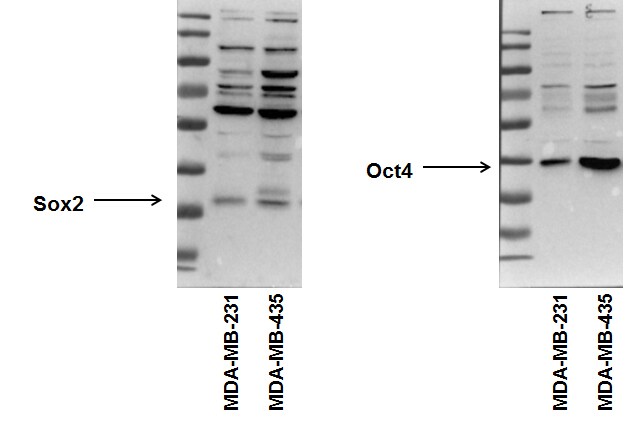
Application: Western BlotSample Tested: Human cancer cell whole cell lysateSpecies: HumanVerified Customer | Posted 09/02/2015Sox2 and Oct4 expression in human breast cancer cell lines. NBP1-42823

There are no reviews that match your criteria.
Protocols
Find general support by application which include: protocols, troubleshooting, illustrated assays, videos and webinars.
- 7-Amino Actinomycin D (7-AAD) Cell Viability Flow Cytometry Protocol
- Antigen Retrieval Protocol (PIER)
- Antigen Retrieval for Frozen Sections Protocol
- Appropriate Fixation of IHC/ICC Samples
- Cellular Response to Hypoxia Protocols
- Chromogenic IHC Staining of Formalin-Fixed Paraffin-Embedded (FFPE) Tissue Protocol
- Chromogenic Immunohistochemistry Staining of Frozen Tissue
- ClariTSA™ Fluorophore Kits
- Detection & Visualization of Antibody Binding
- Extracellular Membrane Flow Cytometry Protocol
- Flow Cytometry Protocol for Cell Surface Markers
- Flow Cytometry Protocol for Staining Membrane Associated Proteins
- Flow Cytometry Staining Protocols
- Flow Cytometry Troubleshooting Guide
- Fluorescent IHC Staining of Frozen Tissue Protocol
- Graphic Protocol for Heat-induced Epitope Retrieval
- Graphic Protocol for the Preparation and Fluorescent IHC Staining of Frozen Tissue Sections
- Graphic Protocol for the Preparation and Fluorescent IHC Staining of Paraffin-embedded Tissue Sections
- Graphic Protocol for the Preparation of Gelatin-coated Slides for Histological Tissue Sections
- ICC Cell Smear Protocol for Suspension Cells
- ICC Immunocytochemistry Protocol Videos
- ICC for Adherent Cells
- IHC Sample Preparation (Frozen sections vs Paraffin)
- Immunocytochemistry (ICC) Protocol
- Immunocytochemistry Troubleshooting
- Immunofluorescence of Organoids Embedded in Cultrex Basement Membrane Extract
- Immunofluorescent IHC Staining of Formalin-Fixed Paraffin-Embedded (FFPE) Tissue Protocol
- Immunohistochemistry (IHC) and Immunocytochemistry (ICC) Protocols
- Immunohistochemistry Frozen Troubleshooting
- Immunohistochemistry Paraffin Troubleshooting
- Intracellular Flow Cytometry Protocol Using Alcohol (Methanol)
- Intracellular Flow Cytometry Protocol Using Detergents
- Intracellular Nuclear Staining Flow Cytometry Protocol Using Detergents
- Intracellular Staining Flow Cytometry Protocol Using Alcohol Permeabilization
- Intracellular Staining Flow Cytometry Protocol Using Detergents to Permeabilize Cells
- Preparing Samples for IHC/ICC Experiments
- Preventing Non-Specific Staining (Non-Specific Binding)
- Primary Antibody Selection & Optimization
- Propidium Iodide Cell Viability Flow Cytometry Protocol
- Protocol for Heat-Induced Epitope Retrieval (HIER)
- Protocol for Liperfluo
- Protocol for Making a 4% Formaldehyde Solution in PBS
- Protocol for VisUCyte™ HRP Polymer Detection Reagent
- Protocol for the Characterization of Human Th22 Cells
- Protocol for the Characterization of Human Th9 Cells
- Protocol for the Fluorescent ICC Staining of Cell Smears - Graphic
- Protocol for the Fluorescent ICC Staining of Cultured Cells on Coverslips - Graphic
- Protocol for the Preparation & Fixation of Cells on Coverslips
- Protocol for the Preparation and Chromogenic IHC Staining of Frozen Tissue Sections
- Protocol for the Preparation and Chromogenic IHC Staining of Frozen Tissue Sections - Graphic
- Protocol for the Preparation and Chromogenic IHC Staining of Paraffin-embedded Tissue Sections
- Protocol for the Preparation and Chromogenic IHC Staining of Paraffin-embedded Tissue Sections - Graphic
- Protocol for the Preparation and Fluorescent ICC Staining of Cells on Coverslips
- Protocol for the Preparation and Fluorescent ICC Staining of Non-adherent Cells
- Protocol for the Preparation and Fluorescent ICC Staining of Stem Cells on Coverslips
- Protocol for the Preparation and Fluorescent IHC Staining of Frozen Tissue Sections
- Protocol for the Preparation and Fluorescent IHC Staining of Paraffin-embedded Tissue Sections
- Protocol for the Preparation of Gelatin-coated Slides for Histological Tissue Sections
- Protocol for the Preparation of a Cell Smear for Non-adherent Cell ICC - Graphic
- Protocol: Annexin V and PI Staining by Flow Cytometry
- Protocol: Annexin V and PI Staining for Apoptosis by Flow Cytometry
- R&D Systems Quality Control Western Blot Protocol
- TUNEL and Active Caspase-3 Detection by IHC/ICC Protocol
- The Importance of IHC/ICC Controls
- Troubleshooting Guide: Fluorokine Flow Cytometry Kits
- Troubleshooting Guide: Immunohistochemistry
- Troubleshooting Guide: Western Blot Figures
- Western Blot Conditions
- Western Blot Protocol
- Western Blot Protocol for Cell Lysates
- Western Blot Troubleshooting
- Western Blot Troubleshooting Guide
- View all Protocols, Troubleshooting, Illustrated assays and Webinars
Loading...