SOX2 Antibody - BSA Free
Novus Biologicals | Catalog # NB110-37235

Key Product Details
Validated by
Species Reactivity
Validated:
Cited:
Predicted:
Applications
Validated:
Cited:
Label
Antibody Source
Format
Product Specifications
Immunogen
Reactivity Notes
Localization
Marker
Clonality
Host
Isotype
Scientific Data Images for SOX2 Antibody - BSA Free
Detection of SOX2 in Mouse Brain Cortex via seqIF™ staining on COMET™
SOX2 Antibody was detected in immersion fixed paraffin-embedded sections of mouse Brain Cortex using Rabbit Anti-Mouse SOX2, Monoclonal Antibody (Catalog # NB110-37235) at 1:125 dilution at 37 ° Celsius for 4 minutes. Before incubation with the primary antibody, tissue underwent an all-in-one dewaxing and antigen retrieval preprocessing using PreTreatment Module (PT Module) and Dewax and HIER Buffer H (pH 9; Epredia Catalog # TA-999-DHBH). Tissue was stained using the Alexa Fluor™ Plus 647 Goat anti-Rabbit IgG Secondary Antibody at 1:200 at 37 ° Celsius for 2 minutes. (Yellow; Lunaphore Catalog # DR647RB) and counterstained with DAPI (blue; Lunaphore Catalog # DR100). Specific staining was localized to the nucleus and cytoplasm. Protocol available in COMET™ Panel Builder.![Immunohistochemistry-Paraffin: SOX2 Antibody - BSA Free [NB110-37235]](https://resources.rndsystems.com/images/products/SOX2-Antibody-Immunohistochemistry-Paraffin-NB110-37235-img0012.jpg "Immunohistochemistry-Paraffin: SOX2 Antibody - BSA Free [NB110-37235]")
Immunohistochemistry-Paraffin: SOX2 Antibody - BSA Free [NB110-37235]
Immunohistochemistry-Paraffin: SOX2 Antibody [NB110-37235] - Staining of human uterus, endometrial glands.![Western Blot: SOX2 AntibodyBSA Free [NB110-37235]](https://resources.rndsystems.com/images/products/SOX2-Antibody-Western-Blot-NB110-37235-img0008.jpg "Western Blot: SOX2 AntibodyBSA Free [NB110-37235]")
Western Blot: SOX2 AntibodyBSA Free [NB110-37235]
Western Blot: SOX2 Antibody [NB110-37235] - Detection of SOX2 in mouse brain lysate using NB110-37235 at 0.5 ug/mL.![Flow Cytometry: SOX2 Antibody - BSA Free [NB110-37235]](https://resources.rndsystems.com/images/products/SOX2-Antibody-Flow-Cytometry-NB110-37235-img0015.jpg "Flow Cytometry: SOX2 Antibody - BSA Free [NB110-37235]")
Flow Cytometry: SOX2 Antibody - BSA Free [NB110-37235]
Flow Cytometry: SOX2 Antibody [NB110-37235] - An intracellular stain was performed on A549 with NB110-37235 and a matched isotype control. Cells were fixed with 4% PFA and then permeablized with 0.1% saponin. Cells were incubated in an antibody dilution of 2.5 ug/mL for 30 minutes at room temperature, followed by Rabbit IgG (H+L) Cross-Adsorbed Secondary Antibody, Dylgiht 550.![Western Blot: SOX2 AntibodyBSA Free [NB110-37235]](https://resources.rndsystems.com/images/products/SOX2-Antibody-Western-Blot-NB110-37235-img0017.jpg "Western Blot: SOX2 AntibodyBSA Free [NB110-37235]")
![Immunocytochemistry/ Immunofluorescence: SOX2 Antibody - BSA Free [NB110-37235]](https://resources.rndsystems.com/images/products/SOX2-Antibody---BSA-Free-Immunocytochemistry-Immunofluorescence-NB110-37235-img0019.jpg "Immunocytochemistry/ Immunofluorescence: SOX2 Antibody - BSA Free [NB110-37235]")
Immunocytochemistry/ Immunofluorescence: SOX2 Antibody - BSA Free [NB110-37235]
Immunocytochemistry/Immunofluorescence: SOX2 Antibody - BSA Free [NB110-37235] - Rat FR cells were fixed in 4% paraformaldehyde for 10 minutes and permeabilized in 0.5% Triton X-100 in PBS for 5 minutes. The cells were incubated with SOX2 Antibody (NB110-37235) at 2ug/ml overnight at 4C and detected with an anti-rabbit DyLight 488 (Green) at a 1:1000 dilution for 60 minutes. Nuclei were counterstained with DAPI (Blue). Cells were imaged using a 40X objective.![Flow Cytometry: SOX2 Antibody - BSA Free [NB110-37235]](https://resources.rndsystems.com/images/products/SOX2-Antibody---BSA-Free-Flow-Cytometry-NB110-37235-img0020.jpg "Flow Cytometry: SOX2 Antibody - BSA Free [NB110-37235]")
Flow Cytometry: SOX2 Antibody - BSA Free [NB110-37235]
Flow Cytometry: SOX2 Antibody - BSA Free [NB110-37235] - An intracellular stain was performed on U-251 MG cells with SOX2 Antibody NB110-37235 (blue) and a matched isotype control NBP2-24891 (orange). Cells were fixed with 4% PFA and then permeabilized with 0.1% saponin. Cells were incubated in an antibody dilution of 2.5 ug/mL for 30 minutes at room temperature, followed by Rabbit IgG (H+L) Cross-Adsorbed Secondary Antibody, Dylight 550 (SA5-10033, Thermo Fisher).![Western Blot: SOX2 AntibodyBSA Free [NB110-37235]](https://resources.rndsystems.com/images/products/SOX2-Antibody-Western-Blot-NB110-37235-img0016.jpg "Western Blot: SOX2 AntibodyBSA Free [NB110-37235]")
Western Blot: SOX2 AntibodyBSA Free [NB110-37235]
Western Blot: SOX2 Antibody [NB110-37235] - Expression of pluripotency factor Sox2 in human head and neck cancer cell lines FaDu and HN5. WB image submitted by a verified customer review.![Immunocytochemistry/ Immunofluorescence: SOX2 Antibody - BSA Free [NB110-37235]](https://resources.rndsystems.com/images/products/SOX2-Antibody-Immunocytochemistry-Immunofluorescence-NB110-37235-img0013.jpg "Immunocytochemistry/ Immunofluorescence: SOX2 Antibody - BSA Free [NB110-37235]")
Immunocytochemistry/ Immunofluorescence: SOX2 Antibody - BSA Free [NB110-37235]
Immunocytochemistry/Immunofluorescence: SOX2 Antibody [NB110-37235] - Staining of SOX2 in NTERA2 cells with Dylight 488 (Green). Alpha-tubulin and nuclei were counterstained with Dylight 550 (Red) and DAPI (Blue), respectively.![Immunocytochemistry/ Immunofluorescence: SOX2 Antibody - BSA Free [NB110-37235]](https://resources.rndsystems.com/images/products/SOX2-Antibody-Immunocytochemistry-Immunofluorescence-NB110-37235-img0014.jpg "Immunocytochemistry/ Immunofluorescence: SOX2 Antibody - BSA Free [NB110-37235]")
Immunocytochemistry/ Immunofluorescence: SOX2 Antibody - BSA Free [NB110-37235]
Immunocytochemistry/Immunofluorescence: SOX2 Antibody [NB110-37235] - HeLa cells were fixed for 10 minutes using 10% formalin and then permeabilized for 5 minutes using 1X TBS + 0.5% Triton X-100. The cells were incubated with anti-SOX2 (NB110-37235) at a 1:200 dilution overnight at 4C and detected with an anti-rabbit Dylight 488 (Green) at a 1:500 dilution. Alpha tubulin was used as a co-stain at a 1:1000 dilution and detected with and anti-mouse Dylight 550 (Red) at a 1:500 dilution. Nuclei were counterstained with DAPI (Blue). Cells were imaged using a 40X objective.![Immunocytochemistry/ Immunofluorescence: SOX2 Antibody - BSA Free [NB110-37235]](https://resources.rndsystems.com/images/products/SOX2-Antibody---BSA-Free-Immunocytochemistry-Immunofluorescence-NB110-37235-img0018.jpg "Immunocytochemistry/ Immunofluorescence: SOX2 Antibody - BSA Free [NB110-37235]")
Immunocytochemistry/ Immunofluorescence: SOX2 Antibody - BSA Free [NB110-37235]
Immunocytochemistry/Immunofluorescence: SOX2 Antibody - BSA Free [NB110-37235] - NIH3T3 cells were fixed in 4% paraformaldehyde for 10 minutes and permeabilized in 0.5% Triton X-100 in PBS for 5 minutes. The cells were incubated with SOX2 Antibody (NB110-37235) at 2ug/ml overnight at 4C and detected with an anti-rabbit DyLight 488 (Green) at a 1:1000 dilution for 60 minutes. Nuclei were counterstained with DAPI (Blue). Cells were imaged using a 40X objective.Applications for SOX2 Antibody - BSA Free
Flow (Intracellular)
Flow Cytometry
Immunocytochemistry/ Immunofluorescence
Immunohistochemistry
Immunohistochemistry-Paraffin
Multiplex Immunofluorescence
Western Blot
Reviewed Applications
Read 4 reviews rated 4.3 using NB110-37235 in the following applications:
Flow Cytometry Panel Builder
Bio-Techne Knows Flow Cytometry
Save time and reduce costly mistakes by quickly finding compatible reagents using the Panel Builder Tool.
Advanced Features
- Spectra Viewer - Custom analysis of spectra from multiple fluorochromes
- Spillover Popups - Visualize the spectra of individual fluorochromes
- Antigen Density Selector - Match fluorochrome brightness with antigen density

Formulation, Preparation, and Storage
Purification
Formulation
Format
Preservative
Concentration
Shipping
Stability & Storage
Background: SOX2
Long Name
Alternate Names
Gene Symbol
UniProt
Additional SOX2 Products
Product Documents for SOX2 Antibody - BSA Free
Certificate of Analysis
To download a Certificate of Analysis, please enter a lot or batch number in the search box below.
Product Specific Notices for SOX2 Antibody - BSA Free
This product is for research use only and is not approved for use in humans or in clinical diagnosis. Primary Antibodies are guaranteed for 1 year from date of receipt.
Citations for SOX2 Antibody - BSA Free
 Powered by Bioz
Customer Reviews for SOX2 Antibody - BSA Free (4)
Have you used SOX2 Antibody - BSA Free?
Submit a review and receive an Amazon gift card!
$25/€18/£15/$25CAN/¥2500 Yen for a review with an image
$10/€7/£6/$10CAN/¥1110 Yen for a review without an image
Submit a review
Customer Images
-
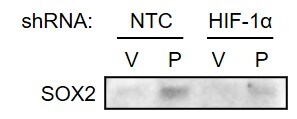
Application: Western BlotSample Tested: Breast cancer cellsSpecies: HumanVerified Customer | Posted 05/18/2020MDA-MB-231 subclones transfected with NTC or HIF-1 alpha shRNA vector were treated with vehicle (V) or paclitaxel (P) and immunoblot assay was performed.

-
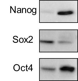
Application: Western BlotSample Tested: Human head and neck cancer cell linesSpecies: HumanVerified Customer | Posted 10/11/2018The expression of pluripotency factors Nanog, Sox2, and Oct4 in human HNSCC cell lines FaDu and HN5.
-
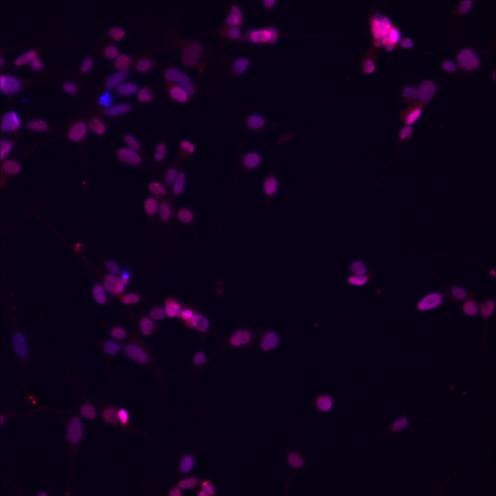
Application: ImmunocytochemistrySample Tested: aptient derived adgerent glioblastoma stem cells and patient-derived glioblastoma stem cells (adherent on laminin coating)Species: HumanVerified Customer | Posted 04/24/2017dilution 1:250 in antibody buffer (1% BSA, 0.1% triton X in PBS)
-
Application: Western BlotSample Tested: Oral cancer cell lineSpecies: HumanVerified Customer | Posted 12/27/2011
There are no reviews that match your criteria.
Protocols
View specific protocols for SOX2 Antibody - BSA Free (NB110-37235):
Culture cells to appropriate density in 35 mm culture dishes or 6-well plates.
1. Remove culture medium and wash the cells briefly in PBS. Add 10% formalin to the dish and fix at room temperature for 10 minutes.
2. Remove the formalin and wash the cells in PBS.
3. Permeablize the cells with 0.1% Triton X100 or other suitable detergent for 10 min.
4. Remove the permeablization buffer and wash three times for 10 minutes each in PBS. Be sure to not let the specimen dry out.
5. To block nonspecific antibody binding, incubate in 10% normal goat serum from 1 hour to overnight at room temperature.
6. Add primary antibody at appropriate dilution and incubate overnight at 4C.
7. Remove primary antibody and replace with PBS. Wash three times for 10 minutes each.
8. Add secondary antibody at appropriate dilution. Incubate for 1 hour at room temperature.
9. Remove secondary antibody and replace with PBS. Wash three times for 10 minutes each.
10. Counter stain DNA with DAPi if required.
Antigen Unmasking:
Bring slides to a boil in 10 mM sodium citrate buffer (pH 6.0) then maintain at a sub-boiling temperature for 10 minutes. Cool slides on bench-top for 30 minutes (keep slides in the sodium citrate buffer all the time).
Staining:
1. Wash sections in deionized water three times for 5 minutes each.
2. Wash sections in PBS for 5 minutes.
3. Block each section with 100-400 ul blocking solution (1% BSA in PBS) for 1 hour at room temperature.
4. Remove blocking solution and add 100-400 ul diluted primary antibody. Incubate overnight at 4 C.
5. Remove antibody solution and wash sections in wash buffer three times for 5 minutes each.
6. Add 100-400 ul HRP polymer conjugated secondary antibody. Incubate 30 minutes at room temperature.
7. Wash sections three times in wash buffer for 5 minutes each.
8. Add 100-400 ul DAB substrate to each section and monitor staining closely.
9. As soon as the sections develop, immerse slides in deionized water.
10. Counterstain sections in hematoxylin.
11. Wash sections in deionized water two times for 5 minutes each.
12. Dehydrate sections.
13. Mount coverslips.
1. Perform SDS-PAGE on samples to be analyzed, loading 10-25 ug of total protein per lane.
2. Transfer proteins to PVDF membrane according to the instructions provided by the manufacturer of the membrane and transfer apparatus.
3. Stain the membrane with Ponceau S (or similar product) to assess transfer success, and mark molecular weight standards where appropriate.
4. Rinse the blot TBS -0.05% Tween 20 (TBST).
5. Block the membrane in 5% Non-fat milk in TBST (blocking buffer) for at least 1 hour.
6. Wash the membrane in TBST three times for 10 minutes each.
7. Dilute primary antibody in blocking buffer and incubate overnight at 4C with gentle rocking.
8. Wash the membrane in TBST three times for 10 minutes each.
9. Incubate the membrane in diluted HRP conjugated secondary antibody in blocking buffer (as per manufacturer's instructions) for 1 hour at room temperature.
10. Wash the blot in TBST three times for 10 minutes each (this step can be repeated as required to reduce background).
11. Apply the detection reagent of choice in accordance with the manufacturers instructions.
Find general support by application which include: protocols, troubleshooting, illustrated assays, videos and webinars.
- 7-Amino Actinomycin D (7-AAD) Cell Viability Flow Cytometry Protocol
- Antigen Retrieval Protocol (PIER)
- Antigen Retrieval for Frozen Sections Protocol
- Appropriate Fixation of IHC/ICC Samples
- Cellular Response to Hypoxia Protocols
- Chromogenic IHC Staining of Formalin-Fixed Paraffin-Embedded (FFPE) Tissue Protocol
- Chromogenic Immunohistochemistry Staining of Frozen Tissue
- ClariTSA™ Fluorophore Kits
- Detection & Visualization of Antibody Binding
- Extracellular Membrane Flow Cytometry Protocol
- Flow Cytometry Protocol for Cell Surface Markers
- Flow Cytometry Protocol for Staining Membrane Associated Proteins
- Flow Cytometry Staining Protocols
- Flow Cytometry Troubleshooting Guide
- Fluorescent IHC Staining of Frozen Tissue Protocol
- Graphic Protocol for Heat-induced Epitope Retrieval
- Graphic Protocol for the Preparation and Fluorescent IHC Staining of Frozen Tissue Sections
- Graphic Protocol for the Preparation and Fluorescent IHC Staining of Paraffin-embedded Tissue Sections
- Graphic Protocol for the Preparation of Gelatin-coated Slides for Histological Tissue Sections
- ICC Cell Smear Protocol for Suspension Cells
- ICC Immunocytochemistry Protocol Videos
- ICC for Adherent Cells
- IHC Sample Preparation (Frozen sections vs Paraffin)
- Immunocytochemistry (ICC) Protocol
- Immunocytochemistry Troubleshooting
- Immunofluorescence of Organoids Embedded in Cultrex Basement Membrane Extract
- Immunofluorescent IHC Staining of Formalin-Fixed Paraffin-Embedded (FFPE) Tissue Protocol
- Immunohistochemistry (IHC) and Immunocytochemistry (ICC) Protocols
- Immunohistochemistry Frozen Troubleshooting
- Immunohistochemistry Paraffin Troubleshooting
- Intracellular Flow Cytometry Protocol Using Alcohol (Methanol)
- Intracellular Flow Cytometry Protocol Using Detergents
- Intracellular Nuclear Staining Flow Cytometry Protocol Using Detergents
- Intracellular Staining Flow Cytometry Protocol Using Alcohol Permeabilization
- Intracellular Staining Flow Cytometry Protocol Using Detergents to Permeabilize Cells
- Preparing Samples for IHC/ICC Experiments
- Preventing Non-Specific Staining (Non-Specific Binding)
- Primary Antibody Selection & Optimization
- Propidium Iodide Cell Viability Flow Cytometry Protocol
- Protocol for Heat-Induced Epitope Retrieval (HIER)
- Protocol for Liperfluo
- Protocol for Making a 4% Formaldehyde Solution in PBS
- Protocol for VisUCyte™ HRP Polymer Detection Reagent
- Protocol for the Characterization of Human Th22 Cells
- Protocol for the Characterization of Human Th9 Cells
- Protocol for the Fluorescent ICC Staining of Cell Smears - Graphic
- Protocol for the Fluorescent ICC Staining of Cultured Cells on Coverslips - Graphic
- Protocol for the Preparation & Fixation of Cells on Coverslips
- Protocol for the Preparation and Chromogenic IHC Staining of Frozen Tissue Sections
- Protocol for the Preparation and Chromogenic IHC Staining of Frozen Tissue Sections - Graphic
- Protocol for the Preparation and Chromogenic IHC Staining of Paraffin-embedded Tissue Sections
- Protocol for the Preparation and Chromogenic IHC Staining of Paraffin-embedded Tissue Sections - Graphic
- Protocol for the Preparation and Fluorescent ICC Staining of Cells on Coverslips
- Protocol for the Preparation and Fluorescent ICC Staining of Non-adherent Cells
- Protocol for the Preparation and Fluorescent ICC Staining of Stem Cells on Coverslips
- Protocol for the Preparation and Fluorescent IHC Staining of Frozen Tissue Sections
- Protocol for the Preparation and Fluorescent IHC Staining of Paraffin-embedded Tissue Sections
- Protocol for the Preparation of Gelatin-coated Slides for Histological Tissue Sections
- Protocol for the Preparation of a Cell Smear for Non-adherent Cell ICC - Graphic
- Protocol: Annexin V and PI Staining by Flow Cytometry
- Protocol: Annexin V and PI Staining for Apoptosis by Flow Cytometry
- R&D Systems Quality Control Western Blot Protocol
- TUNEL and Active Caspase-3 Detection by IHC/ICC Protocol
- The Importance of IHC/ICC Controls
- Troubleshooting Guide: Fluorokine Flow Cytometry Kits
- Troubleshooting Guide: Immunohistochemistry
- Troubleshooting Guide: Western Blot Figures
- Western Blot Conditions
- Western Blot Protocol
- Western Blot Protocol for Cell Lysates
- Western Blot Troubleshooting
- Western Blot Troubleshooting Guide
- View all Protocols, Troubleshooting, Illustrated assays and Webinars
FAQs for SOX2 Antibody - BSA Free
-
Q: As part of an Endometriosis-related project in horses, I would like to ask some information with regard to this. I would like to know about its tested or predicted reactivity with horse.
A:
This antibody has not yet been tested with horse. This means that use of this antibody with horse would not be covered by our guarantee; however, you would be eligible for our Innovator's Reward Program. For an indication of the likely cross-reactivity (how well the antibody is likely to perform with horse), I have run UniProt Blast sequence comparison of the immunogen sequence versus horse proteins. For SOX2 Antibody NB110-37235, the exact immunogen is available to me in our internal notes. The UniProt Blast of this gave a 100% match with the (again, unfortunately unreviewed) horse entries F6PUQ2 and B6VA41. A 100% match indicates that the antibody should be suitable for you. If you would be interested in testing this novel species, please take a look at our Innovators Reward Program.
-
Q: Can you advise me the pH value for the citrate buffer?
A: For the validation of this antibody in IHC-P, we performed antigen retrieval using 0.01 M sodium citrate buffer, pH 6.0. Please note that Domyan et al 2011 from University of Wisconsin-Madison (USA) have cited the use of this antibody in IHC-P application in their experiments published in - Development (2011) 138, 971-981 Biologists site.
-
Q: We are looking for the following markers to use to stain cells derived from both human and mouse embryonic and induced pluripotent stem cells: undifferentiated cells, neurons, oligodendrocytes, and astrocytes.
A: For ES markers some popular targets are OCT4 (NB100-2379), KLF4 (NBP2-17070) and SOX2 (NB110-37235). PEA15 is expressed in Astrocytes and this might be one of interest for you (NBP1-61390). OCT4 is one that can also be expressed in Oligodendrocytes.
-
Q: As part of an Endometriosis-related project in horses, I would like to ask some information with regard to this. I would like to know about its tested or predicted reactivity with horse.
A:
This antibody has not yet been tested with horse. This means that use of this antibody with horse would not be covered by our guarantee; however, you would be eligible for our Innovator's Reward Program. For an indication of the likely cross-reactivity (how well the antibody is likely to perform with horse), I have run UniProt Blast sequence comparison of the immunogen sequence versus horse proteins. For SOX2 Antibody NB110-37235, the exact immunogen is available to me in our internal notes. The UniProt Blast of this gave a 100% match with the (again, unfortunately unreviewed) horse entries F6PUQ2 and B6VA41. A 100% match indicates that the antibody should be suitable for you. If you would be interested in testing this novel species, please take a look at our Innovators Reward Program.
-
Q: Can you advise me the pH value for the citrate buffer?
A: For the validation of this antibody in IHC-P, we performed antigen retrieval using 0.01 M sodium citrate buffer, pH 6.0. Please note that Domyan et al 2011 from University of Wisconsin-Madison (USA) have cited the use of this antibody in IHC-P application in their experiments published in - Development (2011) 138, 971-981 Biologists site.
-
Q: We are looking for the following markers to use to stain cells derived from both human and mouse embryonic and induced pluripotent stem cells: undifferentiated cells, neurons, oligodendrocytes, and astrocytes.
A: For ES markers some popular targets are OCT4 (NB100-2379), KLF4 (NBP2-17070) and SOX2 (NB110-37235). PEA15 is expressed in Astrocytes and this might be one of interest for you (NBP1-61390). OCT4 is one that can also be expressed in Oligodendrocytes.

