PPAR alpha/NR1C1 Antibody (3B6/PPAR) - BSA Free
Novus Biologicals | Catalog # NB300-537
![Flow Cytometry: PPAR alpha/NR1C1 Antibody (3B6/PPAR) - BSA Free [NB300-537]](https://resources.rndsystems.com/images/products/PPAR-alpha-NR1C1-Antibody-3B6-PPAR-Flow-Cytometry-NB300-537-img0008.jpg "Flow Cytometry: PPAR alpha/NR1C1 Antibody (3B6/PPAR) - BSA Free [NB300-537]")
Key Product Details
Validated by
Species Reactivity
Validated:
Cited:
Applications
Validated:
Cited:
Label
Antibody Source
Format
Product Specifications
Immunogen
Localization
Specificity
Clonality
Host
Isotype
Scientific Data Images for PPAR alpha/NR1C1 Antibody (3B6/PPAR) - BSA Free
Flow Cytometry: PPAR alpha/NR1C1 Antibody (3B6/PPAR) - BSA Free [NB300-537]
Flow Cytometry: PPAR alpha/NR1C1 Antibody (3B6/PPAR) [NB300-537] - An intracellular stain was performed on U-87 cells with PPAR alpha/NR1C1 Antibody [3B6/PPAR] NB300-537AF647 (blue) and a matched isotype control (orange). Cells were fixed with 4% PFA and then permeabilized with 0.1% saponin. Cells were incubated in an antibody dilution of 2.5 ug/mL for 30 minutes at room temperature. Both antibodies were conjugated to Alexa Fluor 647.![Western Blot: PPAR alpha/NR1C1 Antibody (3B6/PPAR)BSA Free [NB300-537]](https://resources.rndsystems.com/images/products/PPAR-alpha-NR1C1-Antibody-3B6-PPAR-Western-Blot-NB300-537-img0006.jpg "Western Blot: PPAR alpha/NR1C1 Antibody (3B6/PPAR)BSA Free [NB300-537]")
Western Blot: PPAR alpha/NR1C1 Antibody (3B6/PPAR)BSA Free [NB300-537]
Western Blot: PPAR alpha/NR1C1 Antibody (3B6/PPAR) [NB300-537] - Analysis of 25 ug of Hela (Lane 1), Jurkat (Lane 2), and NIH-3T3 cell lysates (Lane 3) and a molecular weight protein ladder.![Flow Cytometry: PPAR alpha/NR1C1 Antibody (3B6/PPAR) - BSA Free [NB300-537]](https://resources.rndsystems.com/images/products/PPAR-alpha-NR1C1-Antibody-3B6-PPAR-Flow-Cytometry-NB300-537-img0009.jpg "Flow Cytometry: PPAR alpha/NR1C1 Antibody (3B6/PPAR) - BSA Free [NB300-537]")
Flow Cytometry: PPAR alpha/NR1C1 Antibody (3B6/PPAR) - BSA Free [NB300-537]
Flow Cytometry: PPAR alpha/NR1C1 Antibody (3B6/PPAR) [NB300-537] - An intracellular stain was performed on Hek293 cells with PPAR alpha/NR1C1 [3B6/PPAR] Antibody NB300-537AF647 (blue) and a matched isotype control (orange). Cells were fixed with 4% PFA and then permeabilized with 0.1% saponin. Cells were incubated in an antibody dilution of 2.5 ug/mL for 30 minutes at room temperature. Both antibodies were conjugated to Alexa Fluor 647.![Western Blot: PPAR alpha/NR1C1 Antibody (3B6/PPAR)BSA Free [NB300-537]](https://resources.rndsystems.com/images/products/PPAR-alpha-NR1C1-Antibody-3B6-PPAR-Western-Blot-NB300-537-img0002.jpg "Western Blot: PPAR alpha/NR1C1 Antibody (3B6/PPAR)BSA Free [NB300-537]")
Western Blot: PPAR alpha/NR1C1 Antibody (3B6/PPAR)BSA Free [NB300-537]
Western Blot: PPAR alpha/NR1C1 Antibody (3B6/PPAR) [NB300-537] - 293T cell lysate. Image from verified customer review.![Flow Cytometry: PPAR alpha/NR1C1 Antibody (3B6/PPAR) - BSA Free [NB300-537]](https://resources.rndsystems.com/images/products/PPAR-alpha-NR1C1-Antibody-3B6-PPAR-Flow-Cytometry-NB300-537-img0003.jpg "Flow Cytometry: PPAR alpha/NR1C1 Antibody (3B6/PPAR) - BSA Free [NB300-537]")
Flow Cytometry: PPAR alpha/NR1C1 Antibody (3B6/PPAR) - BSA Free [NB300-537]
Flow Cytometry: PPAR alpha/NR1C1 Antibody (3B6/PPAR) [NB300-537] - Analysis of PPAR alpha in Jurkat cells compared to an isotype control (blue).![Flow Cytometry: PPAR alpha/NR1C1 Antibody (3B6/PPAR) - BSA Free [NB300-537]](https://resources.rndsystems.com/images/products/PPAR-alpha-NR1C1-Antibody-3B6-PPAR-Flow-Cytometry-NB300-537-img0004.jpg "Flow Cytometry: PPAR alpha/NR1C1 Antibody (3B6/PPAR) - BSA Free [NB300-537]")
Flow Cytometry: PPAR alpha/NR1C1 Antibody (3B6/PPAR) - BSA Free [NB300-537]
Flow Cytometry: PPAR alpha/NR1C1 Antibody (3B6/PPAR) [NB300-537] - Analysis of PPAR alpha in Hela cells compared to an isotype control (blue).![Flow Cytometry: PPAR alpha/NR1C1 Antibody (3B6/PPAR) - BSA Free [NB300-537]](https://resources.rndsystems.com/images/products/PPAR-alpha-NR1C1-Antibody-3B6-PPAR-Flow-Cytometry-NB300-537-img0005.jpg "Flow Cytometry: PPAR alpha/NR1C1 Antibody (3B6/PPAR) - BSA Free [NB300-537]")
Flow Cytometry: PPAR alpha/NR1C1 Antibody (3B6/PPAR) - BSA Free [NB300-537]
Flow Cytometry: PPAR alpha/NR1C1 Antibody (3B6/PPAR) [NB300-537] - Analysis of PPAR alpha in 3T3 cells compared to an isotype control (blue).![Flow Cytometry: PPAR alpha/NR1C1 Antibody (3B6/PPAR) - BSA Free [NB300-537]](https://resources.rndsystems.com/images/products/PPAR-alpha-NR1C1-Antibody-3B6-PPAR-Flow-Cytometry-NB300-537-img0007.jpg "Flow Cytometry: PPAR alpha/NR1C1 Antibody (3B6/PPAR) - BSA Free [NB300-537]")
Flow Cytometry: PPAR alpha/NR1C1 Antibody (3B6/PPAR) - BSA Free [NB300-537]
Flow Cytometry: PPAR alpha/NR1C1 Antibody (3B6/PPAR) [NB300-537] - An intracellular stain was performed on Jurkat cells with PPAR alpha/NR1C1 Antibody (3B6/PPAR) NB300-537AF647 (blue) and a matched isotype control (orange). Cells were fixed with 4% PFA and then permeabilized with 0.1% saponin. Cells were incubated in an antibody dilution of 5 ug/mL for 30 minutes at room temperature. Both antibodies were conjugated to Alexa Fluor 647.![Knockdown Validated: PPAR alpha/NR1C1 Antibody (3B6/PPAR) - BSA Free [NB300-537]](https://resources.rndsystems.com/images/products/PPAR-alpha-NR1C1-Antibody-3B6-PPAR-BSA-Free-Knockdown-Validated-NB300-537-img0010.jpg "Western Blot: PPAR alpha/NR1C1 Antibody (3B6/PPAR) - BSA Free [NB300-537]")
 - BSA Free [NB300-537] -")
Western Blot: PPAR alpha/NR1C1 Antibody (3B6/PPAR) - BSA Free [NB300-537] -
The metabolic remodeling process in natural kidney aging. A Representative images of Oil Red O staining in the 24-month-old and 6-month-old group (n = 6), scale bar = 50 μm; B, C the downregulated protein levels of FAO markers (i.e., PPAR alpha, ACOX1, and CPT1A) and upregulated protein levels of glycolysis markers (i.e., HK2 and PDK1) by western blot, and their semi-quantitative analyses (n = 6); D immunostaining for GLIS1 (red) and PPAR alpha (green), with DAPI (blue) counterstaining by IF staining in the 24-month-old and 6-month-old group (n = 6), scale bar = 50 μm; E lactate levels in the 24-month-old and 6-month-old group (n = 6); F the downregulated protein levels of PPAR alpha, ACOX1, and CPT1A, and up-regulated protein levels of HK2 and PDK1 in the 24-month-old group by IHC assay and their semi-quantitative analyses (n = 6), scale bar = 50 μm. The data are expressed as the mean +/- SD of three independent experiments. **P <.01 or ***P <.001 versus the 6-month-old group by Student’s t-test Image collected and cropped by CiteAb from the following open publication (https://pubmed.ncbi.nlm.nih.gov/39736678), licensed under a CC-BY license. Not internally tested by Novus Biologicals. - BSA Free [NB300-537] -")
Western Blot: PPAR alpha/NR1C1 Antibody (3B6/PPAR) - BSA Free [NB300-537] -
Bioinformatics analysis of proteomic results.(A). Hierarchical clustering analysis of the differentially expressed proteins in the two groups (si-CD151 vs si-NC) (n = 3) (B) The differentially expressed proteins analyzed by volcano plots between every two groups (si-CD151 vs si-NC) (C) Circus plots of GO Classification Annotation of differential protein. (D) KEGG pathways of differentially expressed proteins. OS: Organismal Systems; M: Metabolism; GIP: Genetic Information Processing; CP: Cellular Processes. (E) Western blot analysis of PPAR-alpha, PCG-1 alpha and GAPDH) in CMs under PE, quantified by Image J (n = 4). (F) Western blot analysis of PPAR-gamma and GAPDH) in CFs under Ang-II, quantified by Image J (n = 4). Data are expressed as mean +/- SEM. Image collected and cropped by CiteAb from the following open publication (https://dx.plos.org/10.1371/journal.pone.0297121), licensed under a CC-BY license. Not internally tested by Novus Biologicals. - BSA Free [NB300-537] -")
Western Blot: PPAR alpha/NR1C1 Antibody (3B6/PPAR) - BSA Free [NB300-537] -
The over-expressed GLIS1 suppressed metabolic remodeling from FAO to glycolysis in the accelerated aging mouse model. A The representative images of mouse kidney tissue in the control, AAV-Vector and AAV-GLIS1 group stained with Oil Red O (n = 6), scale bar = 50 μm; B, C downregulated protein levels of PPAR alpha, ACOX1, and CPT1A in the accelerated aging mouse model were reversed in the presence of AAV-GLIS1, while upregulated protein levels of HK2 and PKD1 in the accelerated aging mouse model were reduced by introducing AAV-GLIS1 (n = 6); D lactate levels in the control, AAV-Vector and AAV-GLIS1 group (n = 6); E the reversed effect of PPAR alpha, ACOX1 and CPT1A levels, as well as HK2 and PDK1 levels in the presence of AAV-GLIS1 by IHC assay, and their semi-quantitative analyses (n = 6), scale bar = 50 μm; F immunostaining for PPAR alpha, ACOX1, CPTA1, HK2, and PDK1 (green), with DAPI (blue) counterstaining by IF staining in accelerated aging mouse model (n = 6), scale bar = 50 μm. The data are expressed as the mean +/- SD of three independent experiments. **P <.01 or ***P <.001 versus the AAV-vector group by one-way ANOVA Image collected and cropped by CiteAb from the following open publication (https://pubmed.ncbi.nlm.nih.gov/39736678), licensed under a CC-BY license. Not internally tested by Novus Biologicals. - BSA Free [NB300-537] -")
Western Blot: PPAR alpha/NR1C1 Antibody (3B6/PPAR) - BSA Free [NB300-537] -
The ablation of METTL3 triggered metabolic remodeling and aggravated cell senescence renal fibrosis. A Protein levels of GLIS1, PPAR alpha, ACOX1, CPT1A, HK2, PDK1, FN, alpha -SMA, and P16INK4A by western blot in the vector and siMETTL3 group, and their semi-quantitative analyses (n = 3); B representative images of Oil Red O staining in the vector and siMETTL3 group (n = 3), scale bar = 50 μm; C double immunostaining for GLS1 and PPAR alpha, ACOX1, CPT1A, HK2, PDK1, and alpha -SMA by IF staining (n = 3) scale bar = 50 μm. The data are expressed as the mean +/- SD of three independent experiments. **P <.01 or ***P <.001 versus the vector group by Student’s t-test Image collected and cropped by CiteAb from the following open publication (https://pubmed.ncbi.nlm.nih.gov/39736678), licensed under a CC-BY license. Not internally tested by Novus Biologicals. - BSA Free [NB300-537] -")
Western Blot: PPAR alpha/NR1C1 Antibody (3B6/PPAR) - BSA Free [NB300-537] -
CCM treatment upregulates AMPK and PPAR-alpha pathways. (a) Western blot analysis of the expression of AMPK. Densitometric analysis is shown in the bar graph. (b) Western blot analysis of the expressions of PPAR-alpha and PGC-1 alpha. Densitometric analysis is shown in the bar graph. Data are expressed as mean values +/- SD (*P < 0.05 compared with the sham group; #P < 0.05 compared with HF group). Image collected and cropped by CiteAb from the following open publication (https://pubmed.ncbi.nlm.nih.gov/35799598), licensed under a CC-BY license. Not internally tested by Novus Biologicals. - BSA Free [NB300-537] -")
Immunohistochemistry: PPAR alpha/NR1C1 Antibody (3B6/PPAR) - BSA Free [NB300-537] -
The metabolic remodeling process in natural kidney aging. A Representative images of Oil Red O staining in the 24-month-old and 6-month-old group (n = 6), scale bar = 50 μm; B, C the downregulated protein levels of FAO markers (i.e., PPAR alpha, ACOX1, and CPT1A) and upregulated protein levels of glycolysis markers (i.e., HK2 and PDK1) by western blot, and their semi-quantitative analyses (n = 6); D immunostaining for GLIS1 (red) and PPAR alpha (green), with DAPI (blue) counterstaining by IF staining in the 24-month-old and 6-month-old group (n = 6), scale bar = 50 μm; E lactate levels in the 24-month-old and 6-month-old group (n = 6); F the downregulated protein levels of PPAR alpha, ACOX1, and CPT1A, and up-regulated protein levels of HK2 and PDK1 in the 24-month-old group by IHC assay and their semi-quantitative analyses (n = 6), scale bar = 50 μm. The data are expressed as the mean +/- SD of three independent experiments. **P <.01 or ***P <.001 versus the 6-month-old group by Student’s t-test Image collected and cropped by CiteAb from the following open publication (https://pubmed.ncbi.nlm.nih.gov/39736678), licensed under a CC-BY license. Not internally tested by Novus Biologicals. - BSA Free [NB300-537] -")
Immunohistochemistry: PPAR alpha/NR1C1 Antibody (3B6/PPAR) - BSA Free [NB300-537] -
The over-expressed GLIS1 suppressed metabolic remodeling from FAO to glycolysis in the accelerated aging mouse model. A The representative images of mouse kidney tissue in the control, AAV-Vector and AAV-GLIS1 group stained with Oil Red O (n = 6), scale bar = 50 μm; B, C downregulated protein levels of PPAR alpha, ACOX1, and CPT1A in the accelerated aging mouse model were reversed in the presence of AAV-GLIS1, while upregulated protein levels of HK2 and PKD1 in the accelerated aging mouse model were reduced by introducing AAV-GLIS1 (n = 6); D lactate levels in the control, AAV-Vector and AAV-GLIS1 group (n = 6); E the reversed effect of PPAR alpha, ACOX1 and CPT1A levels, as well as HK2 and PDK1 levels in the presence of AAV-GLIS1 by IHC assay, and their semi-quantitative analyses (n = 6), scale bar = 50 μm; F immunostaining for PPAR alpha, ACOX1, CPTA1, HK2, and PDK1 (green), with DAPI (blue) counterstaining by IF staining in accelerated aging mouse model (n = 6), scale bar = 50 μm. The data are expressed as the mean +/- SD of three independent experiments. **P <.01 or ***P <.001 versus the AAV-vector group by one-way ANOVA Image collected and cropped by CiteAb from the following open publication (https://pubmed.ncbi.nlm.nih.gov/39736678), licensed under a CC-BY license. Not internally tested by Novus Biologicals.Applications for PPAR alpha/NR1C1 Antibody (3B6/PPAR) - BSA Free
Chromatin Immunoprecipitation (ChIP)
Flow Cytometry
Gel Super Shift Assays
Immunocytochemistry/ Immunofluorescence
Immunohistochemistry
Immunohistochemistry-Paraffin
Immunoprecipitation
Simple Western
Western Blot
See Simple Western Antibody Database for Simple Western validation: Tested in Skin, separated by Size, antibody dilution of 1:50
Reviewed Applications
Read 2 reviews rated 4.5 using NB300-537 in the following applications:
Flow Cytometry Panel Builder
Bio-Techne Knows Flow Cytometry
Save time and reduce costly mistakes by quickly finding compatible reagents using the Panel Builder Tool.
Advanced Features
- Spectra Viewer - Custom analysis of spectra from multiple fluorochromes
- Spillover Popups - Visualize the spectra of individual fluorochromes
- Antigen Density Selector - Match fluorochrome brightness with antigen density

Formulation, Preparation, and Storage
Purification
Formulation
Format
Preservative
Concentration
Shipping
Stability & Storage
Background: PPAR alpha/NR1C1
Long Name
Alternate Names
Gene Symbol
UniProt
Additional PPAR alpha/NR1C1 Products
Product Documents for PPAR alpha/NR1C1 Antibody (3B6/PPAR) - BSA Free
Certificate of Analysis
To download a Certificate of Analysis, please enter a lot or batch number in the search box below.
Product Specific Notices for PPAR alpha/NR1C1 Antibody (3B6/PPAR) - BSA Free
This product is for research use only and is not approved for use in humans or in clinical diagnosis. Primary Antibodies are guaranteed for 1 year from date of receipt.
Related Research Areas
Citations for PPAR alpha/NR1C1 Antibody (3B6/PPAR) - BSA Free
 Powered by Bioz
Customer Reviews for PPAR alpha/NR1C1 Antibody (3B6/PPAR) - BSA Free (2)
Have you used PPAR alpha/NR1C1 Antibody (3B6/PPAR) - BSA Free?
Submit a review and receive an Amazon gift card!
$25/€18/£15/$25CAN/¥2500 Yen for a review with an image
$10/€7/£6/$10CAN/¥1110 Yen for a review without an image
Submit a review
Customer Images
-(01-mg)_NB300-537_10656.jpg)
-
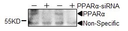
Application: Western BlotSample Tested: primary neonatal ventricular cardiomyocytesSpecies: Rat primary cultured cardiomyocytes and RatVerified Customer | Posted 08/24/2022PPARa was knocked down with siRNA in primary cultured rat cardiomyocytes.Endogenous PPARa was knocked down with siRNA.
 Bio-Techne ResponseThis review was submitted through the legacy Novus Innovators Program, reflecting a new species or application tested on a primary antibody.
 -
Application: Western BlotSample Tested: Whole cell lysate from 293T cells, in lysis bufferSpecies: HumanVerified Customer | Posted 10/02/2014WB for PPARalpha in 293T cells
There are no reviews that match your criteria.
Protocols
Find general support by application which include: protocols, troubleshooting, illustrated assays, videos and webinars.
- 7-Amino Actinomycin D (7-AAD) Cell Viability Flow Cytometry Protocol
- Antigen Retrieval Protocol (PIER)
- Antigen Retrieval for Frozen Sections Protocol
- Appropriate Fixation of IHC/ICC Samples
- Cellular Response to Hypoxia Protocols
- ChIP Protocol Video
- Chromatin Immunoprecipitation (ChIP) Protocol
- Chromatin Immunoprecipitation Protocol
- Chromogenic IHC Staining of Formalin-Fixed Paraffin-Embedded (FFPE) Tissue Protocol
- Chromogenic Immunohistochemistry Staining of Frozen Tissue
- ClariTSA™ Fluorophore Kits
- Detection & Visualization of Antibody Binding
- Extracellular Membrane Flow Cytometry Protocol
- Flow Cytometry Protocol for Cell Surface Markers
- Flow Cytometry Protocol for Staining Membrane Associated Proteins
- Flow Cytometry Staining Protocols
- Flow Cytometry Troubleshooting Guide
- Fluorescent IHC Staining of Frozen Tissue Protocol
- Graphic Protocol for Heat-induced Epitope Retrieval
- Graphic Protocol for the Preparation and Fluorescent IHC Staining of Frozen Tissue Sections
- Graphic Protocol for the Preparation and Fluorescent IHC Staining of Paraffin-embedded Tissue Sections
- Graphic Protocol for the Preparation of Gelatin-coated Slides for Histological Tissue Sections
- ICC Cell Smear Protocol for Suspension Cells
- ICC Immunocytochemistry Protocol Videos
- ICC for Adherent Cells
- IHC Sample Preparation (Frozen sections vs Paraffin)
- Immunocytochemistry (ICC) Protocol
- Immunocytochemistry Troubleshooting
- Immunofluorescence of Organoids Embedded in Cultrex Basement Membrane Extract
- Immunofluorescent IHC Staining of Formalin-Fixed Paraffin-Embedded (FFPE) Tissue Protocol
- Immunohistochemistry (IHC) and Immunocytochemistry (ICC) Protocols
- Immunohistochemistry Frozen Troubleshooting
- Immunohistochemistry Paraffin Troubleshooting
- Immunoprecipitation Protocol
- Intracellular Flow Cytometry Protocol Using Alcohol (Methanol)
- Intracellular Flow Cytometry Protocol Using Detergents
- Intracellular Nuclear Staining Flow Cytometry Protocol Using Detergents
- Intracellular Staining Flow Cytometry Protocol Using Alcohol Permeabilization
- Intracellular Staining Flow Cytometry Protocol Using Detergents to Permeabilize Cells
- Preparing Samples for IHC/ICC Experiments
- Preventing Non-Specific Staining (Non-Specific Binding)
- Primary Antibody Selection & Optimization
- Propidium Iodide Cell Viability Flow Cytometry Protocol
- Protocol for Heat-Induced Epitope Retrieval (HIER)
- Protocol for Liperfluo
- Protocol for Making a 4% Formaldehyde Solution in PBS
- Protocol for VisUCyte™ HRP Polymer Detection Reagent
- Protocol for the Characterization of Human Th22 Cells
- Protocol for the Characterization of Human Th9 Cells
- Protocol for the Fluorescent ICC Staining of Cell Smears - Graphic
- Protocol for the Fluorescent ICC Staining of Cultured Cells on Coverslips - Graphic
- Protocol for the Preparation & Fixation of Cells on Coverslips
- Protocol for the Preparation and Chromogenic IHC Staining of Frozen Tissue Sections
- Protocol for the Preparation and Chromogenic IHC Staining of Frozen Tissue Sections - Graphic
- Protocol for the Preparation and Chromogenic IHC Staining of Paraffin-embedded Tissue Sections
- Protocol for the Preparation and Chromogenic IHC Staining of Paraffin-embedded Tissue Sections - Graphic
- Protocol for the Preparation and Fluorescent ICC Staining of Cells on Coverslips
- Protocol for the Preparation and Fluorescent ICC Staining of Non-adherent Cells
- Protocol for the Preparation and Fluorescent ICC Staining of Stem Cells on Coverslips
- Protocol for the Preparation and Fluorescent IHC Staining of Frozen Tissue Sections
- Protocol for the Preparation and Fluorescent IHC Staining of Paraffin-embedded Tissue Sections
- Protocol for the Preparation of Gelatin-coated Slides for Histological Tissue Sections
- Protocol for the Preparation of a Cell Smear for Non-adherent Cell ICC - Graphic
- Protocol: Annexin V and PI Staining by Flow Cytometry
- Protocol: Annexin V and PI Staining for Apoptosis by Flow Cytometry
- R&D Systems Quality Control Western Blot Protocol
- TUNEL and Active Caspase-3 Detection by IHC/ICC Protocol
- The Importance of IHC/ICC Controls
- Troubleshooting Guide: Fluorokine Flow Cytometry Kits
- Troubleshooting Guide: Immunohistochemistry
- Troubleshooting Guide: Western Blot Figures
- Western Blot Conditions
- Western Blot Protocol
- Western Blot Protocol for Cell Lysates
- Western Blot Troubleshooting
- Western Blot Troubleshooting Guide
- View all Protocols, Troubleshooting, Illustrated assays and Webinars
FAQs for PPAR alpha/NR1C1 Antibody (3B6/PPAR) - BSA Free
-
Q: Please differentiate to me between PPAR and PGC clearly. I am confused with the difference between these two
A:
Thank you very much for contacting Novus Biologicals technical support team and sharing your query on the differences between PGC-1 alpha and PPAR. These are two different proteins encoded by their respective genes and serves different functions. PGC-1 alpha (PGC1A or PPAR gamma coactivator 1-alpha) is a transcriptional co-activator for steroid receptors and nuclear receptors, and it regulates diverse aspects of cellular physiology. It up-regulates the transcriptional activity of PPAR-gamma /thyroid hormone receptor on the uncoupling protein promoter; regulates the key mitochondrial genes involved in adaptive thermogenesis; implicates in the metabolic reprogramming in response to nutrients availability through the coordination of the expression of a wide array of genes involved in the regulation of glucose and fatty acid metabolism. Among our PGC-1 alpha antibodies, NBP1-04676 is our best selling product with nice customer feedback and citations in at least 13 research publications. PPAR (PPAR alpha) on the other hand is a ligand-activated transcription factor which gets activated by the endogenous ligand 1-palmitoyl-2-oleoyl-sn-glycerol-3-phosphocholine, and oleylethanolamide (a naturally occurring lipid that regulates satiety), and acts as a key regulator of lipid metabolism. It also acts as a receptor for peroxisome proliferators such as hypolipidemic drugs and fatty acids. It regulates the peroxisomal beta-oxidation pathway of fatty acids, and also functions as transcription activator for the ACOX1 and P450 genes. We have a variety of PPAR alpha antibodies. I hope you will find this information helpful but please let me know if I can support you with anything else from my end. Thank you very much for choosing Novus Biologicals as your quality reagent supplier and we wish you the best with your research projects.
-
Q: Would you so kind to let me have protocol of product which cat have cat number NB300-537?
A:
We do not have any protocols specifically for product NB300-537. However, we do have some publications listed for this product on the datasheet under the "Images, Reviews & Publications" tab. Furthermore, our general protocols for a range of antibody applications can be found using this link.
-
Q: Please differentiate to me between PPAR and PGC clearly. I am confused with the difference between these two
A:
Thank you very much for contacting Novus Biologicals technical support team and sharing your query on the differences between PGC-1 alpha and PPAR. These are two different proteins encoded by their respective genes and serves different functions. PGC-1 alpha (PGC1A or PPAR gamma coactivator 1-alpha) is a transcriptional co-activator for steroid receptors and nuclear receptors, and it regulates diverse aspects of cellular physiology. It up-regulates the transcriptional activity of PPAR-gamma /thyroid hormone receptor on the uncoupling protein promoter; regulates the key mitochondrial genes involved in adaptive thermogenesis; implicates in the metabolic reprogramming in response to nutrients availability through the coordination of the expression of a wide array of genes involved in the regulation of glucose and fatty acid metabolism. Among our PGC-1 alpha antibodies, NBP1-04676 is our best selling product with nice customer feedback and citations in at least 13 research publications. PPAR (PPAR alpha) on the other hand is a ligand-activated transcription factor which gets activated by the endogenous ligand 1-palmitoyl-2-oleoyl-sn-glycerol-3-phosphocholine, and oleylethanolamide (a naturally occurring lipid that regulates satiety), and acts as a key regulator of lipid metabolism. It also acts as a receptor for peroxisome proliferators such as hypolipidemic drugs and fatty acids. It regulates the peroxisomal beta-oxidation pathway of fatty acids, and also functions as transcription activator for the ACOX1 and P450 genes. We have a variety of PPAR alpha antibodies. I hope you will find this information helpful but please let me know if I can support you with anything else from my end. Thank you very much for choosing Novus Biologicals as your quality reagent supplier and we wish you the best with your research projects.
-
Q: Would you so kind to let me have protocol of product which cat have cat number NB300-537?
A:
We do not have any protocols specifically for product NB300-537. However, we do have some publications listed for this product on the datasheet under the "Images, Reviews & Publications" tab. Furthermore, our general protocols for a range of antibody applications can be found using this link.