Prox1 Antibody - Azide and BSA Free
Novus Biologicals | Catalog # NBP1-18605
![Western Blot: Prox1 Antibody [NBP1-18605]](https://resources.rndsystems.com/images/products/Prox1-Antibody-Western-Blot-NBP1-18605-img0007.jpg "Western Blot: Prox1 Antibody [NBP1-18605]")
Loading...
Key Product Details
Species Reactivity
Validated:
Human, Mouse
Cited:
Human, Mouse
Applications
Validated:
Immunohistochemistry, Immunohistochemistry-Paraffin, Immunohistochemistry-Frozen, Western Blot, Immunocytochemistry/ Immunofluorescence
Cited:
Immunohistochemistry-Paraffin, Immunohistochemistry-Frozen, Immunocytochemistry/ Immunofluorescence
Label
Unconjugated
Antibody Source
Polyclonal Rabbit IgG
Format
Azide and BSA Free
Loading...
Product Specifications
Immunogen
Recombinant human Prox-1
Reactivity Notes
Mouse reactivity reported in scientific literature (PMID: 27122034).
Localization
Nucleus
Clonality
Polyclonal
Host
Rabbit
Isotype
IgG
Scientific Data Images for Prox1 Antibody - Azide and BSA Free
Western Blot: Prox1 Antibody [NBP1-18605]
Western Blot: Prox1 Antibody [NBP1-18605] - Analysis with anti-human Prox-1![Immunocytochemistry/ Immunofluorescence: Prox1 Antibody [NBP1-18605]](https://resources.rndsystems.com/images/products/Prox1-Antibody-Immunocytochemistry-Immunofluorescence-NBP1-18605-img0008.jpg "Immunocytochemistry/ Immunofluorescence: Prox1 Antibody [NBP1-18605]")
Immunocytochemistry/ Immunofluorescence: Prox1 Antibody [NBP1-18605]
Prox1-Antibody-Immunocytochemistry-Immunofluorescence-NBP1-18605-img0008.jpg![Immunocytochemistry/ Immunofluorescence: Prox1 Antibody [NBP1-18605]](https://resources.rndsystems.com/images/products/Prox1-Antibody-Immunocytochemistry-Immunofluorescence-NBP1-18605-img0006.jpg "Immunocytochemistry/ Immunofluorescence: Prox1 Antibody [NBP1-18605]")
Immunocytochemistry/ Immunofluorescence: Prox1 Antibody [NBP1-18605]
Immunocytochemistry/Immunofluorescence: Prox1 Antibody [NBP1-18605] - PROX1 Antibody [NBP1-18605] - human skin stained with PROX1 (red), VEGFR3 (green) and TO-PRO (nuclei). ICC image submitted by a verified customer review.
Immunocytochemistry/ Immunofluorescence: Prox1 Antibody [NBP1-18605] -
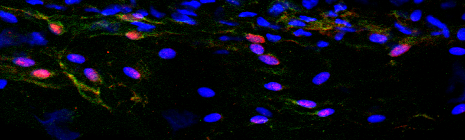
Immunocytochemistry/ Immunofluorescence: Prox1 Antibody [NBP1-18605] - Mouse LLECs develop independent of the transcription factor PU.1. a–d As detected by IHC, E15.5 wild-type mice have cells that co-express LYVE1 (a, red) PROX1 (b, green) & MRC1 (c, white) in the developing meninges (white bracket). DAPI labels nuclei blue in the merged image (d). White arrows indicate cells with these three markers. Scale = 50 µm. n = 2 brains. e–h) The meninges (white bracket) of E15.5 PU.1 knockout siblings contain many cells (white arrows) that co-express LYVE1 (e, red), PROX1 (f, green), & MRC1 (g, white). Scale = 50 µm. n = 3 brains Image collected & cropped by CiteAb from the following publication (https://pubmed.ncbi.nlm.nih.gov/31696318), licensed under a CC-BY license. Not internally tested by Novus Biologicals.Applications for Prox1 Antibody - Azide and BSA Free
Application
Recommended Usage
Immunocytochemistry/ Immunofluorescence
1:750 - 1:1000
Western Blot
1-5 ug/ml
Application Notes
Use in IHC-Frozen reported in scientific literature (PMID 27122034). Use in Immunohistochemistry-paraffin reported in scientific literature (PMID 31696318).
Reviewed Applications
Read 1 review rated 3 using NBP1-18605 in the following applications:
Formulation, Preparation, and Storage
Purification
Protein A purified
Reconstitution
Centrifuge vial prior to opening. Reconstitute in sterile water to a concentration of 0.1-1.0 mg/ml. Please note the sample size of this product will be provided in reconstituted liquid form.
Formulation
0.5X PBS, pH 7.2
Format
Azide and BSA Free
Preservative
No Preservative
Concentration
LYOPH mg/ml
Shipping
The product is shipped with polar packs. Upon receipt, store it immediately at the temperature recommended below.
Stability & Storage
Store at 4C short term. Aliquot and store at -20C long term. Avoid freeze-thaw cycles.
Calculators
Background: Prox1
Long Name
Prospero-related Homeobox 1
Alternate Names
Homeobox prospero-like protein PROX1, prospero homeobox 1, prospero homeobox protein 1, prospero-related homeobox 1, PROX-1
Entrez Gene IDs
5629 (Human)
Gene Symbol
PROX1
UniProt
Additional Prox1 Products
Product Documents for Prox1 Antibody - Azide and BSA Free
Certificate of Analysis
To download a Certificate of Analysis, please enter a lot or batch number in the search box below.
Product Specific Notices for Prox1 Antibody - Azide and BSA Free
This product is for research use only and is not approved for use in humans or in clinical diagnosis. Primary Antibodies are guaranteed for 1 year from date of receipt.
Related Research Areas
Citations for Prox1 Antibody - Azide and BSA Free
 Powered by Bioz
Customer Reviews for Prox1 Antibody - Azide and BSA Free (1)
3 out of 5
1 Customer Rating
Have you used Prox1 Antibody - Azide and BSA Free?
Submit a review and receive an Amazon gift card!
$25/€18/£15/$25CAN/¥2500 Yen for a review with an image
$10/€7/£6/$10CAN/¥1110 Yen for a review without an image
Submit a review
Customer Images
Showing
1
-
1 of
1 review
Showing All
Filter By:
-
Application: ImmunofluorescenceSample Tested: human skinSpecies: HumanVerified Customer | Posted 11/06/2014Human skin stained with prox1 (red), VEGFR3 (green) and Topro (nuclei)

There are no reviews that match your criteria.
Protocols
Find general support by application which include: protocols, troubleshooting, illustrated assays, videos and webinars.
- Antigen Retrieval Protocol (PIER)
- Antigen Retrieval for Frozen Sections Protocol
- Appropriate Fixation of IHC/ICC Samples
- Cellular Response to Hypoxia Protocols
- Chromogenic IHC Staining of Formalin-Fixed Paraffin-Embedded (FFPE) Tissue Protocol
- Chromogenic Immunohistochemistry Staining of Frozen Tissue
- ClariTSA™ Fluorophore Kits
- Detection & Visualization of Antibody Binding
- Fluorescent IHC Staining of Frozen Tissue Protocol
- Graphic Protocol for Heat-induced Epitope Retrieval
- Graphic Protocol for the Preparation and Fluorescent IHC Staining of Frozen Tissue Sections
- Graphic Protocol for the Preparation and Fluorescent IHC Staining of Paraffin-embedded Tissue Sections
- Graphic Protocol for the Preparation of Gelatin-coated Slides for Histological Tissue Sections
- ICC Cell Smear Protocol for Suspension Cells
- ICC Immunocytochemistry Protocol Videos
- ICC for Adherent Cells
- IHC Sample Preparation (Frozen sections vs Paraffin)
- Immunocytochemistry (ICC) Protocol
- Immunocytochemistry Troubleshooting
- Immunofluorescence of Organoids Embedded in Cultrex Basement Membrane Extract
- Immunofluorescent IHC Staining of Formalin-Fixed Paraffin-Embedded (FFPE) Tissue Protocol
- Immunohistochemistry (IHC) and Immunocytochemistry (ICC) Protocols
- Immunohistochemistry Frozen Troubleshooting
- Immunohistochemistry Paraffin Troubleshooting
- Preparing Samples for IHC/ICC Experiments
- Preventing Non-Specific Staining (Non-Specific Binding)
- Primary Antibody Selection & Optimization
- Protocol for Heat-Induced Epitope Retrieval (HIER)
- Protocol for Making a 4% Formaldehyde Solution in PBS
- Protocol for VisUCyte™ HRP Polymer Detection Reagent
- Protocol for the Fluorescent ICC Staining of Cell Smears - Graphic
- Protocol for the Fluorescent ICC Staining of Cultured Cells on Coverslips - Graphic
- Protocol for the Preparation & Fixation of Cells on Coverslips
- Protocol for the Preparation and Chromogenic IHC Staining of Frozen Tissue Sections
- Protocol for the Preparation and Chromogenic IHC Staining of Frozen Tissue Sections - Graphic
- Protocol for the Preparation and Chromogenic IHC Staining of Paraffin-embedded Tissue Sections
- Protocol for the Preparation and Chromogenic IHC Staining of Paraffin-embedded Tissue Sections - Graphic
- Protocol for the Preparation and Fluorescent ICC Staining of Cells on Coverslips
- Protocol for the Preparation and Fluorescent ICC Staining of Non-adherent Cells
- Protocol for the Preparation and Fluorescent ICC Staining of Stem Cells on Coverslips
- Protocol for the Preparation and Fluorescent IHC Staining of Frozen Tissue Sections
- Protocol for the Preparation and Fluorescent IHC Staining of Paraffin-embedded Tissue Sections
- Protocol for the Preparation of Gelatin-coated Slides for Histological Tissue Sections
- Protocol for the Preparation of a Cell Smear for Non-adherent Cell ICC - Graphic
- R&D Systems Quality Control Western Blot Protocol
- TUNEL and Active Caspase-3 Detection by IHC/ICC Protocol
- The Importance of IHC/ICC Controls
- Troubleshooting Guide: Immunohistochemistry
- Troubleshooting Guide: Western Blot Figures
- Western Blot Conditions
- Western Blot Protocol
- Western Blot Protocol for Cell Lysates
- Western Blot Troubleshooting
- Western Blot Troubleshooting Guide
- View all Protocols, Troubleshooting, Illustrated assays and Webinars
Loading...
Associated Pathways
