PSD-95 Antibody (6G6-1C9) - BSA Free
Novus Biologicals | Catalog # NB300-556
![Western Blot: PSD-95 Antibody (6G6-1C9)BSA Free [NB300-556]](https://resources.rndsystems.com/images/products/PSD-95-Antibody-6G6-1C9-Western-Blot-NB300-556-img0020.jpg "Western Blot: PSD-95 Antibody (6G6-1C9)BSA Free [NB300-556]")
Key Product Details
Species Reactivity
Validated:
Human, Mouse, Rat, Invertebrate, Primate
Cited:
Human, Mouse, Rat, Invertebrate, Mollusc
Applications
Validated:
Immunohistochemistry, Immunohistochemistry-Paraffin, Immunohistochemistry-Frozen, Western Blot, Block/Neutralize, Flow Cytometry, Immunocytochemistry/ Immunofluorescence, Immunoprecipitation, Chromatin Immunoprecipitation (ChIP)
Cited:
Immunohistochemistry-Frozen, Western Blot, Flow Cytometry, Immunocytochemistry/ Immunofluorescence
Label
Unconjugated
Antibody Source
Monoclonal Mouse IgG2A Clone # 6G6-1C9
Format
BSA Free
Loading...
Product Specifications
Immunogen
Purified recombinant rat PSD-95.
Reactivity Notes
Invertebrate reactivity reported in scientific literature (PMID: 18182049). Primate reactivity reported in scientific literature (PMID: 20519524). Please note that this antibody is reactive to Mouse and derived from the same host, Mouse. Additional Mouse on Mouse blocking steps may be required for IHC and ICC experiments. Please contact Technical Support for more information.
Localization
Cytoplasmic
Marker
post-Synaptic Marker
Specificity
Detects Post Synaptic Density 95 kDa (PSD-95) from rat tissues.
Clonality
Monoclonal
Host
Mouse
Isotype
IgG2A
Scientific Data Images for PSD-95 Antibody (6G6-1C9) - BSA Free
Western Blot: PSD-95 Antibody (6G6-1C9)BSA Free [NB300-556]
Western Blot: PSD-95 Antibody (6G6-1C9) [NB300-556] - Analysis of 25 ug of mouse brain (lane 1) and rat brain (lane 2) cell lysates.![Immunocytochemistry/ Immunofluorescence: PSD-95 Antibody (6G6-1C9) - BSA Free [NB300-556]](https://resources.rndsystems.com/images/products/PSD-95-Antibody-6G6-1C9-Immunocytochemistry-Immunofluorescence-NB300-556-img0025.jpg "Immunocytochemistry/ Immunofluorescence: PSD-95 Antibody (6G6-1C9) - BSA Free [NB300-556]")
Immunocytochemistry/ Immunofluorescence: PSD-95 Antibody (6G6-1C9) - BSA Free [NB300-556]
PSD-95-Antibody-6G6-1C9-Immunocytochemistry-Immunofluorescence-NB300-556-img0025.jpg![Immunohistochemistry-Paraffin: PSD-95 Antibody (6G6-1C9) - BSA Free [NB300-556]](https://resources.rndsystems.com/images/products/PSD-95-Antibody-6G6-1C9-Immunohistochemistry-Paraffin-NB300-556-img0024.jpg "Immunohistochemistry-Paraffin: PSD-95 Antibody (6G6-1C9) - BSA Free [NB300-556]")
Immunohistochemistry-Paraffin: PSD-95 Antibody (6G6-1C9) - BSA Free [NB300-556]
PSD-95-Antibody-6G6-1C9-Immunohistochemistry-Paraffin-NB300-556-img0024.jpg![Flow Cytometry: PSD-95 Antibody (6G6-1C9) - BSA Free [NB300-556]](https://resources.rndsystems.com/images/products/PSD-95-Antibody-6G6-1C9-Flow-Cytometry-NB300-556-img0023.jpg "Flow Cytometry: PSD-95 Antibody (6G6-1C9) - BSA Free [NB300-556]")
Flow Cytometry: PSD-95 Antibody (6G6-1C9) - BSA Free [NB300-556]
Flow Cytometry: PSD-95 Antibody (6G6-1C9) [NB300-556] - An intracellular stain was performed on HeLa cells with PSD-95 Antibody (6G6-1C9) NB300-556AF488 (blue) and a matched isotype control (orange). Cells were fixed with 4% PFA and then permeablized with 0.1% saponin. Cells were incubated in an antibody dilution of 10 ug/mL for 30 minutes at room temperature. Both antibodies were conjugated to Alexa Fluor 488.![Immunocytochemistry/ Immunofluorescence: PSD-95 Antibody (6G6-1C9) - BSA Free [NB300-556]](https://resources.rndsystems.com/images/products/PSD-95-Antibody-6G6-1C9-Immunocytochemistry-Immunofluorescence-NB300-556-img0011.jpg "Immunocytochemistry/ Immunofluorescence: PSD-95 Antibody (6G6-1C9) - BSA Free [NB300-556]")
Immunocytochemistry/ Immunofluorescence: PSD-95 Antibody (6G6-1C9) - BSA Free [NB300-556]
Immunocytochemistry/Immunofluorescence: PSD-95 Antibody (6G6-1C9) [NB300-556] - Analysis of PSD95 using PSD95 Monoclonal antibody (6G6-1C9) shows staining in HeLa cells. PSD95 staining (green), F-Actin staining with Phalloidin (red) and nuclei with DAPI (blue) is shown. Cells were grown on chamber slides and fixed with formaldehyde prior to staining. Cells were probed without (control) or with or an antibody recognizing PSD95 at a dilution of 1:100-1:200 over night at 4C, washed with PBS and incubated with a DyLight-488 conjugated.![Immunocytochemistry/ Immunofluorescence: PSD-95 Antibody (6G6-1C9) - BSA Free [NB300-556]](https://resources.rndsystems.com/images/products/PSD-95-Antibody-6G6-1C9-Immunocytochemistry-Immunofluorescence-NB300-556-img0017.jpg "Immunocytochemistry/ Immunofluorescence: PSD-95 Antibody (6G6-1C9) - BSA Free [NB300-556]")
Immunocytochemistry/ Immunofluorescence: PSD-95 Antibody (6G6-1C9) - BSA Free [NB300-556]
Immunocytochemistry/Immunofluorescence: PSD-95 Antibody (6G6-1C9) [NB300-556] - Analysis of PSD95 using PSD95 Monoclonal antibody (6G6-1C9) shows staining in U251 glioma cells. PSD95 staining (green), F-Actin staining with Phalloidin (red) and nuclei with DAPI (blue) is shown. Cells were grown on chamber slides and fixed with formaldehyde prior to staining. Cells were probed without (control) or with or an antibody recognizing PSD95 at a dilution of 1:100-1:200 over night at 4C, washed with PBS and incubated with a DyLight-488 conjugated.![Immunocytochemistry/ Immunofluorescence: PSD-95 Antibody (6G6-1C9) - BSA Free [NB300-556]](https://resources.rndsystems.com/images/products/PSD-95-Antibody-6G6-1C9-Immunocytochemistry-Immunofluorescence-NB300-556-img0018.jpg "Immunocytochemistry/ Immunofluorescence: PSD-95 Antibody (6G6-1C9) - BSA Free [NB300-556]")
Immunocytochemistry/ Immunofluorescence: PSD-95 Antibody (6G6-1C9) - BSA Free [NB300-556]
Immunocytochemistry/Immunofluorescence: PSD-95 Antibody (6G6-1C9) [NB300-556] - Analysis of PSD95 using PSD95 Monoclonal antibody (6G6-1C9) shows staining in C6 glioma cells. PSD95 staining (green), F-Actin staining with Phalloidin (red) and nuclei with DAPI (blue) is shown. Cells were grown on chamber slides and fixed with formaldehyde prior to staining. Cells were probed without (control) or with or an antibody recognizing PSD95 at a dilution of 1:100-1:200 over night at 4C, washed with PBS and incubated with a DyLight-488 conjugated.![Immunocytochemistry/ Immunofluorescence: PSD-95 Antibody (6G6-1C9) - BSA Free [NB300-556]](https://resources.rndsystems.com/images/products/PSD-95-Antibody-6G6-1C9-Immunocytochemistry-Immunofluorescence-NB300-556-img0019.jpg "Immunocytochemistry/ Immunofluorescence: PSD-95 Antibody (6G6-1C9) - BSA Free [NB300-556]")
Immunocytochemistry/ Immunofluorescence: PSD-95 Antibody (6G6-1C9) - BSA Free [NB300-556]
Immunocytochemistry/Immunofluorescence: PSD-95 Antibody (6G6-1C9) [NB300-556] - Analysis of Post Synaptic Density 95kD protein (PSD95, green) in cultured primary cortical neurons. Primary cortical neurons are isolated and cultured using the Primary Neuron Isolation Kit. At day 28, neurons were fixed with 4% paraformaldehyde, permeablilized with 0.1% triton X-100 in HBSS for 10 minutes at room temperature, and blocked with 3% BSA in PBS for 30 minutes at room temperature. Cells were probed with a PSD95 monoclonal antibody at a dilution of 1:500 for 2 hours at room temperature or overnight at 4C, washed with HBSS, and incubated with DyLight 488 goat anti-mouse IgG secondary antibody at dilution of 1:500 for 1 hour at room temperature.![Immunocytochemistry/ Immunofluorescence: PSD-95 Antibody (6G6-1C9) - BSA Free [NB300-556]](https://resources.rndsystems.com/images/products/PSD-95-Antibody-6G6-1C9-Immunocytochemistry-Immunofluorescence-NB300-556-img0021.jpg "Immunocytochemistry/ Immunofluorescence: PSD-95 Antibody (6G6-1C9) - BSA Free [NB300-556]")
Immunocytochemistry/ Immunofluorescence: PSD-95 Antibody (6G6-1C9) - BSA Free [NB300-556]
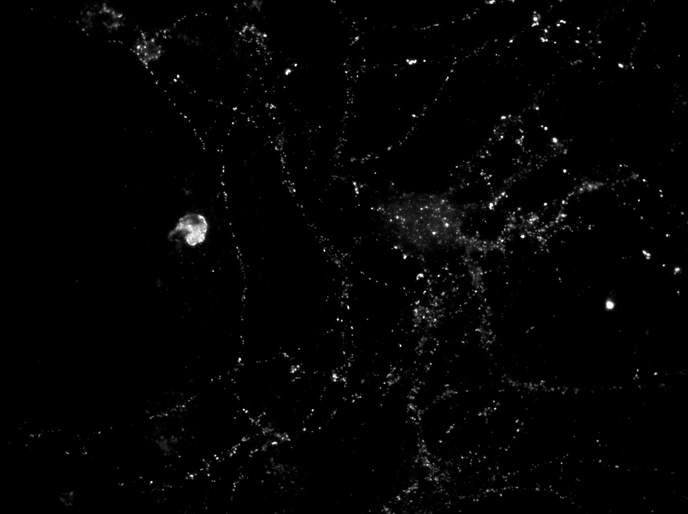
Immunocytochemistry/Immunofluorescence: PSD-95 Antibody (6G6-1C9) [NB300-556] - Mixed 3 week old rat cortical cultures (21 DIV) were labelled with anti-PSD95 (1:650) and visualised with anti-mouse Alexa647 (1:2000). The antibody labels small punctae along neuronal processes as expected. This image was submitted via customer Review.![Flow Cytometry: PSD-95 Antibody (6G6-1C9) - BSA Free [NB300-556]](https://resources.rndsystems.com/images/products/PSD-95-Antibody-6G6-1C9-Flow-Cytometry-NB300-556-img0012.jpg "Flow Cytometry: PSD-95 Antibody (6G6-1C9) - BSA Free [NB300-556]")
Flow Cytometry: PSD-95 Antibody (6G6-1C9) - BSA Free [NB300-556]
Flow Cytometry: PSD-95 Antibody (6G6-1C9) [NB300-556] - Analysis of PSD95 in Neuro-2a cells compared to an isotype control (blue).![Flow Cytometry: PSD-95 Antibody (6G6-1C9) - BSA Free [NB300-556]](https://resources.rndsystems.com/images/products/PSD-95-Antibody-6G6-1C9-Flow-Cytometry-NB300-556-img0013.jpg "Flow Cytometry: PSD-95 Antibody (6G6-1C9) - BSA Free [NB300-556]")
Flow Cytometry: PSD-95 Antibody (6G6-1C9) - BSA Free [NB300-556]
Flow Cytometry: PSD-95 Antibody (6G6-1C9) [NB300-556] - Analysis of PSD95 in SH-SY5Y cells compared to an isotype control (blue).![Flow Cytometry: PSD-95 Antibody (6G6-1C9) - BSA Free [NB300-556]](https://resources.rndsystems.com/images/products/PSD-95-Antibody-6G6-1C9-Flow-Cytometry-NB300-556-img0014.jpg "Flow Cytometry: PSD-95 Antibody (6G6-1C9) - BSA Free [NB300-556]")
Flow Cytometry: PSD-95 Antibody (6G6-1C9) - BSA Free [NB300-556]
Flow Cytometry: PSD-95 Antibody (6G6-1C9) [NB300-556] - Analysis of PSD95 in U87-MG cells compared to an isotype control (blue). - BSA Free [NB300-556] -")
Immunocytochemistry/ Immunofluorescence: PSD-95 Antibody (6G6-1C9) - BSA Free [NB300-556] -
Immunocytochemistry/ Immunofluorescence: PSD-95 Antibody (6G6-1C9) - BSA Free [NB300-556] - Confocal images of dissociated hippocampal cultures. Double labeling with anti-syntaxin-1 (A) & anti-Tuj1 (B) demonstrates that syntaxin-1 is located along the dendrites & gives characteristic punctate labeling (C). Double labeling with antibodies against syntaxin-1 (D) & synaptophysin (E) indicates that syntaxin-1 is colocalized with presynaptic marker (F). Double labeling for syntaxin-1 (G) & PSD-95 (H) shows partial colocalization postsynaptic (I). Scale bars: 10 μm. (J–L) Western blot analysis of synaptosomes (Syn), synaptic cytosolic fraction (Cy), active zone (AZ) & post synaptic density (PSD). Labeled with anti-synaptophysin (J) anti-PSD95 (K) & anti-syntaxin-1 (L). Image collected & cropped by CiteAb from the following publication (http://journal.frontiersin.org/Article/10.3389/fnmol.2016.00010/abstract), licensed under a CC-BY license. Not internally tested by Novus Biologicals. - BSA Free [NB300-556] -")
Western Blot: PSD-95 Antibody (6G6-1C9) - BSA Free [NB300-556] -
Western Blot: PSD-95 Antibody (6G6-1C9) - BSA Free [NB300-556] - Confocal images of dissociated hippocampal cultures. Double labeling with anti-syntaxin-1 (A) & anti-Tuj1 (B) demonstrates that syntaxin-1 is located along the dendrites & gives characteristic punctate labeling (C). Double labeling with antibodies against syntaxin-1 (D) & synaptophysin (E) indicates that syntaxin-1 is colocalized with presynaptic marker (F). Double labeling for syntaxin-1 (G) & PSD-95 (H) shows partial colocalization postsynaptic (I). Scale bars: 10 μm. (J–L) Western blot analysis of synaptosomes (Syn), synaptic cytosolic fraction (Cy), active zone (AZ) & post synaptic density (PSD). Labeled with anti-synaptophysin (J) anti-PSD95 (K) & anti-syntaxin-1 (L). Image collected & cropped by CiteAb from the following publication (http://journal.frontiersin.org/Article/10.3389/fnmol.2016.00010/abstract), licensed under a CC-BY license. Not internally tested by Novus Biologicals. - BSA Free [NB300-556] -")
Immunocytochemistry/ Immunofluorescence: PSD-95 Antibody (6G6-1C9) - BSA Free [NB300-556] -
Immunocytochemistry/ Immunofluorescence: PSD-95 Antibody (6G6-1C9) - BSA Free [NB300-556] - Confocal images of dissociated hippocampal cultures.Double labeling with anti-TUJ1 (A) & anti-VAMP2 (B) demonstrates that VAMP2 is located along the dendrites & gives characteristic punctate labeling (C). Double labeling with antibodies against synaptophysin (P38) (D) & VAMP2 (E) indicates that VAMP2 is colocalized with the presynaptic marker (F). Double labeling for PSD-95 (G) & VAMP2 (H) shows partial colocalization postsynaptic (I). Small, high-resolution pictures of single synapses from (F) & (I) are shown in the right part of these images, respectively. Scale bar: 20 μm. Image collected & cropped by CiteAb from the following publication (https://pubmed.ncbi.nlm.nih.gov/26488171), licensed under a CC-BY license. Not internally tested by Novus Biologicals. - BSA Free [NB300-556] -")
Immunocytochemistry/ Immunofluorescence: PSD-95 Antibody (6G6-1C9) - BSA Free [NB300-556] -
Immunocytochemistry/ Immunofluorescence: PSD-95 Antibody (6G6-1C9) - BSA Free [NB300-556] - Control immunolocalization of plasma membrane AMPA receptor subunits (external epitopes) in hippocampal neuronal cultures.(A) Beta-tubulin (green, anti-TuJ1) labeling of dendrite, after anti-GluA1 external epitope [66] labeling & subsequent plasma membrane permeabilization. Note punctate GluA1 [66] labeling along the dendrite. (B) Synaptophysin (P38, green) & GluA1 [66]. (C) Synaptophysin (green) & GluA2 [66]. (D) Labeling with anti-GluA1. (E) Labeling with anti-GluA1, but the antibody was preincubated with the peptide antigen before staining. (F) Labeling with anti-GluA2. (G) Labeling with anti-GluA2, but the antibody was preincubated with the peptide antigen before staining. (H) Immunolabeling of non-permeabilized neuronal cultures with ani-GluA1 (green). (I) Immunolabeling with the postsynaptic marker anti-PSD95 [66] after permeabilization of neuronal cultures. (J) Merging of (H) & (I) showing postsynaptic labeling of GluA1. Scale bar (in A, valid for all images): 10 μm. Image collected & cropped by CiteAb from the following publication (https://pubmed.ncbi.nlm.nih.gov/26488171), licensed under a CC-BY license. Not internally tested by Novus Biologicals.Applications for PSD-95 Antibody (6G6-1C9) - BSA Free
Application
Recommended Usage
Chromatin Immunoprecipitation (ChIP)
1:10-1:500
Flow Cytometry
2 ug / test (100ul)
Immunocytochemistry/ Immunofluorescence
1:100 - 1:2000
Immunohistochemistry
1:10 - 1:500
Immunohistochemistry-Frozen
1:10 - 1:500
Immunohistochemistry-Paraffin
1:10 - 1:500
Immunoprecipitation
1:10 - 1:500
Western Blot
1:2000
Application Notes
WB: Detects an approx. 95 kDa protein and a slightly larger band in rat brain extracts. Flow usage was reported in scientific literature (PMID: 19187438). IHC usage was reported in scientific literature (PMID: 21289286). IHC-Fr usage was reported in scientific literature (PMID: 24875483).
Reviewed Applications
Read 1 review rated 5 using NB300-556 in the following applications:
Flow Cytometry Panel Builder
Bio-Techne Knows Flow Cytometry
Save time and reduce costly mistakes by quickly finding compatible reagents using the Panel Builder Tool.
Advanced Features
- Spectra Viewer - Custom analysis of spectra from multiple fluorochromes
- Spillover Popups - Visualize the spectra of individual fluorochromes
- Antigen Density Selector - Match fluorochrome brightness with antigen density

Formulation, Preparation, and Storage
Purification
Protein A purified
Formulation
PBS
Format
BSA Free
Preservative
0.05% Sodium Azide
Concentration
3.3 mg/ml
Shipping
The product is shipped with polar packs. Upon receipt, store it immediately at the temperature recommended below.
Stability & Storage
Store at -20C. Avoid freeze-thaw cycles.
Background: PSD-95
Long Name
Postsynaptic Density Protein 95/Disks Large Homolog 4
Alternate Names
DLG4, PSD95, SAP90
Gene Symbol
DLG4
Additional PSD-95 Products
Product Documents for PSD-95 Antibody (6G6-1C9) - BSA Free
Certificate of Analysis
To download a Certificate of Analysis, please enter a lot or batch number in the search box below.
Product Specific Notices for PSD-95 Antibody (6G6-1C9) - BSA Free
This product is for research use only and is not approved for use in humans or in clinical diagnosis. Primary Antibodies are guaranteed for 1 year from date of receipt.
Related Research Areas
Citations for PSD-95 Antibody (6G6-1C9) - BSA Free
 Powered by Bioz
Customer Reviews for PSD-95 Antibody (6G6-1C9) - BSA Free (1)
5 out of 5
1 Customer Rating
Have you used PSD-95 Antibody (6G6-1C9) - BSA Free?
Submit a review and receive an Amazon gift card!
$25/€18/£15/$25CAN/¥2500 Yen for a review with an image
$10/€7/£6/$10CAN/¥1110 Yen for a review without an image
Submit a review
Customer Images
Showing
1
-
1 of
1 review
Showing All
Filter By:
-
Application: ImmunocytochemistrySample Tested: rat cortical culture (3 weeks old)Species: RatVerified Customer | Posted 06/27/2017Mixed rat cortical cultures (21 DIV) were labelled with anti-PSD95 (1:650) and visualised with anti-mouse Alexa647 (1:2000). The antibody labels small punctae along neuronal processes as expected.Fixation Solution and Conditions: 4% paraformaldehyde in PBS, 15 minutes RT Blocking Solution & Duration: 10% donkey serum in PBS with 0.1% triton, 30 minutes RT Primary Antibody Diluent and Dilutions Tested: 1:650 in 5% donkey serum in PBS, over night 4°C Secondary Antibody Manufacturer, Host Species, Dilution, & Diluent: Thermo donkey anti-mouse Alexa647, 1:1250 in PBS, 2 hours, RT

There are no reviews that match your criteria.
Protocols
Find general support by application which include: protocols, troubleshooting, illustrated assays, videos and webinars.
- 7-Amino Actinomycin D (7-AAD) Cell Viability Flow Cytometry Protocol
- Antigen Retrieval Protocol (PIER)
- Antigen Retrieval for Frozen Sections Protocol
- Appropriate Fixation of IHC/ICC Samples
- Cellular Response to Hypoxia Protocols
- ChIP Protocol Video
- Chromatin Immunoprecipitation (ChIP) Protocol
- Chromatin Immunoprecipitation Protocol
- Chromogenic IHC Staining of Formalin-Fixed Paraffin-Embedded (FFPE) Tissue Protocol
- Chromogenic Immunohistochemistry Staining of Frozen Tissue
- ClariTSA™ Fluorophore Kits
- Detection & Visualization of Antibody Binding
- Extracellular Membrane Flow Cytometry Protocol
- Flow Cytometry Protocol for Cell Surface Markers
- Flow Cytometry Protocol for Staining Membrane Associated Proteins
- Flow Cytometry Staining Protocols
- Flow Cytometry Troubleshooting Guide
- Fluorescent IHC Staining of Frozen Tissue Protocol
- Graphic Protocol for Heat-induced Epitope Retrieval
- Graphic Protocol for the Preparation and Fluorescent IHC Staining of Frozen Tissue Sections
- Graphic Protocol for the Preparation and Fluorescent IHC Staining of Paraffin-embedded Tissue Sections
- Graphic Protocol for the Preparation of Gelatin-coated Slides for Histological Tissue Sections
- ICC Cell Smear Protocol for Suspension Cells
- ICC Immunocytochemistry Protocol Videos
- ICC for Adherent Cells
- IHC Sample Preparation (Frozen sections vs Paraffin)
- Immunocytochemistry (ICC) Protocol
- Immunocytochemistry Troubleshooting
- Immunofluorescence of Organoids Embedded in Cultrex Basement Membrane Extract
- Immunofluorescent IHC Staining of Formalin-Fixed Paraffin-Embedded (FFPE) Tissue Protocol
- Immunohistochemistry (IHC) and Immunocytochemistry (ICC) Protocols
- Immunohistochemistry Frozen Troubleshooting
- Immunohistochemistry Paraffin Troubleshooting
- Immunoprecipitation Protocol
- Intracellular Flow Cytometry Protocol Using Alcohol (Methanol)
- Intracellular Flow Cytometry Protocol Using Detergents
- Intracellular Nuclear Staining Flow Cytometry Protocol Using Detergents
- Intracellular Staining Flow Cytometry Protocol Using Alcohol Permeabilization
- Intracellular Staining Flow Cytometry Protocol Using Detergents to Permeabilize Cells
- Preparing Samples for IHC/ICC Experiments
- Preventing Non-Specific Staining (Non-Specific Binding)
- Primary Antibody Selection & Optimization
- Propidium Iodide Cell Viability Flow Cytometry Protocol
- Protocol for Heat-Induced Epitope Retrieval (HIER)
- Protocol for Liperfluo
- Protocol for Making a 4% Formaldehyde Solution in PBS
- Protocol for VisUCyte™ HRP Polymer Detection Reagent
- Protocol for the Characterization of Human Th22 Cells
- Protocol for the Characterization of Human Th9 Cells
- Protocol for the Fluorescent ICC Staining of Cell Smears - Graphic
- Protocol for the Fluorescent ICC Staining of Cultured Cells on Coverslips - Graphic
- Protocol for the Preparation & Fixation of Cells on Coverslips
- Protocol for the Preparation and Chromogenic IHC Staining of Frozen Tissue Sections
- Protocol for the Preparation and Chromogenic IHC Staining of Frozen Tissue Sections - Graphic
- Protocol for the Preparation and Chromogenic IHC Staining of Paraffin-embedded Tissue Sections
- Protocol for the Preparation and Chromogenic IHC Staining of Paraffin-embedded Tissue Sections - Graphic
- Protocol for the Preparation and Fluorescent ICC Staining of Cells on Coverslips
- Protocol for the Preparation and Fluorescent ICC Staining of Non-adherent Cells
- Protocol for the Preparation and Fluorescent ICC Staining of Stem Cells on Coverslips
- Protocol for the Preparation and Fluorescent IHC Staining of Frozen Tissue Sections
- Protocol for the Preparation and Fluorescent IHC Staining of Paraffin-embedded Tissue Sections
- Protocol for the Preparation of Gelatin-coated Slides for Histological Tissue Sections
- Protocol for the Preparation of a Cell Smear for Non-adherent Cell ICC - Graphic
- Protocol: Annexin V and PI Staining by Flow Cytometry
- Protocol: Annexin V and PI Staining for Apoptosis by Flow Cytometry
- R&D Systems Quality Control Western Blot Protocol
- TUNEL and Active Caspase-3 Detection by IHC/ICC Protocol
- The Importance of IHC/ICC Controls
- Troubleshooting Guide: Fluorokine Flow Cytometry Kits
- Troubleshooting Guide: Immunohistochemistry
- Troubleshooting Guide: Western Blot Figures
- Western Blot Conditions
- Western Blot Protocol
- Western Blot Protocol for Cell Lysates
- Western Blot Troubleshooting
- Western Blot Troubleshooting Guide
- View all Protocols, Troubleshooting, Illustrated assays and Webinars
Loading...
Associated Pathways
