PSD-95 Antibody (7E3) - BSA Free
Novus Biologicals | Catalog # NBP2-12872
![Western Blot: PSD-95 Antibody (7E3) [NBP2-12872]](https://resources.rndsystems.com/images/products/PSD-95-Antibody-7E3-Western-Blot-NBP2-12872-img0013.jpg "Western Blot: PSD-95 Antibody (7E3) [NBP2-12872]")
Key Product Details
Species Reactivity
Validated:
Human, Mouse, Rat, Bovine
Cited:
Mouse, Rat
Applications
Validated:
Immunohistochemistry, Immunohistochemistry-Paraffin, Immunohistochemistry-Frozen, Western Blot, Immunocytochemistry/ Immunofluorescence, Microarray
Cited:
Western Blot, Immunocytochemistry/ Immunofluorescence
Label
Unconjugated
Antibody Source
Monoclonal Mouse IgG1 Clone # 7E3
Format
BSA Free
Loading...
Product Specifications
Immunogen
Recombinant rat PSD-95
Localization
Cell Membrane, Cell Junction, Synapse, Postsynaptic Cell Membrane, Postsynaptic Density, Cell Projection, Axon
Marker
post-Synaptic Marker
Specificity
Detects ~100 kDa. An additional protein of >100 kDa is also detected. Additional cross-reactive bands are detected at ~75 kDa and 50 kDa in rat and mouse samples.
Clonality
Monoclonal
Host
Mouse
Isotype
IgG1
Scientific Data Images for PSD-95 Antibody (7E3) - BSA Free
Western Blot: PSD-95 Antibody (7E3) [NBP2-12872]
Western Blot: PSD-95 Antibody (7E3) [NBP2-12872] - Western Blot analysis of Rat brain membrane lysate showing detection of PSD-95 protein using Mouse Anti-PSD-95 Monoclonal Antibody, Clone 7E3 (NBP2-12872). Primary Antibody: Mouse Anti-PSD-95 Monoclonal Antibody (NBP2-12872) at 1:1000.![Immunocytochemistry/ Immunofluorescence: PSD-95 Antibody (7E3) [NBP2-12872]](https://resources.rndsystems.com/images/products/PSD-95-Antibody-7E3-Immunocytochemistry-Immunofluorescence-NBP2-12872-img0015.jpg "Immunocytochemistry/ Immunofluorescence: PSD-95 Antibody (7E3) [NBP2-12872]")
Immunocytochemistry/ Immunofluorescence: PSD-95 Antibody (7E3) [NBP2-12872]
Immunocytochemistry/Immunofluorescence: PSD-95 Antibody (7E3) [NBP2-12872] - Immunocytochemistry/Immunofluorescence analysis using Mouse Anti-PSD-95 Monoclonal Antibody, Clone 7E3 (NBP2-12872). Tissue: HaCaT cells. Species: Human. Fixation: Cold 100% methanol for 10 minutes at -20C. Primary Antibody: Mouse Anti-PSD-95 Monoclonal Antibody (NBP2-12872) at 1:100 for 1 hour at RT. Secondary Antibody: FITC Goat Anti-Mouse (green) at 1:50 for 1 hour at RT. Localization: Filamentous-like staining.![Immunohistochemistry: PSD-95 Antibody (7E3) [NBP2-12872]](https://resources.rndsystems.com/images/products/PSD-95-Antibody-7E3-Immunohistochemistry-NBP2-12872-img0014.jpg "Immunohistochemistry: PSD-95 Antibody (7E3) [NBP2-12872]")
Immunohistochemistry: PSD-95 Antibody (7E3) [NBP2-12872]
Immunohistochemistry: PSD-95 Antibody (7E3) [NBP2-12872] - Immunohistochemistry analysis using Mouse Anti-PSD-95 Monoclonal Antibody, Clone 7E3 (NBP2-12872). Tissue: backskin. Species: Mouse. Fixation: Bouin's Fixative and paraffin-embedded. Primary Antibody: Mouse Anti-PSD-95 Monoclonal Antibody (NBP2-12872) at 1:100 for 1 hour at RT. Secondary Antibody: FITC Goat Anti-Mouse (green) at 1:50 for 1 hour at RT. Localization: Basal cell staining in the epidermis, some hair follicle staining, dermal staining. Backskin obtained from transgenic mice..![Immunocytochemistry/ Immunofluorescence: PSD-95 Antibody (7E3) [NBP2-12872]](https://resources.rndsystems.com/images/products/PSD-95-Antibody-7E3-Immunocytochemistry-Immunofluorescence-NBP2-12872-img0012.jpg "Immunocytochemistry/ Immunofluorescence: PSD-95 Antibody (7E3) [NBP2-12872]")
Immunocytochemistry/ Immunofluorescence: PSD-95 Antibody (7E3) [NBP2-12872]
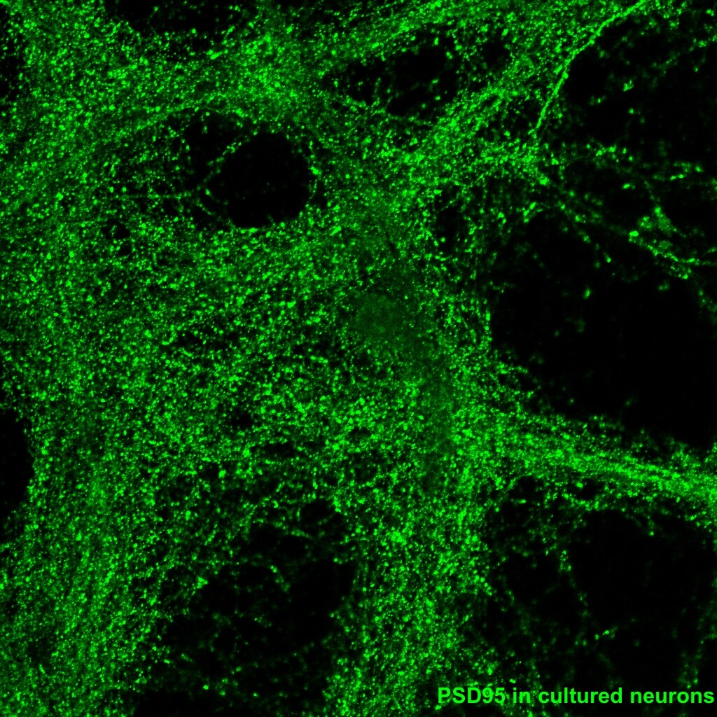
Immunocytochemistry/Immunofluorescence: PSD-95 Antibody (7E3) [NBP2-12872] - Mouse brain cultured neurons. Image from verified customer review.![Immunohistochemistry: PSD-95 Antibody (7E3) [NBP2-12872]](https://resources.rndsystems.com/images/products/PSD-95-Antibody-7E3-Immunohistochemistry-NBP2-12872-img0010.jpg "Immunohistochemistry: PSD-95 Antibody (7E3) [NBP2-12872]")
Immunohistochemistry: PSD-95 Antibody (7E3) [NBP2-12872]
Immunohistochemistry: PSD-95 Antibody (7E3) [NBP2-12872] - Tissue: Neocortex. Species: Rat. Primary Antibody: Mouse Anti-PSD95 Monoclonal Antibody at 1:1000.![Immunohistochemistry-Frozen: PSD-95 Antibody (7E3) [NBP2-12872]](https://resources.rndsystems.com/images/products/PSD-95-Antibody-7E3-Immunohistochemistry-Frozen-NBP2-12872-img0011.jpg "Immunohistochemistry-Frozen: PSD-95 Antibody (7E3) [NBP2-12872]")
Immunohistochemistry-Frozen: PSD-95 Antibody (7E3) [NBP2-12872]
Immunohistochemistry-Frozen: PSD-95 Antibody (7E3) [NBP2-12872] - Mouse brain section (hippocampus). Image from verified customer review. - BSA Free [NBP2-12872] -")
Western Blot: PSD-95 Antibody (7E3) - BSA Free [NBP2-12872] -
Microglial cells and SYN/PSD95 protein in the hippocampal region of HO rats. (A) Immunofluorescence staining of Iba1+ cells. Iba1 (green) and DAPI (blue). Scale bars, 100 μm for low magnification images and 50 μm for high magnification images; quantification of Iba1+ cells and the cell body area in the hippocampus, n = 4–6, **P < 0.01 vs. sham-operated group. (B) Immunofluorescence staining of Iba1+ and SYN in the hippocampus: SYN (red), Iba1 (green), and DAPI (blue); scale bars, 50 μm, n = 6, **P < 0.01 vs. sham-operated group. (C) Immunofluorescence staining of Iba1+ and PSD95 in the hippocampus: PSD95 (red), Iba1 (green), and DAPI (blue); scale bars, 50 μm, n = 6, **P < 0.01 vs. sham-operated group. (D–F) The protein levels of SYN and PSD95 in the hippocampus, n = 3, *P < 0.05 vs. sham-operated group. All data are presented as the M +/- SEM of each group. Image collected and cropped by CiteAb from the following open publication (https://pubmed.ncbi.nlm.nih.gov/36072565), licensed under a CC-BY license. Not internally tested by Novus Biologicals.Applications for PSD-95 Antibody (7E3) - BSA Free
Application
Recommended Usage
Immunocytochemistry/ Immunofluorescence
1:100
Immunohistochemistry
1:1000
Immunohistochemistry-Paraffin
1:10-1:500
Western Blot
1:1000
Application Notes
1 ug/mL was sufficient for detection of PSD-95 on 20 ug rat brain tissue extract by ECL immunoblot analysis using Goat Anti-Mouse IgG: HRP as the secondary. Use in IHC-Fr reported by customer review.
Reviewed Applications
Read 1 review rated 5 using NBP2-12872 in the following applications:
Formulation, Preparation, and Storage
Purification
Protein G purified
Formulation
PBS (pH 7.4), 50% Glycerol
Format
BSA Free
Preservative
0.09% Sodium Azide
Concentration
1 mg/ml
Shipping
The product is shipped with polar packs. Upon receipt, store it immediately at the temperature recommended below.
Stability & Storage
Store at 4C short term. Aliquot and store at -20C long term. Avoid freeze-thaw cycles.
Background: PSD-95
Long Name
Postsynaptic Density Protein 95/Disks Large Homolog 4
Alternate Names
DLG4, PSD95, SAP90
Gene Symbol
DLG4
UniProt
Additional PSD-95 Products
Product Documents for PSD-95 Antibody (7E3) - BSA Free
Certificate of Analysis
To download a Certificate of Analysis, please enter a lot or batch number in the search box below.
Product Specific Notices for PSD-95 Antibody (7E3) - BSA Free
This product is for research use only and is not approved for use in humans or in clinical diagnosis. Primary Antibodies are guaranteed for 1 year from date of receipt.
Related Research Areas
Citations for PSD-95 Antibody (7E3) - BSA Free
 Powered by Bioz
Customer Reviews for PSD-95 Antibody (7E3) - BSA Free (1)
5 out of 5
1 Customer Rating
Have you used PSD-95 Antibody (7E3) - BSA Free?
Submit a review and receive an Amazon gift card!
$25/€18/£15/$25CAN/¥2500 Yen for a review with an image
$10/€7/£6/$10CAN/¥1110 Yen for a review without an image
Submit a review
Customer Images
Showing
1
-
1 of
1 review
Showing All
Filter By:
-
Application: Immunohistochemistry-FrozenSample Tested: Mouse brain and Cell cultureSpecies: MouseVerified Customer | Posted 05/12/2019

There are no reviews that match your criteria.
Protocols
Find general support by application which include: protocols, troubleshooting, illustrated assays, videos and webinars.
- Antigen Retrieval Protocol (PIER)
- Antigen Retrieval for Frozen Sections Protocol
- Appropriate Fixation of IHC/ICC Samples
- Cellular Response to Hypoxia Protocols
- Chromogenic IHC Staining of Formalin-Fixed Paraffin-Embedded (FFPE) Tissue Protocol
- Chromogenic Immunohistochemistry Staining of Frozen Tissue
- ClariTSA™ Fluorophore Kits
- Detection & Visualization of Antibody Binding
- Fluorescent IHC Staining of Frozen Tissue Protocol
- Graphic Protocol for Heat-induced Epitope Retrieval
- Graphic Protocol for the Preparation and Fluorescent IHC Staining of Frozen Tissue Sections
- Graphic Protocol for the Preparation and Fluorescent IHC Staining of Paraffin-embedded Tissue Sections
- Graphic Protocol for the Preparation of Gelatin-coated Slides for Histological Tissue Sections
- ICC Cell Smear Protocol for Suspension Cells
- ICC Immunocytochemistry Protocol Videos
- ICC for Adherent Cells
- IHC Sample Preparation (Frozen sections vs Paraffin)
- Immunocytochemistry (ICC) Protocol
- Immunocytochemistry Troubleshooting
- Immunofluorescence of Organoids Embedded in Cultrex Basement Membrane Extract
- Immunofluorescent IHC Staining of Formalin-Fixed Paraffin-Embedded (FFPE) Tissue Protocol
- Immunohistochemistry (IHC) and Immunocytochemistry (ICC) Protocols
- Immunohistochemistry Frozen Troubleshooting
- Immunohistochemistry Paraffin Troubleshooting
- Preparing Samples for IHC/ICC Experiments
- Preventing Non-Specific Staining (Non-Specific Binding)
- Primary Antibody Selection & Optimization
- Protocol for Heat-Induced Epitope Retrieval (HIER)
- Protocol for Making a 4% Formaldehyde Solution in PBS
- Protocol for VisUCyte™ HRP Polymer Detection Reagent
- Protocol for the Fluorescent ICC Staining of Cell Smears - Graphic
- Protocol for the Fluorescent ICC Staining of Cultured Cells on Coverslips - Graphic
- Protocol for the Preparation & Fixation of Cells on Coverslips
- Protocol for the Preparation and Chromogenic IHC Staining of Frozen Tissue Sections
- Protocol for the Preparation and Chromogenic IHC Staining of Frozen Tissue Sections - Graphic
- Protocol for the Preparation and Chromogenic IHC Staining of Paraffin-embedded Tissue Sections
- Protocol for the Preparation and Chromogenic IHC Staining of Paraffin-embedded Tissue Sections - Graphic
- Protocol for the Preparation and Fluorescent ICC Staining of Cells on Coverslips
- Protocol for the Preparation and Fluorescent ICC Staining of Non-adherent Cells
- Protocol for the Preparation and Fluorescent ICC Staining of Stem Cells on Coverslips
- Protocol for the Preparation and Fluorescent IHC Staining of Frozen Tissue Sections
- Protocol for the Preparation and Fluorescent IHC Staining of Paraffin-embedded Tissue Sections
- Protocol for the Preparation of Gelatin-coated Slides for Histological Tissue Sections
- Protocol for the Preparation of a Cell Smear for Non-adherent Cell ICC - Graphic
- R&D Systems Quality Control Western Blot Protocol
- TUNEL and Active Caspase-3 Detection by IHC/ICC Protocol
- The Importance of IHC/ICC Controls
- Troubleshooting Guide: Immunohistochemistry
- Troubleshooting Guide: Western Blot Figures
- Western Blot Conditions
- Western Blot Protocol
- Western Blot Protocol for Cell Lysates
- Western Blot Troubleshooting
- Western Blot Troubleshooting Guide
- View all Protocols, Troubleshooting, Illustrated assays and Webinars
Loading...
Associated Pathways
