PSGL-1/CD162 Antibody (HECA-452) - BSA Free
Novus Biologicals | Catalog # NB100-78039
![Western Blot: PSGL-1/CD162 Antibody (HECA-452) [NB100-78039]](https://resources.rndsystems.com/images/products/Cutaneous-Lymphocyte-Antigen-CLA-Antibody-HECA-452-Western-Blot-NB100-78039-img0006.jpg "Western Blot: PSGL-1/CD162 Antibody (HECA-452) [NB100-78039]")
Key Product Details
Species Reactivity
Validated:
Human, Mouse
Cited:
Mouse
Applications
Validated:
Immunohistochemistry, Immunohistochemistry-Paraffin, Immunohistochemistry-Frozen, Western Blot, ELISA, Flow Cytometry, Flow (Cell Surface), Immunocytochemistry/ Immunofluorescence, CyTOF-ready
Cited:
Immunohistochemistry, Western Blot, ELISA, Flow Cytometry, IF/IHC
Label
Unconjugated
Antibody Source
Monoclonal Rat IgM Kappa Clone # HECA-452
Format
BSA Free
Loading...
Product Specifications
Immunogen
Human tonsil stroma
Specificity
CLA antibody clone HECA-452 is known to recognize the cutaneous lymphocyte-associated antigen / CLA which is a specialized glycosylated form of P-selectin glycoprotein ligand-1/PSGL-1. Besides lymphocytes, this clone has been shown to react with normal endometrium as well as breast cancer tissues (PMID 19625313 and 22970241).
Clonality
Monoclonal
Host
Rat
Isotype
IgM Kappa
Scientific Data Images for PSGL-1/CD162 Antibody (HECA-452) - BSA Free
Western Blot: PSGL-1/CD162 Antibody (HECA-452) [NB100-78039]
Western Blot: PSGL-1/CD162 Antibody (HECA-452) [NB100-78039] - Cutaneous Lymphocyte Antigen (CLA) Antibody (HECA-452) [NB100-78039] - Analysis of human tonsil (1), human spleen (2), and human lymph node (3) tissue using CD162 antibody at a concentration of 2 ug/ml.![Immunohistochemistry-Paraffin: PSGL-1/CD162 Antibody (HECA-452) [NB100-78039]](https://resources.rndsystems.com/images/products/PSGL-1-CD162-Antibody-HECA-452-Immunohistochemistry-Paraffin-NB100-78039-img0014.jpg "Immunohistochemistry-Paraffin: PSGL-1/CD162 Antibody (HECA-452) [NB100-78039]")
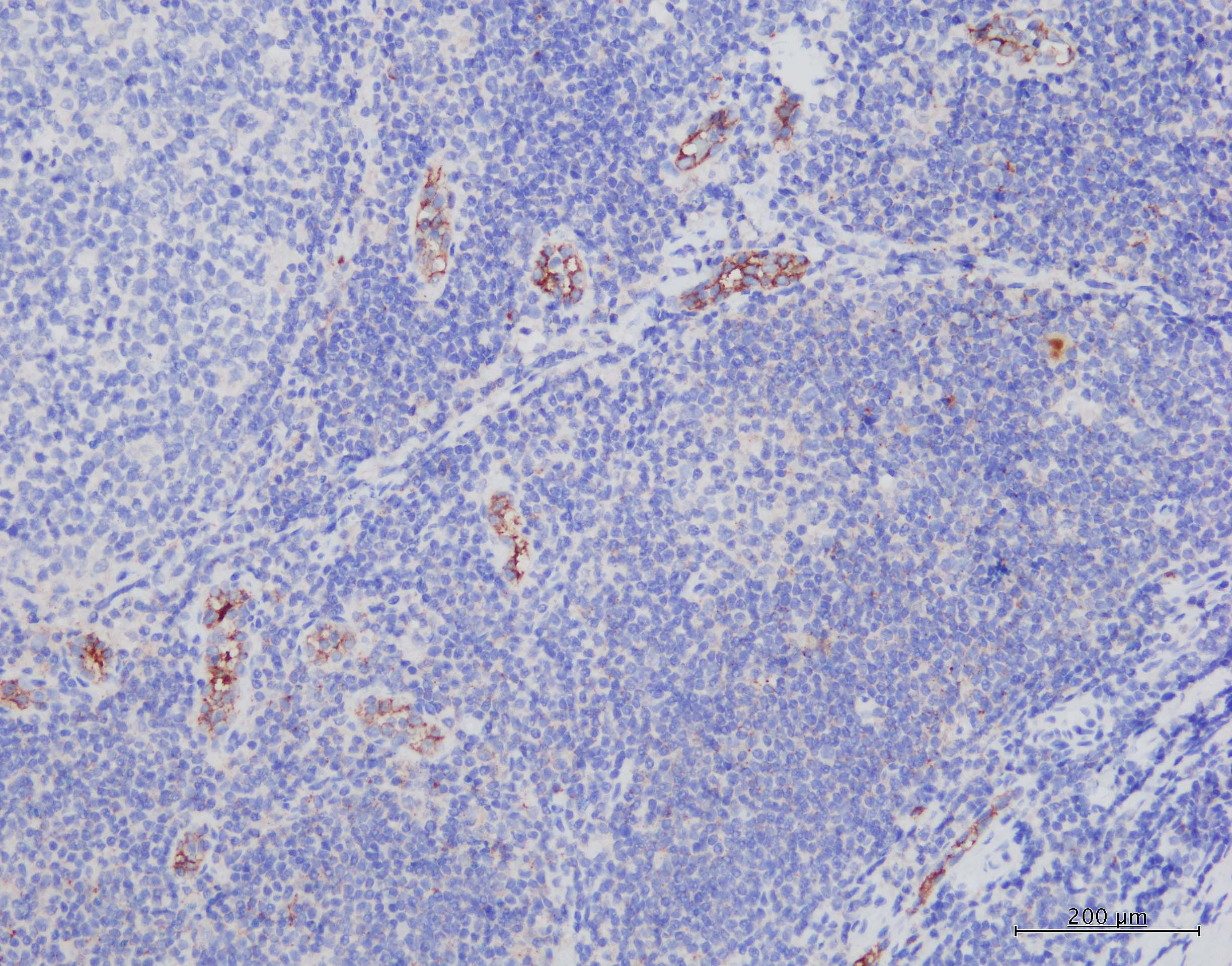
Immunohistochemistry-Paraffin: PSGL-1/CD162 Antibody (HECA-452) [NB100-78039]
Immunohistochemistry-Paraffin: PSGL-1/CD162 Antibody (HECA-452) [NB100-78039] - Human tonsil tissue stained with PSGL-1/CD162 Antibody (HECA-452) at 5ug/mL, and Leica Refine DAB kit. IHC-P image submitted by a verified customer review.![Flow (Cell Surface): PSGL-1/CD162 Antibody (HECA-452) [NB100-78039]](https://resources.rndsystems.com/images/products/Cutaneous-Lymphocyte-Antigen-CLA-Antibody-HECA-452-Flow-Cell-Surface-NB100-78039-img0008.jpg "Flow (Cell Surface): PSGL-1/CD162 Antibody (HECA-452) [NB100-78039]")
Flow (Cell Surface): PSGL-1/CD162 Antibody (HECA-452) [NB100-78039]
Flow (Cell Surface): PSGL-1/CD162 Antibody (HECA-452) [NB100-78039] - Cutaneous Lymphocyte Antigen (CLA) Antibody (HECA-452) [NB100-78039] - A cell surface stain was performed on CD3+ hPBMCs with Cutaneous Lymphocyte Antigen (CLA) antibody (HECA-452) NB100-78039 and a matched isotype control NBP2-31379. Cells were incubated in an antibody dilution of 1 ug/mL for 20 minutes at room temperature, followed by mouse F(ab)2 IgG (H+L) APC-conjugated secondary antibody [F0101B, R&D Systems]. A co-stain was performed using CD3 antibody NBP2-25186APC.![Immunohistochemistry-Paraffin: PSGL-1/CD162 Antibody (HECA-452) [NB100-78039]](https://resources.rndsystems.com/images/products/Cutaneous-Lymphocyte-Antigen-CLA-Antibody-HECA-452-Immunohistochemistry-Paraffin-NB100-78039-img0004.jpg "Immunohistochemistry-Paraffin: PSGL-1/CD162 Antibody (HECA-452) [NB100-78039]")
Immunohistochemistry-Paraffin: PSGL-1/CD162 Antibody (HECA-452) [NB100-78039]
Immunohistochemistry-Paraffin: PSGL-1/CD162 Antibody (HECA-452) [NB100-78039] - Cutaneous Lymphocyte Antigen (CLA) Antibody (HECA-452) [NB100-78039] - IHC analysis of formalin-fixed paraffin-embedded tissue section of human breast adenocarcinoma using CLA antibody (clone HECA-452) at 5 ug/ml concentration. The antibody generated a membrane-cytoplasmic staining in the cancerous cells of breast adenocarcinoma. Note:- Besides lymphocytes, CLA has been shown to be present on cells of normal endometrium as well as breast cancer tissues (PMID 19625313 and 22970241).![Flow Cytometry: PSGL-1/CD162 Antibody (HECA-452) [NB100-78039]](https://resources.rndsystems.com/images/products/Cutaneous-Lymphocyte-Antigen-CLA-Antibody-HECA-452-Flow-Cytometry-NB100-78039-img0005.jpg "Flow Cytometry: PSGL-1/CD162 Antibody (HECA-452) [NB100-78039]")
Flow Cytometry: PSGL-1/CD162 Antibody (HECA-452) [NB100-78039]
Flow Cytometry: PSGL-1/CD162 Antibody (HECA-452) [NB100-78039] - Cutaneous Lymphocyte Antigen (CLA) Antibody (HECA-452) [NB100-78039] - Human peripheral blood lymphocytes stained with purified HECA-452, followed by anti-mouse IgG FITC![Flow (Cell Surface): PSGL-1/CD162 Antibody (HECA-452) [NB100-78039]](https://resources.rndsystems.com/images/products/Cutaneous-Lymphocyte-Antigen-CLA-Antibody-HECA-452-Flow-Cell-Surface-NB100-78039-img0007.jpg "Flow (Cell Surface): PSGL-1/CD162 Antibody (HECA-452) [NB100-78039]")
Flow (Cell Surface): PSGL-1/CD162 Antibody (HECA-452) [NB100-78039]
Flow (Cell Surface): PSGL-1/CD162 Antibody (HECA-452) [NB100-78039] - Cutaneous Lymphocyte Antigen (CLA) Antibody (HECA-452) [NB100-78039] - A cell surface stain was performed on CD14+ hPBMCs with Cutaneous Lymphocyte Antigen (CLA) antibody (HECA-452) NB100-78039 and a matched isotype control NBP2-31379. Cells were incubated in an antibody dilution of 1 ug/mL for 20 minutes at room temperature, followed by rat-Dylight 488 conjugated secondary antibody. A co-stain was performed using CD14 antibody NB100-77758AF488. - BSA Free [NB100-78039] -")
Western Blot: PSGL-1/CD162 Antibody (HECA-452) - BSA Free [NB100-78039] -
High level of P-selectin leads to increased PSGL-1 expression and NETs formation in human neutrophils. Human peripheral blood neutrophils were isolated and treated with P-selectin recombinant protein at concentrations of 10, 20 and 50 nM. (A) Representative images of neutrophil stained with PSGL-1 (yellow) and DAPI (blue). The inset box from each group is magnified. Scale bar: 5 μm and 20 μm, respectively. (B) Cell lysates were collected and subjected to Western blot analysis for PSGL-1. GAPDH was used as a loading control. Data from one representative experiment are shown. (C) Relative intensities of PSGL-1 against GAPDH. (n=3, **P < 0.01) (D) Representative images of neutrophils stained with MPO (red), CitH3 (Green) and DAPI (blue). The inset box from each group is magnified. Scale bar: 50 μm and 20 μm, respectively. The arrows indicate NETs. (E) NET-forming cells per field are quantified. (n=6, ****P < 0.0001) The neutrophil supernatants were assessed for cf-DNA (F) and MPO-DNA complexes (G). (n=3, *P < 0.05, **P < 0.01, ***P < 0.001). Image collected and cropped by CiteAb from the following open publication (https://pubmed.ncbi.nlm.nih.gov/37841279), licensed under a CC-BY license. Not internally tested by Novus Biologicals.Applications for PSGL-1/CD162 Antibody (HECA-452) - BSA Free
Application
Recommended Usage
Flow Cytometry
< = 2 ug/10^6 cells in 100 ul
Immunocytochemistry/ Immunofluorescence
reported in scientific literature (PMID 1693467)
Immunohistochemistry
1:10-1:500
Immunohistochemistry-Frozen
1:10-1:500
Immunohistochemistry-Paraffin
1:10-1:500
Western Blot
2 ug/ml
Reviewed Applications
Read 1 review rated 5 using NB100-78039 in the following applications:
Flow Cytometry Panel Builder
Bio-Techne Knows Flow Cytometry
Save time and reduce costly mistakes by quickly finding compatible reagents using the Panel Builder Tool.
Advanced Features
- Spectra Viewer - Custom analysis of spectra from multiple fluorochromes
- Spillover Popups - Visualize the spectra of individual fluorochromes
- Antigen Density Selector - Match fluorochrome brightness with antigen density

Formulation, Preparation, and Storage
Purification
IgM purified
Formulation
PBS
Format
BSA Free
Preservative
0.02% Sodium Azide
Concentration
1.0 mg/ml
Shipping
The product is shipped with polar packs. Upon receipt, store it immediately at the temperature recommended below.
Stability & Storage
Store at 4C short term. Aliquot and store at -20C long term. Avoid freeze-thaw cycles.
Background: PSGL-1/CD162
Long Name
P-Selectin Glycoprotein Ligand 1
Alternate Names
CD162, PSGL1, SELPLG
Gene Symbol
SELPLG
Additional PSGL-1/CD162 Products
Product Documents for PSGL-1/CD162 Antibody (HECA-452) - BSA Free
Certificate of Analysis
To download a Certificate of Analysis, please enter a lot or batch number in the search box below.
Product Specific Notices for PSGL-1/CD162 Antibody (HECA-452) - BSA Free
This product is for research use only and is not approved for use in humans or in clinical diagnosis. Primary Antibodies are guaranteed for 1 year from date of receipt.
Citations for PSGL-1/CD162 Antibody (HECA-452) - BSA Free
 Powered by Bioz
Customer Reviews for PSGL-1/CD162 Antibody (HECA-452) - BSA Free (1)
5 out of 5
1 Customer Rating
Have you used PSGL-1/CD162 Antibody (HECA-452) - BSA Free?
Submit a review and receive an Amazon gift card!
$25/€18/£15/$25CAN/¥2500 Yen for a review with an image
$10/€7/£6/$10CAN/¥1110 Yen for a review without an image
Submit a review
Customer Images
Showing
1
-
1 of
1 review
Showing All
Filter By:
-
Application: Immunohistochemistry-ParaffinSample Tested: human tonsilSpecies: HumanVerified Customer | Posted 10/13/20215ug/mL, stained with Leica Refine DAB kit

There are no reviews that match your criteria.
Protocols
Find general support by application which include: protocols, troubleshooting, illustrated assays, videos and webinars.
- 7-Amino Actinomycin D (7-AAD) Cell Viability Flow Cytometry Protocol
- Antigen Retrieval Protocol (PIER)
- Antigen Retrieval for Frozen Sections Protocol
- Appropriate Fixation of IHC/ICC Samples
- Cellular Response to Hypoxia Protocols
- Chromogenic IHC Staining of Formalin-Fixed Paraffin-Embedded (FFPE) Tissue Protocol
- Chromogenic Immunohistochemistry Staining of Frozen Tissue
- ClariTSA™ Fluorophore Kits
- Detection & Visualization of Antibody Binding
- ELISA Sample Preparation & Collection Guide
- ELISA Troubleshooting Guide
- Extracellular Membrane Flow Cytometry Protocol
- Flow Cytometry Protocol for Cell Surface Markers
- Flow Cytometry Protocol for Staining Membrane Associated Proteins
- Flow Cytometry Staining Protocols
- Flow Cytometry Troubleshooting Guide
- Fluorescent IHC Staining of Frozen Tissue Protocol
- Graphic Protocol for Heat-induced Epitope Retrieval
- Graphic Protocol for the Preparation and Fluorescent IHC Staining of Frozen Tissue Sections
- Graphic Protocol for the Preparation and Fluorescent IHC Staining of Paraffin-embedded Tissue Sections
- Graphic Protocol for the Preparation of Gelatin-coated Slides for Histological Tissue Sections
- How to Run an R&D Systems DuoSet ELISA
- How to Run an R&D Systems Quantikine ELISA
- How to Run an R&D Systems Quantikine™ QuicKit™ ELISA
- ICC Cell Smear Protocol for Suspension Cells
- ICC Immunocytochemistry Protocol Videos
- ICC for Adherent Cells
- IHC Sample Preparation (Frozen sections vs Paraffin)
- Immunocytochemistry (ICC) Protocol
- Immunocytochemistry Troubleshooting
- Immunofluorescence of Organoids Embedded in Cultrex Basement Membrane Extract
- Immunofluorescent IHC Staining of Formalin-Fixed Paraffin-Embedded (FFPE) Tissue Protocol
- Immunohistochemistry (IHC) and Immunocytochemistry (ICC) Protocols
- Immunohistochemistry Frozen Troubleshooting
- Immunohistochemistry Paraffin Troubleshooting
- Intracellular Flow Cytometry Protocol Using Alcohol (Methanol)
- Intracellular Flow Cytometry Protocol Using Detergents
- Intracellular Nuclear Staining Flow Cytometry Protocol Using Detergents
- Intracellular Staining Flow Cytometry Protocol Using Alcohol Permeabilization
- Intracellular Staining Flow Cytometry Protocol Using Detergents to Permeabilize Cells
- Preparing Samples for IHC/ICC Experiments
- Preventing Non-Specific Staining (Non-Specific Binding)
- Primary Antibody Selection & Optimization
- Propidium Iodide Cell Viability Flow Cytometry Protocol
- Protocol for Heat-Induced Epitope Retrieval (HIER)
- Protocol for Liperfluo
- Protocol for Making a 4% Formaldehyde Solution in PBS
- Protocol for VisUCyte™ HRP Polymer Detection Reagent
- Protocol for the Characterization of Human Th22 Cells
- Protocol for the Characterization of Human Th9 Cells
- Protocol for the Fluorescent ICC Staining of Cell Smears - Graphic
- Protocol for the Fluorescent ICC Staining of Cultured Cells on Coverslips - Graphic
- Protocol for the Preparation & Fixation of Cells on Coverslips
- Protocol for the Preparation and Chromogenic IHC Staining of Frozen Tissue Sections
- Protocol for the Preparation and Chromogenic IHC Staining of Frozen Tissue Sections - Graphic
- Protocol for the Preparation and Chromogenic IHC Staining of Paraffin-embedded Tissue Sections
- Protocol for the Preparation and Chromogenic IHC Staining of Paraffin-embedded Tissue Sections - Graphic
- Protocol for the Preparation and Fluorescent ICC Staining of Cells on Coverslips
- Protocol for the Preparation and Fluorescent ICC Staining of Non-adherent Cells
- Protocol for the Preparation and Fluorescent ICC Staining of Stem Cells on Coverslips
- Protocol for the Preparation and Fluorescent IHC Staining of Frozen Tissue Sections
- Protocol for the Preparation and Fluorescent IHC Staining of Paraffin-embedded Tissue Sections
- Protocol for the Preparation of Gelatin-coated Slides for Histological Tissue Sections
- Protocol for the Preparation of a Cell Smear for Non-adherent Cell ICC - Graphic
- Protocol: Annexin V and PI Staining by Flow Cytometry
- Protocol: Annexin V and PI Staining for Apoptosis by Flow Cytometry
- Quantikine HS ELISA Kit Assay Principle, Alkaline Phosphatase
- Quantikine HS ELISA Kit Principle, Streptavidin-HRP Polymer
- R&D Systems Quality Control Western Blot Protocol
- Sandwich ELISA (Colorimetric) – Biotin/Streptavidin Detection Protocol
- Sandwich ELISA (Colorimetric) – Direct Detection Protocol
- TUNEL and Active Caspase-3 Detection by IHC/ICC Protocol
- The Importance of IHC/ICC Controls
- Troubleshooting Guide: ELISA
- Troubleshooting Guide: Fluorokine Flow Cytometry Kits
- Troubleshooting Guide: Immunohistochemistry
- Troubleshooting Guide: Western Blot Figures
- Western Blot Conditions
- Western Blot Protocol
- Western Blot Protocol for Cell Lysates
- Western Blot Troubleshooting
- Western Blot Troubleshooting Guide
- View all Protocols, Troubleshooting, Illustrated assays and Webinars
Loading...