Rab5a Antibody - BSA Free
Novus Biologicals | Catalog # NB120-13253
![Western Blot: Rab5a Antibody [NB120-13253]](https://resources.rndsystems.com/images/products/Rab5a-Antibody-Western-Blot-NB120-13253-img0012.jpg "Western Blot: Rab5a Antibody [NB120-13253]")
Loading...
Key Product Details
Species Reactivity
Validated:
Human, Mouse, Rat, Bovine, Monkey
Cited:
Human, Mouse, Rat
Applications
Validated:
Immunohistochemistry, Immunohistochemistry-Paraffin, Immunohistochemistry Free-Floating, Western Blot, Immunocytochemistry/ Immunofluorescence, Immunoprecipitation
Cited:
Immunohistochemistry Free-Floating, Western Blot, Immunocytochemistry/ Immunofluorescence, Immunoprecipitation, Functional
Label
Unconjugated
Antibody Source
Polyclonal Rabbit IgG
Format
BSA Free
Loading...
Product Specifications
Immunogen
Human Rab5 synthetic peptide conjugated to KLH; identical to dog Rab5 sequence over the residues
Reactivity Notes
Mouse reactivity reported in scientific literature (PMID: 23954637).
Localization
Cell Membrane, Endosomone, Early Endosome Membrane, Melanosome
Marker
Early Endosome Marker
Specificity
Detects approx 26kDa.
Clonality
Polyclonal
Host
Rabbit
Isotype
IgG
Scientific Data Images for Rab5a Antibody - BSA Free
Western Blot: Rab5a Antibody [NB120-13253]
Western Blot: Rab5a Antibody [NB120-13253] - Western blot analysis of Human Cell line lysates showing detection of Rab5aprotein using Rabbit Anti-Rab5aPolyclonal Antibody (NB120-13253). Load: 15 ugprotein. Block: 1.5% BSA for 30 minutes at RT. Primary Antibody: Rabbit Anti-Rab5aPolyclonal Antibody (NB120-13253) at 1:1000 for 2 hours at RT. Secondary Antibody: Donkey Anti-Rabbit IgG: HRP for 1 hour at RT.![Immunocytochemistry/ Immunofluorescence: Rab5a Antibody [NB120-13253]](https://resources.rndsystems.com/images/products/Rab5a-Antibody-Immunocytochemistry-Immunofluorescence-NB120-13253-img0010.jpg "Immunocytochemistry/ Immunofluorescence: Rab5a Antibody [NB120-13253]")
Immunocytochemistry/ Immunofluorescence: Rab5a Antibody [NB120-13253]
Immunocytochemistry/Immunofluorescence: Rab5a Antibody [NB120-13253] - Tissue: HeLa Cells. Species: Human. Fixation: 2% Formaldehyde for 20 min at RT. Primary Antibody: Rabbit Anti-Rab5 Polyclonal Antibody at 1:80 for 12 hours at 4C. Secondary Antibody: R-PE Goat Anti-Rabbit (yellow) at 1:200 for 2 hours at RT. Counterstain: DAPI (blue) nuclear stain at 1:40000 for 2 hours at RT. Localization: Cytoplasm. Melanosome. Nucleus. Magnification: 100x.![Immunohistochemistry: Rab5a Antibody [NB120-13253]](https://resources.rndsystems.com/images/products/Rab5a-Antibody-Immunohistochemistry-NB120-13253-img0013.jpg "Immunohistochemistry: Rab5a Antibody [NB120-13253]")
Immunohistochemistry: Rab5a Antibody [NB120-13253]
Immunohistochemistry: Rab5a Antibody [NB120-13253] - Immunohistochemistry analysis using Rabbit Anti-Rab5aPolyclonal Antibody (NB120-13253). Tissue: backskin. Species: Mouse. Fixation: Bouin's Fixative Solution. Primary Antibody: Rabbit Anti-Rab5aPolyclonal Antibody (NB120-13253) at 1:100 for 1 hour at RT. Secondary Antibody: FITC Goat Anti-Rabbit (green) at 1:50 for 1 hour at RT. Localization: Cytoplasm.![Immunocytochemistry/ Immunofluorescence: Rab5a Antibody [NB120-13253]](https://resources.rndsystems.com/images/products/Rab5a-Antibody-Immunocytochemistry-Immunofluorescence-NB120-13253-img0004.jpg "Immunocytochemistry/ Immunofluorescence: Rab5a Antibody [NB120-13253]")
Immunocytochemistry/ Immunofluorescence: Rab5a Antibody [NB120-13253]
Immunocytochemistry/Immunofluorescence: Rab5a Antibody [NB120-13253] - IF staining of Rab5a in hepatocytes. Image from verified customer review.![Immunocytochemistry/ Immunofluorescence: Rab5a Antibody [NB120-13253]](https://resources.rndsystems.com/images/products/Rab5a-Antibody-Immunocytochemistry-Immunofluorescence-NB120-13253-img0011.jpg "Immunocytochemistry/ Immunofluorescence: Rab5a Antibody [NB120-13253]")
Immunocytochemistry/ Immunofluorescence: Rab5a Antibody [NB120-13253]
Immunocytochemistry/Immunofluorescence: Rab5a Antibody [NB120-13253] - Tissue: HeLa Cells. Species: Human. Fixation: 2% Formaldehyde for 20 min at RT. Primary Antibody: Rabbit Anti-Rab5 Polyclonal Antibody at 1:80 for 12 hours at 4C. Secondary Antibody: FITC Goat Anti-Rabbit (green) at 1:200 for 2 hours at RT. Counterstain: DAPI (blue) nuclear stain at 1:40000 for 2 hours at RT. Localization: Cytoplasm. Melanosome. Nucleus. Magnification: 20x.![Immunohistochemistry-Paraffin: Rab5a Antibody [NB120-13253]](https://resources.rndsystems.com/images/products/Rab5a-Antibody-Immunohistochemistry-Paraffin-NB120-13253-img0008.jpg "Immunohistochemistry-Paraffin: Rab5a Antibody [NB120-13253]")
Immunohistochemistry-Paraffin: Rab5a Antibody [NB120-13253]
Immunohistochemistry-Paraffin: Rab5a Antibody [NB120-13253] - analysis using Rabbit Anti-Rab5 Polyclonal Antibody. Tissue: backskin. Species: Mouse. Fixation: Bouins Fixative Solution. Primary Antibody: Rabbit Anti-Rab5 Polyclonal Antibody at 1:100 for 1 hour at RT. Secondary Antibody: FITC Goat Anti-Rabbit (green) at 1:50 for 1 hour at RT. Localization: Cytoplasm.Applications for Rab5a Antibody - BSA Free
Application
Recommended Usage
Immunocytochemistry/ Immunofluorescence
1:80
Immunohistochemistry
1:100
Immunohistochemistry-Paraffin
1:10-1:500
Western Blot
1:1000
Application Notes
1 ul/ml of Rab5 Antibody was sufficient for detection of Rab5 in 15 ug of HeLa lysate by ECL immunoblot analysis using Donkey anti-rabbit IgG:HRP as the secondary Antibody. Use in Immunoprecipitation reported in scientific literature (PMID 23954637). Use in Immunohistochemistry free floating reported in scientific literature (PMID: 31618685).
Reviewed Applications
Read 2 reviews rated 4 using NB120-13253 in the following applications:
Formulation, Preparation, and Storage
Purification
Protein A purified
Formulation
PBS, 50% Glycerol
Format
BSA Free
Preservative
0.09% Sodium Azide
Concentration
1 mg/ml
Shipping
The product is shipped with polar packs. Upon receipt, store it immediately at the temperature recommended below.
Stability & Storage
Store at 4C short term. Aliquot and store at -20C long term. Avoid freeze-thaw cycles.
Background: Rab5a
Alternate Names
RAB5A, member RAS oncogene family, RAB5RAS-associated protein RAB5A, ras-related protein Rab-5A
Gene Symbol
RAB5A
UniProt
Additional Rab5a Products
Product Documents for Rab5a Antibody - BSA Free
Certificate of Analysis
To download a Certificate of Analysis, please enter a lot or batch number in the search box below.
Product Specific Notices for Rab5a Antibody - BSA Free
This product is for research use only and is not approved for use in humans or in clinical diagnosis. Primary Antibodies are guaranteed for 1 year from date of receipt.
Citations for Rab5a Antibody - BSA Free
 Powered by Bioz
Customer Reviews for Rab5a Antibody - BSA Free (2)
4 out of 5
2 Customer Ratings
Have you used Rab5a Antibody - BSA Free?
Submit a review and receive an Amazon gift card!
$25/€18/£15/$25CAN/¥2500 Yen for a review with an image
$10/€7/£6/$10CAN/¥1110 Yen for a review without an image
Submit a review
Customer Images
-(01-mg)_NB120-13253_9456.jpg)
Showing
1
-
2 of
2 reviews
Showing All
Filter By:
-
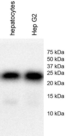
Application: Western BlotSample Tested: Human primary Colon Cancer cell line and human primary hepatocytes and Hep G2Species: HumanVerified Customer | Posted 12/15/2017human primary hepatocytes and Hep G2 cellshuman primary hepatocytes and Hep G2 cells Name: Anti-Rab5a antibody (NB120-13253) Catalog #: Anti-Rab5a antibody (NB120-13253) Lot Number: Anti-Rab5a antibody (NB120-13253, Lot # 140819) PO/Order Number: Click here to enter text.. WB Image Description (Please provide labels for all lanes): lane 1: human primary hepatocytes; lane 2: Hep G2 Sample Information: Cell Line or Tissue: human primary hepatocytes, Hep G2 Species: human Treatment: No treatment Lysate Preparation: Date of lysate preparation: December 11, 2017 Lysis buffer used: 1X lysis buffer from Cell Signaling by adding PMSF Reducing agent: beta-mercaptoethanol, DTT If boiled (temperature/time): Yes Controls: Positive Control: No Negative Control: No Loading Control (please attach additional images if applicable): No Protein Amount Loaded per lane: 20 ug Antibody Storage Conditions: -20℃ Electrophoresis: Gel Percentage: 15% Electrophoresis Conditions: Tris-Glycine-SDS at room temperature Voltage: 120V Time: 2 hours Membrane Transfer: Method (Submersion/Semi-dry): wet transfer Membrane Type (PVDF/Nitrocellulose): Nitrocellulose Time: 2 hours Voltage: 100V Blocking: Blocking Solution: 5% milk in 1X TBST Time: 1 hour at room temperature Primary Antibody: Dilution: 1/1000 Diluent Buffer: 2.5% BSA Incubation Time: overnight Incubation Temperature: 4℃ Washing Conditions: Wash Solution: 1X TBST Time and Repetitions: 5 min each for 3 times Secondary Antibody Manufacturer and Catalog #: Promega, W401B, Lot # 0000187662 Secondary description: goat anti-rabbit secondary antibody Dilution: 1/2000 Diluent Buffer: 3% milk Incubation Time: 1 hour Incubation Temperature: room temperature Detection Method: Detection: ECL (GE, cat # RPN2209, lot # 9838243) Procedure: Add equal volume of A and B, mix and apply on the membranes for 3-5 min before exposure Development Time: 20 seconds Molecular weight of band(s): ~ 23 kDa Experimental Concerns and Observations: A specific band around 23 kDa was observed

-
Application: ImmunofluorescenceSample Tested:Species: MouseVerified Customer | Posted 08/19/2014An immunofluorescent staining review for Rab5a antibody (NB120-13253)
There are no reviews that match your criteria.
Protocols
Find general support by application which include: protocols, troubleshooting, illustrated assays, videos and webinars.
- Antigen Retrieval Protocol (PIER)
- Antigen Retrieval for Frozen Sections Protocol
- Appropriate Fixation of IHC/ICC Samples
- Cellular Response to Hypoxia Protocols
- Chromogenic IHC Staining of Formalin-Fixed Paraffin-Embedded (FFPE) Tissue Protocol
- Chromogenic Immunohistochemistry Staining of Frozen Tissue
- ClariTSA™ Fluorophore Kits
- Detection & Visualization of Antibody Binding
- Fluorescent IHC Staining of Frozen Tissue Protocol
- Graphic Protocol for Heat-induced Epitope Retrieval
- Graphic Protocol for the Preparation and Fluorescent IHC Staining of Frozen Tissue Sections
- Graphic Protocol for the Preparation and Fluorescent IHC Staining of Paraffin-embedded Tissue Sections
- Graphic Protocol for the Preparation of Gelatin-coated Slides for Histological Tissue Sections
- ICC Cell Smear Protocol for Suspension Cells
- ICC Immunocytochemistry Protocol Videos
- ICC for Adherent Cells
- IHC Sample Preparation (Frozen sections vs Paraffin)
- Immunocytochemistry (ICC) Protocol
- Immunocytochemistry Troubleshooting
- Immunofluorescence of Organoids Embedded in Cultrex Basement Membrane Extract
- Immunofluorescent IHC Staining of Formalin-Fixed Paraffin-Embedded (FFPE) Tissue Protocol
- Immunohistochemistry (IHC) and Immunocytochemistry (ICC) Protocols
- Immunohistochemistry Frozen Troubleshooting
- Immunohistochemistry Paraffin Troubleshooting
- Immunoprecipitation Protocol
- Preparing Samples for IHC/ICC Experiments
- Preventing Non-Specific Staining (Non-Specific Binding)
- Primary Antibody Selection & Optimization
- Protocol for Heat-Induced Epitope Retrieval (HIER)
- Protocol for Making a 4% Formaldehyde Solution in PBS
- Protocol for VisUCyte™ HRP Polymer Detection Reagent
- Protocol for the Fluorescent ICC Staining of Cell Smears - Graphic
- Protocol for the Fluorescent ICC Staining of Cultured Cells on Coverslips - Graphic
- Protocol for the Preparation & Fixation of Cells on Coverslips
- Protocol for the Preparation and Chromogenic IHC Staining of Frozen Tissue Sections
- Protocol for the Preparation and Chromogenic IHC Staining of Frozen Tissue Sections - Graphic
- Protocol for the Preparation and Chromogenic IHC Staining of Paraffin-embedded Tissue Sections
- Protocol for the Preparation and Chromogenic IHC Staining of Paraffin-embedded Tissue Sections - Graphic
- Protocol for the Preparation and Fluorescent ICC Staining of Cells on Coverslips
- Protocol for the Preparation and Fluorescent ICC Staining of Non-adherent Cells
- Protocol for the Preparation and Fluorescent ICC Staining of Stem Cells on Coverslips
- Protocol for the Preparation and Fluorescent IHC Staining of Frozen Tissue Sections
- Protocol for the Preparation and Fluorescent IHC Staining of Paraffin-embedded Tissue Sections
- Protocol for the Preparation of Gelatin-coated Slides for Histological Tissue Sections
- Protocol for the Preparation of a Cell Smear for Non-adherent Cell ICC - Graphic
- R&D Systems Quality Control Western Blot Protocol
- TUNEL and Active Caspase-3 Detection by IHC/ICC Protocol
- The Importance of IHC/ICC Controls
- Troubleshooting Guide: Immunohistochemistry
- Troubleshooting Guide: Western Blot Figures
- Western Blot Conditions
- Western Blot Protocol
- Western Blot Protocol for Cell Lysates
- Western Blot Troubleshooting
- Western Blot Troubleshooting Guide
- View all Protocols, Troubleshooting, Illustrated assays and Webinars
Loading...