RAB6A Antibody - BSA Free
Novus Biologicals | Catalog # NBP1-33110
![Western Blot: RAB6A Antibody [NBP1-33110]](https://resources.rndsystems.com/images/products/RAB6A-Antibody-Western-Blot-NBP1-33110-img0016.jpg "Western Blot: RAB6A Antibody [NBP1-33110]")
Loading...
Key Product Details
Validated by
Knockout/Knockdown
Species Reactivity
Validated:
Human, Mouse, Rat
Cited:
Mouse
Predicted:
Bovine (98%), Chicken (97%), Porcine (97%), Rhesus Macaque (98%), Xenopus (96%), Zebrafish (98%). Backed by our 100% Guarantee.
Applications
Validated:
Knockout Validated, Immunohistochemistry, Immunohistochemistry-Paraffin, Western Blot, Immunocytochemistry/ Immunofluorescence
Cited:
Immunohistochemistry, Western Blot
Label
Unconjugated
Antibody Source
Polyclonal Rabbit IgG
Format
BSA Free
Loading...
Product Specifications
Immunogen
Full length human RAB6A Recombinant protein.
Reactivity Notes
Xenopus laevis (96%).
Localization
Golgi apparatus membrane, Lipid-anchor
Marker
Golgi Apparatus Marker
Clonality
Polyclonal
Host
Rabbit
Isotype
IgG
Theoretical MW
24 kDa.
Disclaimer note: The observed molecular weight of the protein may vary from the listed predicted molecular weight due to post translational modifications, post translation cleavages, relative charges, and other experimental factors.
Disclaimer note: The observed molecular weight of the protein may vary from the listed predicted molecular weight due to post translational modifications, post translation cleavages, relative charges, and other experimental factors.
Scientific Data Images for RAB6A Antibody - BSA Free
Western Blot: RAB6A Antibody [NBP1-33110]
Western Blot: RAB6A Antibody [NBP1-33110] - A. 50 ug mouse brain lysate/extract 12 % SDS-PAGE RAB6A antibody dilution: :1000![Immunocytochemistry/ Immunofluorescence: RAB6A Antibody [NBP1-33110]](https://resources.rndsystems.com/images/products/RAB6A-Antibody-Immunocytochemistry-Immunofluorescence-NBP1-33110-img0017.jpg "Immunocytochemistry/ Immunofluorescence: RAB6A Antibody [NBP1-33110]")
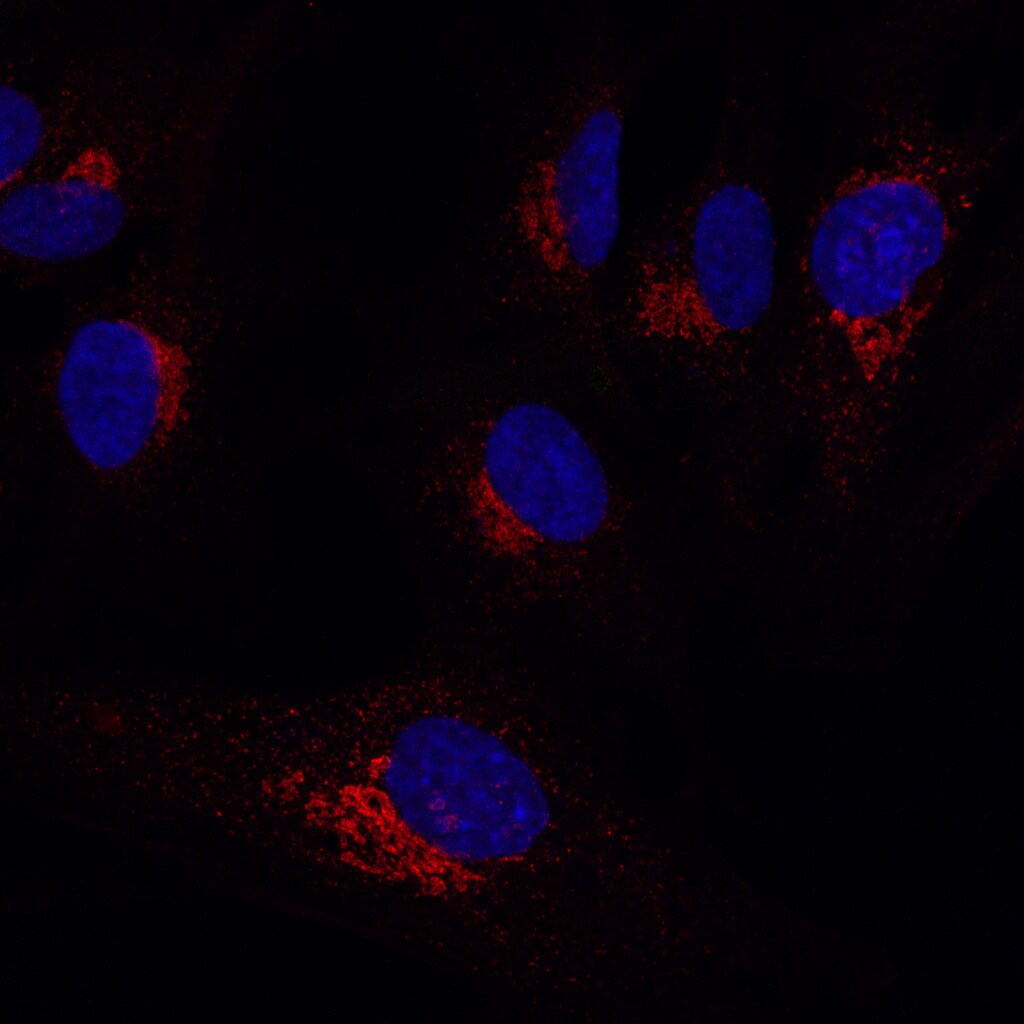
Immunocytochemistry/ Immunofluorescence: RAB6A Antibody [NBP1-33110]
Immunocytochemistry/Immunofluorescence: RAB6A Antibody [NBP1-33110] - Analysis of Human RPE1 cells using RAB6A antibody. Image from verified customer review.![Immunohistochemistry-Paraffin: RAB6A Antibody [NBP1-33110]](https://resources.rndsystems.com/images/products/RAB6A-Antibody-Immunohistochemistry-Paraffin-NBP1-33110-img0005.jpg "Immunohistochemistry-Paraffin: RAB6A Antibody [NBP1-33110]")
Immunohistochemistry-Paraffin: RAB6A Antibody [NBP1-33110]
Immunohistochemistry-Paraffin: RAB6A Antibody [NBP1-33110] - MDA-MB-468 xenograft, using RAB6A antibody at 1:500 dilution. Antigen Retrieval: Trilogy™ (EDTA based, pH 8.0) buffer, 15min.![Western Blot: RAB6A Antibody [NBP1-33110]](https://resources.rndsystems.com/images/products/RAB6A-Antibody-NBP1-33110-img0015.jpg "Western Blot: RAB6A Antibody [NBP1-33110]")
Western Blot: RAB6A Antibody [NBP1-33110]
Western Blot: RAB6A Antibody [NBP1-33110] - A. 50 ug rat brain lysate/extract 12 % SDS-PAGE RAB6A antibody dilution: 1:1000![Knockout Validated: RAB6A Antibody [NBP1-33110]](https://resources.rndsystems.com/images/products/RAB6A-Antibody-Western-Blot-NBP1-33110-img0014.jpg "Western Blot: RAB6A Antibody [NBP1-33110]")
Western Blot: RAB6A Antibody [NBP1-33110]
Western Blot: RAB6A Antibody [NBP1-33110] - Wild-type (WT) and RAB6A knockout (KO) 293T cell extracts (30 ug) were separated by 12% SDS-PAGE, and the membrane was blotted with RAB6A antibody diluted at 1:500. HRP-conjugated anti-rabbit IgG antibody was used to detect the primary antibody.
Western Blot Shows Human RAB6A Specificity Using Knockout Cell Line.
Western blot shows lysates of HAP1 cell line and RAB6A knockout HAP1 cell line (KO). Nitrocellulose membrane was probed with RAB6A Antibody (Catalog # NBP1-33110) followed by HRP-conjugated secondary antibody. A specific band was detected for RAB6A at approximately 24 kDa (as indicated) in the parental HAP1 cell line, but is not detectable in knockout HAP1 cell line. Primary antibody dilution used: 1/1000. The Ponceau stained transfer of the blot is shown. This experiment was conducted under reducing conditions. Image, protocol, and testing courtesy of YCharOS Inc. See ycharos.com for additional details.
RAB6A Specificity is Shown by Immunocytochemistry in Knockout Cell Line.
HAP1 WT and RAB6A KO cells were labelled with a green or a far-red fluorescent dye, respectively. Cells were stained with RAB6A Antibody (Catalog # NBP1-33110) and with an Alexa-fluor 555 coupled secondary antibody including DAPI. Acquisition of the blue (nucleus-DAPI), green (identification of WT cells), red (antibody staining) and far-red (identification of KO cells) channels was performed. Representative images of the blue and red (grayscale) channels are shown. WT and KO cells are outlined with green and magenta dashed line, respectively. Primary antibody dilution used: 1/800. Image, protocol and testing courtesy of YCharOS Inc. (ycharos.com).
Western Blot: RAB6A Antibody [NBP1-33110] -
Western Blot: RAB6A Antibody [NBP1-33110] - Various whole cell extracts (30 ug) were separated by 12% SDS-PAGE, and the membrane was blotted with RAB6A antibody (NBP1-33110) diluted at 1:500. The HRP-conjugated anti-rabbit IgG antibody was used to detect the primary antibody.Applications for RAB6A Antibody - BSA Free
Application
Recommended Usage
Immunocytochemistry/ Immunofluorescence
1:100-1:1000
Immunohistochemistry
1:100-1:1000
Immunohistochemistry-Paraffin
1:100-1:1000
Western Blot
1:500-1:3000
Reviewed Applications
Read 1 review rated 5 using NBP1-33110 in the following applications:
Formulation, Preparation, and Storage
Purification
Antigen Affinity-purified
Formulation
PBS, 1% BSA, 20% Glycerol
Format
BSA Free
Preservative
0.025% Proclin 300
Concentration
Concentrations vary lot to lot. See vial label for concentration. If unlisted please contact technical services.
Shipping
The product is shipped with polar packs. Upon receipt, store it immediately at the temperature recommended below.
Stability & Storage
Aliquot and store at -20C or -80C. Avoid freeze-thaw cycles.
Background: RAB6A
Alternate Names
GTP-binding protein RAB6A', GTP-binding protein RAB6B, Oncogene RAB6, Rab GTPase, Rab-6, RAB6, member RAS oncogene family, RAB6A', Rab6A variant 3, RAB6A, member RAS oncogene family, RAB6B, RAB6rab-6, ras-related protein Rab-6A
Gene Symbol
RAB6A
Additional RAB6A Products
Product Documents for RAB6A Antibody - BSA Free
Certificate of Analysis
To download a Certificate of Analysis, please enter a lot or batch number in the search box below.
Product Specific Notices for RAB6A Antibody - BSA Free
This product is for research use only and is not approved for use in humans or in clinical diagnosis. Primary Antibodies are guaranteed for 1 year from date of receipt.
Citations for RAB6A Antibody - BSA Free
 Powered by Bioz
Customer Reviews for RAB6A Antibody - BSA Free (1)
5 out of 5
1 Customer Rating
Have you used RAB6A Antibody - BSA Free?
Submit a review and receive an Amazon gift card!
$25/€18/£15/$25CAN/¥2500 Yen for a review with an image
$10/€7/£6/$10CAN/¥1110 Yen for a review without an image
Submit a review
Customer Images
Showing
1
-
1 of
1 review
Showing All
Filter By:
-
Application: ImmunocytochemistrySample Tested: RPE1 cellsSpecies: HumanVerified Customer | Posted 08/23/2022Very good for detecting endogenous Rab6 protein in the Golgi

There are no reviews that match your criteria.
Protocols
Find general support by application which include: protocols, troubleshooting, illustrated assays, videos and webinars.
- Antigen Retrieval Protocol (PIER)
- Antigen Retrieval for Frozen Sections Protocol
- Appropriate Fixation of IHC/ICC Samples
- Cellular Response to Hypoxia Protocols
- Chromogenic IHC Staining of Formalin-Fixed Paraffin-Embedded (FFPE) Tissue Protocol
- Chromogenic Immunohistochemistry Staining of Frozen Tissue
- ClariTSA™ Fluorophore Kits
- Detection & Visualization of Antibody Binding
- Fluorescent IHC Staining of Frozen Tissue Protocol
- Graphic Protocol for Heat-induced Epitope Retrieval
- Graphic Protocol for the Preparation and Fluorescent IHC Staining of Frozen Tissue Sections
- Graphic Protocol for the Preparation and Fluorescent IHC Staining of Paraffin-embedded Tissue Sections
- Graphic Protocol for the Preparation of Gelatin-coated Slides for Histological Tissue Sections
- ICC Cell Smear Protocol for Suspension Cells
- ICC Immunocytochemistry Protocol Videos
- ICC for Adherent Cells
- IHC Sample Preparation (Frozen sections vs Paraffin)
- Immunocytochemistry (ICC) Protocol
- Immunocytochemistry Troubleshooting
- Immunofluorescence of Organoids Embedded in Cultrex Basement Membrane Extract
- Immunofluorescent IHC Staining of Formalin-Fixed Paraffin-Embedded (FFPE) Tissue Protocol
- Immunohistochemistry (IHC) and Immunocytochemistry (ICC) Protocols
- Immunohistochemistry Frozen Troubleshooting
- Immunohistochemistry Paraffin Troubleshooting
- Preparing Samples for IHC/ICC Experiments
- Preventing Non-Specific Staining (Non-Specific Binding)
- Primary Antibody Selection & Optimization
- Protocol for Heat-Induced Epitope Retrieval (HIER)
- Protocol for Making a 4% Formaldehyde Solution in PBS
- Protocol for VisUCyte™ HRP Polymer Detection Reagent
- Protocol for the Fluorescent ICC Staining of Cell Smears - Graphic
- Protocol for the Fluorescent ICC Staining of Cultured Cells on Coverslips - Graphic
- Protocol for the Preparation & Fixation of Cells on Coverslips
- Protocol for the Preparation and Chromogenic IHC Staining of Frozen Tissue Sections
- Protocol for the Preparation and Chromogenic IHC Staining of Frozen Tissue Sections - Graphic
- Protocol for the Preparation and Chromogenic IHC Staining of Paraffin-embedded Tissue Sections
- Protocol for the Preparation and Chromogenic IHC Staining of Paraffin-embedded Tissue Sections - Graphic
- Protocol for the Preparation and Fluorescent ICC Staining of Cells on Coverslips
- Protocol for the Preparation and Fluorescent ICC Staining of Non-adherent Cells
- Protocol for the Preparation and Fluorescent ICC Staining of Stem Cells on Coverslips
- Protocol for the Preparation and Fluorescent IHC Staining of Frozen Tissue Sections
- Protocol for the Preparation and Fluorescent IHC Staining of Paraffin-embedded Tissue Sections
- Protocol for the Preparation of Gelatin-coated Slides for Histological Tissue Sections
- Protocol for the Preparation of a Cell Smear for Non-adherent Cell ICC - Graphic
- R&D Systems Quality Control Western Blot Protocol
- TUNEL and Active Caspase-3 Detection by IHC/ICC Protocol
- The Importance of IHC/ICC Controls
- Troubleshooting Guide: Immunohistochemistry
- Troubleshooting Guide: Western Blot Figures
- Western Blot Conditions
- Western Blot Protocol
- Western Blot Protocol for Cell Lysates
- Western Blot Troubleshooting
- Western Blot Troubleshooting Guide
- View all Protocols, Troubleshooting, Illustrated assays and Webinars
Loading...