Rab7a Antibody - BSA Free
Novus Biologicals | Catalog # NBP2-24591
![Western Blot: Rab7a AntibodyBSA Free [NBP2-24591]](https://resources.rndsystems.com/images/products/Rab7a-Antibody-Western-Blot-NBP2-24591-img0004.jpg "Western Blot: Rab7a AntibodyBSA Free [NBP2-24591]")
Key Product Details
Species Reactivity
Validated:
Human, Mouse
Predicted:
Bovine (98%), Canine (98%), Fish (98%), Primate (98%), Rabbit (96%), Rat (98%). Backed by our 100% Guarantee.
Applications
Validated:
Immunohistochemistry, Immunohistochemistry-Paraffin, Western Blot, Immunocytochemistry/ Immunofluorescence
Cited:
Western Blot
Label
Unconjugated
Antibody Source
Polyclonal Rabbit IgG
Format
BSA Free
Loading...
Product Specifications
Immunogen
A portion of amino acids 90-140 of human Rab7 protein was used as the immunogen for this antibody.
Marker
Late Endosome Marker
Clonality
Polyclonal
Host
Rabbit
Isotype
IgG
Scientific Data Images for Rab7a Antibody - BSA Free
Western Blot: Rab7a AntibodyBSA Free [NBP2-24591]
Western Blot: Rab7a Antibody [NBP2-24591] - Analysis of human Rab7 in HeLa lysate in the 1) absence and 2) presence of immunizing peptide 3) mouse NIH 3T3 and 4) RAW using Rab7 at 0.25 ug/ml.![Immunocytochemistry/ Immunofluorescence: Rab7a Antibody - BSA Free [NBP2-24591]](https://resources.rndsystems.com/images/products/Rab7a-Antibody-Immunocytochemistry-Immunofluorescence-NBP2-24591-img0006.jpg "Immunocytochemistry/ Immunofluorescence: Rab7a Antibody - BSA Free [NBP2-24591]")
Immunocytochemistry/ Immunofluorescence: Rab7a Antibody - BSA Free [NBP2-24591]
Immunocytochemistry/Immunofluorescence: Rab7a Antibody [NBP2-24591] - NIH3T3 cells were fixed for 10 minutes using 10% formalin and then permeabilized for 5 minutes using 1X PBS + 0.05% Triton-X100. The cells were incubated with anti-Rab7A at 2ug/ml overnight at 4C and detected with an anti-rabbit Dylight 488 (Green) at a 1:500 dilution. Alpha tubulin (DM1A) NB100-690 was used as a co-stain at a 1:1000 dilution and detected with an anti-mouse Dylight 550 (Red) at a 1:500 dilution. Nuclei were counterstained with DAPI (Blue). Cells were imaged using a 40X objective.![Immunohistochemistry-Paraffin: Rab7a Antibody - BSA Free [NBP2-24591]](https://resources.rndsystems.com/images/products/Rab7a-Antibody-Immunohistochemistry-Paraffin-NBP2-24591-img0003.jpg "Immunohistochemistry-Paraffin: Rab7a Antibody - BSA Free [NBP2-24591]")
Immunohistochemistry-Paraffin: Rab7a Antibody - BSA Free [NBP2-24591]
Immunohistochemistry-Paraffin: Rab7a Antibody [NBP2-24591] - Analysis of human skeletal muscle using RAB7A antibody at 10 ug/ml.![Immunocytochemistry/ Immunofluorescence: Rab7a Antibody - BSA Free [NBP2-24591]](https://resources.rndsystems.com/images/products/Rab7a-Antibody-Immunocytochemistry-Immunofluorescence-NBP2-24591-img0005.jpg "Immunocytochemistry/ Immunofluorescence: Rab7a Antibody - BSA Free [NBP2-24591]")
Immunocytochemistry/ Immunofluorescence: Rab7a Antibody - BSA Free [NBP2-24591]
Immunocytochemistry/Immunofluorescence: Rab7a Antibody [NBP2-24591] - A431 cells were fixed for 10 minutes using 10% formalin and then permeabilized for 5 minutes using 1X PBS + 0.05% Triton-X100. The cells were incubated with anti-Rab7A at 2 ug/ml overnight at 4C and detected with an anti-rabbit Dylight 488 (Green) at a 1:500 dilution. Alpha tubulin (DM1A) NB100-690 was used as a co-stain at a 1:1000 dilution and detected with an anti-mouse Dylight 550 (Red) at a 1:500 dilution. Nuclei were counterstained with DAPI (Blue). Cells were imaged using a 40X objective.Applications for Rab7a Antibody - BSA Free
Application
Recommended Usage
Immunocytochemistry/ Immunofluorescence
2-5 ug/ml
Immunohistochemistry-Paraffin
10 ug/ml
Western Blot
0.5-1 ug/ml
Reviewed Applications
Read 1 review rated 3 using NBP2-24591 in the following applications:
Formulation, Preparation, and Storage
Purification
Immunogen affinity purified
Formulation
PBS
Format
BSA Free
Preservative
0.02% Sodium Azide
Concentration
1.0 mg/ml
Shipping
The product is shipped with polar packs. Upon receipt, store it immediately at the temperature recommended below.
Stability & Storage
Store at -20C. Avoid freeze-thaw cycles.
Background: Rab7a
Long Name
RAs Genes from Brain Protein 7a
Alternate Names
PRO2706, RAB7
Gene Symbol
RAB7A
UniProt
Additional Rab7a Products
Product Documents for Rab7a Antibody - BSA Free
Certificate of Analysis
To download a Certificate of Analysis, please enter a lot or batch number in the search box below.
Product Specific Notices for Rab7a Antibody - BSA Free
This product is for research use only and is not approved for use in humans or in clinical diagnosis. Primary Antibodies are guaranteed for 1 year from date of receipt.
Related Research Areas
Citations for Rab7a Antibody - BSA Free
 Powered by Bioz
Customer Reviews for Rab7a Antibody - BSA Free (1)
3 out of 5
1 Customer Rating
Have you used Rab7a Antibody - BSA Free?
Submit a review and receive an Amazon gift card!
$25/€18/£15/$25CAN/¥2500 Yen for a review with an image
$10/€7/£6/$10CAN/¥1110 Yen for a review without an image
Submit a review
Customer Images
Showing
1
-
1 of
1 review
Showing All
Filter By:
-
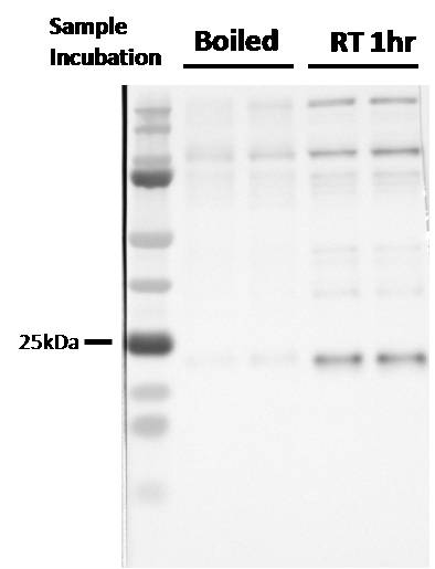
Application: Western BlotSample Tested: Human microglia cell lysateSpecies: HumanVerified Customer | Posted 02/08/2019Do not boil samples, leave in loading dye 1hr at RT. Antibody picks up multiple non-specific bands

There are no reviews that match your criteria.
Protocols
Find general support by application which include: protocols, troubleshooting, illustrated assays, videos and webinars.
- Antigen Retrieval Protocol (PIER)
- Antigen Retrieval for Frozen Sections Protocol
- Appropriate Fixation of IHC/ICC Samples
- Cellular Response to Hypoxia Protocols
- Chromogenic IHC Staining of Formalin-Fixed Paraffin-Embedded (FFPE) Tissue Protocol
- Chromogenic Immunohistochemistry Staining of Frozen Tissue
- ClariTSA™ Fluorophore Kits
- Detection & Visualization of Antibody Binding
- Fluorescent IHC Staining of Frozen Tissue Protocol
- Graphic Protocol for Heat-induced Epitope Retrieval
- Graphic Protocol for the Preparation and Fluorescent IHC Staining of Frozen Tissue Sections
- Graphic Protocol for the Preparation and Fluorescent IHC Staining of Paraffin-embedded Tissue Sections
- Graphic Protocol for the Preparation of Gelatin-coated Slides for Histological Tissue Sections
- ICC Cell Smear Protocol for Suspension Cells
- ICC Immunocytochemistry Protocol Videos
- ICC for Adherent Cells
- IHC Sample Preparation (Frozen sections vs Paraffin)
- Immunocytochemistry (ICC) Protocol
- Immunocytochemistry Troubleshooting
- Immunofluorescence of Organoids Embedded in Cultrex Basement Membrane Extract
- Immunofluorescent IHC Staining of Formalin-Fixed Paraffin-Embedded (FFPE) Tissue Protocol
- Immunohistochemistry (IHC) and Immunocytochemistry (ICC) Protocols
- Immunohistochemistry Frozen Troubleshooting
- Immunohistochemistry Paraffin Troubleshooting
- Preparing Samples for IHC/ICC Experiments
- Preventing Non-Specific Staining (Non-Specific Binding)
- Primary Antibody Selection & Optimization
- Protocol for Heat-Induced Epitope Retrieval (HIER)
- Protocol for Making a 4% Formaldehyde Solution in PBS
- Protocol for VisUCyte™ HRP Polymer Detection Reagent
- Protocol for the Fluorescent ICC Staining of Cell Smears - Graphic
- Protocol for the Fluorescent ICC Staining of Cultured Cells on Coverslips - Graphic
- Protocol for the Preparation & Fixation of Cells on Coverslips
- Protocol for the Preparation and Chromogenic IHC Staining of Frozen Tissue Sections
- Protocol for the Preparation and Chromogenic IHC Staining of Frozen Tissue Sections - Graphic
- Protocol for the Preparation and Chromogenic IHC Staining of Paraffin-embedded Tissue Sections
- Protocol for the Preparation and Chromogenic IHC Staining of Paraffin-embedded Tissue Sections - Graphic
- Protocol for the Preparation and Fluorescent ICC Staining of Cells on Coverslips
- Protocol for the Preparation and Fluorescent ICC Staining of Non-adherent Cells
- Protocol for the Preparation and Fluorescent ICC Staining of Stem Cells on Coverslips
- Protocol for the Preparation and Fluorescent IHC Staining of Frozen Tissue Sections
- Protocol for the Preparation and Fluorescent IHC Staining of Paraffin-embedded Tissue Sections
- Protocol for the Preparation of Gelatin-coated Slides for Histological Tissue Sections
- Protocol for the Preparation of a Cell Smear for Non-adherent Cell ICC - Graphic
- R&D Systems Quality Control Western Blot Protocol
- TUNEL and Active Caspase-3 Detection by IHC/ICC Protocol
- The Importance of IHC/ICC Controls
- Troubleshooting Guide: Immunohistochemistry
- Troubleshooting Guide: Western Blot Figures
- Western Blot Conditions
- Western Blot Protocol
- Western Blot Protocol for Cell Lysates
- Western Blot Troubleshooting
- Western Blot Troubleshooting Guide
- View all Protocols, Troubleshooting, Illustrated assays and Webinars
Loading...