Rad50 Antibody (13B3) - Azide and BSA Free
Novus Biologicals | Catalog # NB100-147
![Immunohistochemistry-Paraffin: Rad50 Antibody (13B3) [NB100-147]](https://resources.rndsystems.com/images/products/Rad50-Antibody-13B3-Immunohistochemistry-Paraffin-NB100-147-img0015.jpg "Immunohistochemistry-Paraffin: Rad50 Antibody (13B3) [NB100-147]")
Loading...
Key Product Details
Validated by
Knockout/Knockdown
Species Reactivity
Validated:
Human, Mouse, Rat, Monkey
Cited:
Human, Mouse
Applications
Validated:
Immunohistochemistry, Immunohistochemistry-Paraffin, Western Blot, Immunocytochemistry/ Immunofluorescence, Immunoprecipitation, In vitro assay, Knockdown Validated
Cited:
Immunohistochemistry-Paraffin, Western Blot, Immunocytochemistry/ Immunofluorescence, IF/IHC
Label
Unconjugated
Antibody Source
Monoclonal Mouse IgG1 Clone # 13B3
Format
Azide and BSA Free
Loading...
Product Specifications
Immunogen
Amino acids 1-425 of Rad50 expressed in E. coli.
Reactivity Notes
Please note that this antibody is reactive to Mouse and derived from the same host, Mouse. Mouse-On-Mouse blocking reagent may be needed for IHC and ICC experiments to reduce high background signal. You can find these reagents under catalog numbers PK-2200-NB and MP-2400-NB. Please contact Technical Support if you have any questions.
Localization
Nuclear
Specificity
This is specific for Rad50.
Clonality
Monoclonal
Host
Mouse
Isotype
IgG1
Theoretical MW
153 kDa.
Disclaimer note: The observed molecular weight of the protein may vary from the listed predicted molecular weight due to post translational modifications, post translation cleavages, relative charges, and other experimental factors.
Disclaimer note: The observed molecular weight of the protein may vary from the listed predicted molecular weight due to post translational modifications, post translation cleavages, relative charges, and other experimental factors.
Scientific Data Images for Rad50 Antibody (13B3) - Azide and BSA Free
Immunohistochemistry-Paraffin: Rad50 Antibody (13B3) [NB100-147]
Immunohistochemistry-Paraffin: Rad50 Antibody (13B3) [NB100-147] - Human lung cancer. Rad50 stained by Rad50 antibody [13B3] diluted at 1:100.Antigen Retrieval: Citrate buffer, pH 6.0, 15 min![Western Blot: Rad50 Antibody (13B3) [NB100-147]](https://resources.rndsystems.com/images/products/Rad50-Antibody-13B3-Western-Blot-NB100-147-img0007.jpg "Western Blot: Rad50 Antibody (13B3) [NB100-147]")
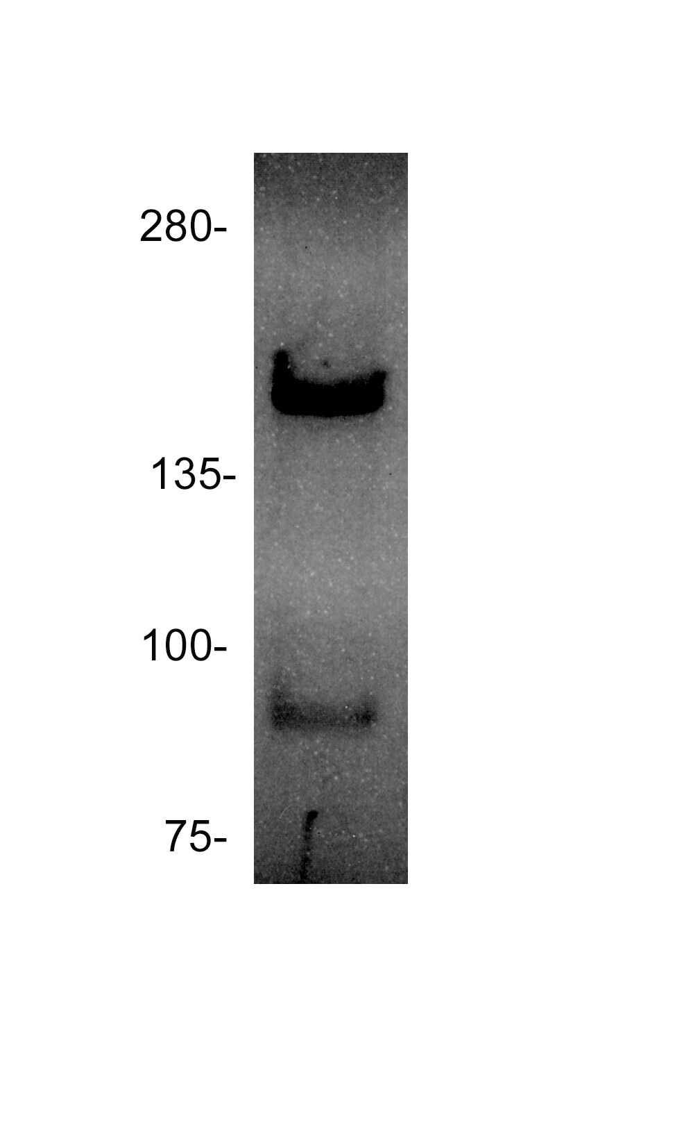
Western Blot: Rad50 Antibody (13B3) [NB100-147]
Western Blot: Rad50 Antibody (13B3) [NB100-147] - Analysis of RAD50 in 293T cell lysate using anti-RAD50 antibody.Image submitted by a verified customer review.![Immunohistochemistry-Paraffin: Rad50 Antibody (13B3) [NB100-147]](https://resources.rndsystems.com/images/products/Rad50-Antibody-13B3-Immunohistochemistry-Paraffin-NB100-147-img0009.jpg "Immunohistochemistry-Paraffin: Rad50 Antibody (13B3) [NB100-147]")
Immunohistochemistry-Paraffin: Rad50 Antibody (13B3) [NB100-147]
Immunohistochemistry-Paraffin: Rad50 Antibody (13B3) [NB100-147] - CAL 27 xenograft. Rad50 antibody [13B3] diluted at 1:200. Antigen Retrieval: Citrate buffer, pH 6.0, 15 min![Immunohistochemistry-Paraffin: Rad50 Antibody (13B3) [NB100-147]](https://resources.rndsystems.com/images/products/Rad50-Antibody-13B3-Immunohistochemistry-Paraffin-NB100-147-img0010.jpg "Immunohistochemistry-Paraffin: Rad50 Antibody (13B3) [NB100-147]")
Immunohistochemistry-Paraffin: Rad50 Antibody (13B3) [NB100-147]
Immunohistochemistry-Paraffin: Rad50 Antibody (13B3) [NB100-147] - PC-3 xenograft. Rad50 antibody [13B3] diluted at 1:200. Antigen Retrieval: Citrate buffer, pH 6.0, 15 min![Immunohistochemistry-Paraffin: Rad50 Antibody (13B3) [NB100-147]](https://resources.rndsystems.com/images/products/Rad50-Antibody-13B3-Immunohistochemistry-Paraffin-NB100-147-img0014.jpg "Immunohistochemistry-Paraffin: Rad50 Antibody (13B3) [NB100-147]")
Immunohistochemistry-Paraffin: Rad50 Antibody (13B3) [NB100-147]
Immunohistochemistry-Paraffin: Rad50 Antibody (13B3) [NB100-147] - Human lung cancer. Rad50 stained by Rad50 antibody [13B3] diluted at 1:100.Antigen Retrieval: Citrate buffer, pH 6.0, 15 min![Knockdown Validated: Rad50 Antibody (13B3) [NB100-147]](https://resources.rndsystems.com/images/products/Rad50-Antibody-13B3-Western-Blot-NB100-147-img0013.jpg "Western Blot: Rad50 Antibody (13B3) [NB100-147]")
 [NB100-147] -")
Western Blot: Rad50 Antibody (13B3) [NB100-147] -
Western Blot: Rad50 Antibody (13B3) [NB100-147] - Various whole cell extracts (30 ug) were separated by 5% SDS-PAGE, and the membrane was blotted with Rad50 antibody [13B3] (NB100-147) diluted at 1:1000. The HRP-conjugated anti-mouse IgG antibody was used to detect the primary antibody. [NB100-147] -")
Immunocytochemistry/ Immunofluorescence: Rad50 Antibody (13B3) [NB100-147] -
Rad50 antibody [13B3] detects Rad50 protein at nucleus by immunofluorescent analysis.Sample: HeLa cells were fixed in 4% paraformaldehyde at RT for 15 min.
Green: Rad50 protein stained by Rad50 antibody [13B3] (NB100-147) diluted at 1:200.
Red: phalloidin, a cytoskeleton marker, diluted at 1:200.
Scale bar = 10 ?m.
 [NB100-147] -")
Western Blot: Rad50 Antibody (13B3) [NB100-147] -
HeLa whole cell and nuclear extracts (30 ug) were separated by 5% SDS-PAGE, and the membrane was blotted with Rad50 antibody [13B3] (NB100-147) diluted at 1:1000. The HRP-conjugated anti-mouse IgG antibody was used to detect the primary antibody.Applications for Rad50 Antibody (13B3) - Azide and BSA Free
Application
Recommended Usage
Immunocytochemistry/ Immunofluorescence
1:100-1:1000
Immunohistochemistry
1:100 - 1:1000
Immunohistochemistry-Paraffin
1:100-1:1000
Western Blot
1:500-1:3000
Application Notes
IHC usage reported in scientific literature (PMID: 16431910). in vitro Assay dependent, IP Assay dependent. Rad50 antibody validated for WB from a verified customer review.
Reviewed Applications
Read 1 review rated 4 using NB100-147 in the following applications:
Formulation, Preparation, and Storage
Purification
Protein G purified
Formulation
PBS
Format
Azide and BSA Free
Preservative
No Preservative
Concentration
Concentrations vary lot to lot. See vial label for concentration. If unlisted please contact technical services.
Shipping
The product is shipped with polar packs. Upon receipt, store it immediately at the temperature recommended below.
Stability & Storage
Store at 4C short term. Aliquot and store at -20C long term. Avoid freeze-thaw cycles.
Background: Rad50
Alternate Names
DNA repair protein RAD50, EC 3.6, EC 3.6.1.15, EC 3.6.3, EC 3.6.3.27, hRAD50, NBSLD, RAD50 (S. cerevisiae) homolog, RAD50 homolog (S. cerevisiae), RAD502, RAD50-2
Entrez Gene IDs
10111 (Human)
Gene Symbol
RAD50
Additional Rad50 Products
Product Documents for Rad50 Antibody (13B3) - Azide and BSA Free
Certificate of Analysis
To download a Certificate of Analysis, please enter a lot or batch number in the search box below.
Product Specific Notices for Rad50 Antibody (13B3) - Azide and BSA Free
This product is for research use only and is not approved for use in humans or in clinical diagnosis. Primary Antibodies are guaranteed for 1 year from date of receipt.
Citations for Rad50 Antibody (13B3) - Azide and BSA Free
 Powered by Bioz
Customer Reviews for Rad50 Antibody (13B3) - Azide and BSA Free (1)
4 out of 5
1 Customer Rating
Have you used Rad50 Antibody (13B3) - Azide and BSA Free?
Submit a review and receive an Amazon gift card!
$25/€18/£15/$25CAN/¥2500 Yen for a review with an image
$10/€7/£6/$10CAN/¥1110 Yen for a review without an image
Submit a review
Customer Images
Showing
1
-
1 of
1 review
Showing All
Filter By:
-
Application: Western BlotSample Tested: 293T lysateSpecies: HumanVerified Customer | Posted 12/17/2014RAD50 WB in 293T cells

There are no reviews that match your criteria.
Protocols
Find general support by application which include: protocols, troubleshooting, illustrated assays, videos and webinars.
- Antigen Retrieval Protocol (PIER)
- Antigen Retrieval for Frozen Sections Protocol
- Appropriate Fixation of IHC/ICC Samples
- Cellular Response to Hypoxia Protocols
- Chromogenic IHC Staining of Formalin-Fixed Paraffin-Embedded (FFPE) Tissue Protocol
- Chromogenic Immunohistochemistry Staining of Frozen Tissue
- ClariTSA™ Fluorophore Kits
- Detection & Visualization of Antibody Binding
- Fluorescent IHC Staining of Frozen Tissue Protocol
- Graphic Protocol for Heat-induced Epitope Retrieval
- Graphic Protocol for the Preparation and Fluorescent IHC Staining of Frozen Tissue Sections
- Graphic Protocol for the Preparation and Fluorescent IHC Staining of Paraffin-embedded Tissue Sections
- Graphic Protocol for the Preparation of Gelatin-coated Slides for Histological Tissue Sections
- ICC Cell Smear Protocol for Suspension Cells
- ICC Immunocytochemistry Protocol Videos
- ICC for Adherent Cells
- IHC Sample Preparation (Frozen sections vs Paraffin)
- Immunocytochemistry (ICC) Protocol
- Immunocytochemistry Troubleshooting
- Immunofluorescence of Organoids Embedded in Cultrex Basement Membrane Extract
- Immunofluorescent IHC Staining of Formalin-Fixed Paraffin-Embedded (FFPE) Tissue Protocol
- Immunohistochemistry (IHC) and Immunocytochemistry (ICC) Protocols
- Immunohistochemistry Frozen Troubleshooting
- Immunohistochemistry Paraffin Troubleshooting
- Immunoprecipitation Protocol
- Preparing Samples for IHC/ICC Experiments
- Preventing Non-Specific Staining (Non-Specific Binding)
- Primary Antibody Selection & Optimization
- Protocol for Heat-Induced Epitope Retrieval (HIER)
- Protocol for Making a 4% Formaldehyde Solution in PBS
- Protocol for VisUCyte™ HRP Polymer Detection Reagent
- Protocol for the Fluorescent ICC Staining of Cell Smears - Graphic
- Protocol for the Fluorescent ICC Staining of Cultured Cells on Coverslips - Graphic
- Protocol for the Preparation & Fixation of Cells on Coverslips
- Protocol for the Preparation and Chromogenic IHC Staining of Frozen Tissue Sections
- Protocol for the Preparation and Chromogenic IHC Staining of Frozen Tissue Sections - Graphic
- Protocol for the Preparation and Chromogenic IHC Staining of Paraffin-embedded Tissue Sections
- Protocol for the Preparation and Chromogenic IHC Staining of Paraffin-embedded Tissue Sections - Graphic
- Protocol for the Preparation and Fluorescent ICC Staining of Cells on Coverslips
- Protocol for the Preparation and Fluorescent ICC Staining of Non-adherent Cells
- Protocol for the Preparation and Fluorescent ICC Staining of Stem Cells on Coverslips
- Protocol for the Preparation and Fluorescent IHC Staining of Frozen Tissue Sections
- Protocol for the Preparation and Fluorescent IHC Staining of Paraffin-embedded Tissue Sections
- Protocol for the Preparation of Gelatin-coated Slides for Histological Tissue Sections
- Protocol for the Preparation of a Cell Smear for Non-adherent Cell ICC - Graphic
- R&D Systems Quality Control Western Blot Protocol
- TUNEL and Active Caspase-3 Detection by IHC/ICC Protocol
- The Importance of IHC/ICC Controls
- Troubleshooting Guide: Immunohistochemistry
- Troubleshooting Guide: Western Blot Figures
- Western Blot Conditions
- Western Blot Protocol
- Western Blot Protocol for Cell Lysates
- Western Blot Troubleshooting
- Western Blot Troubleshooting Guide
- View all Protocols, Troubleshooting, Illustrated assays and Webinars
Loading...