Rad51 Antibody (14B4) - Azide and BSA Free
Novus Biologicals | Catalog # NB100-148
![Western Blot: Rad51 Antibody (14B4) [NB100-148]](https://resources.rndsystems.com/images/products/Rad51-Antibody-14B4-Western-Blot-NB100-148-img0020.jpg "Western Blot: Rad51 Antibody (14B4) [NB100-148]")
Loading...
Key Product Details
Validated by
Knockout/Knockdown, Biological Validation
Species Reactivity
Validated:
Human, Mouse, Rat, Chicken
Cited:
Human, Mouse, Nematode - Caenorhabditis elegans
Applications
Validated:
Immunohistochemistry, Immunohistochemistry-Paraffin, Immunohistochemistry-Frozen, Western Blot, Immunocytochemistry/ Immunofluorescence, Immunoprecipitation, Chromatin Immunoprecipitation (ChIP), In vitro assay, Proximity Ligation Assay, Knockdown Validated
Cited:
Immunohistochemistry-Paraffin, Western Blot, Immunocytochemistry/ Immunofluorescence, Immunoprecipitation, Chromatin Immunoprecipitation (ChIP), Chemotaxis, In vitro assay, IF/IHC
Label
Unconjugated
Antibody Source
Monoclonal Mouse IgG2B Clone # 14B4
Format
Azide and BSA Free
Loading...
Product Specifications
Immunogen
Full length (amino acids 1-338) Rad51 expressed in E. coli.
Reactivity Notes
C. elegans reactivity reported in scientific literature (PMID: 23942865). Please note that this antibody is reactive to Mouse and derived from the same host, Mouse. Mouse-On-Mouse blocking reagent may be needed for IHC and ICC experiments to reduce high background signal. You can find these reagents under catalog numbers PK-2200-NB and MP-2400-NB. Please contact Technical Support if you have any questions.
Clonality
Monoclonal
Host
Mouse
Isotype
IgG2B
Theoretical MW
37 kDa.
Disclaimer note: The observed molecular weight of the protein may vary from the listed predicted molecular weight due to post translational modifications, post translation cleavages, relative charges, and other experimental factors.
Disclaimer note: The observed molecular weight of the protein may vary from the listed predicted molecular weight due to post translational modifications, post translation cleavages, relative charges, and other experimental factors.
Scientific Data Images for Rad51 Antibody (14B4) - Azide and BSA Free
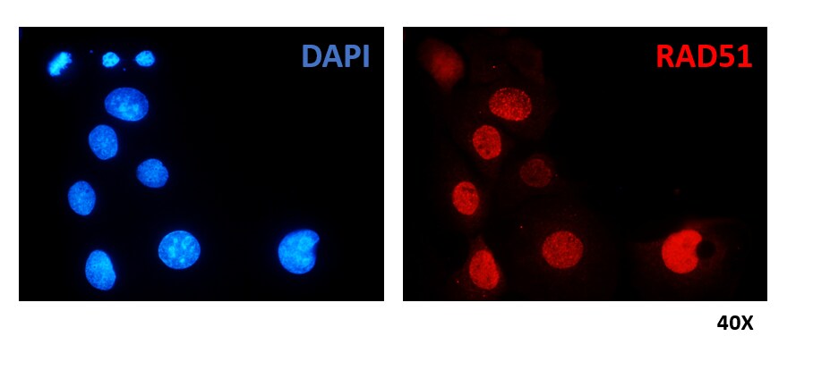
![Immunocytochemistry/ Immunofluorescence: Rad51 Antibody (14B4) [NB100-148]](https://resources.rndsystems.com/images/products/Rad51-Antibody-14B4-Immunocytochemistry-Immunofluorescence-NB100-148-img0021.jpg "Immunocytochemistry/ Immunofluorescence: Rad51 Antibody (14B4) [NB100-148]")
Immunocytochemistry/ Immunofluorescence: Rad51 Antibody (14B4) [NB100-148]
Immunocytochemistry/Immunofluorescence: Rad51 Antibody (14B4) [NB100-148] - HCC1937 (human immortalized breast cancer) cells treated with PARPi for 96 h. Nuclei are stained in blue (DAPI) and RAD51 in red. ICC/IF image submitted by a verified customer review.![Immunohistochemistry-Paraffin: Rad51 Antibody (14B4) [NB100-148]](https://resources.rndsystems.com/images/products/Rad51-Antibody-14B4-Immunohistochemistry-Paraffin-NB100-148-img0003.jpg "Immunohistochemistry-Paraffin: Rad51 Antibody (14B4) [NB100-148]")
Immunohistochemistry-Paraffin: Rad51 Antibody (14B4) [NB100-148]
Immunohistochemistry-Paraffin: Rad51 Antibody (14B4) [NB100-148] - BT483 xenograft. RAD51 antibody 14B4 dilution at 1:200. Antigen Retrieval: Trilogy(TM) (EDTA based, pH 8.0) buffer, 15 min.![Western Blot: Rad51 Antibody (14B4) [NB100-148]](https://resources.rndsystems.com/images/products/Rad51-Antibody-14B4-Western-Blot-NB100-148-img0009.jpg "Western Blot: Rad51 Antibody (14B4) [NB100-148]")
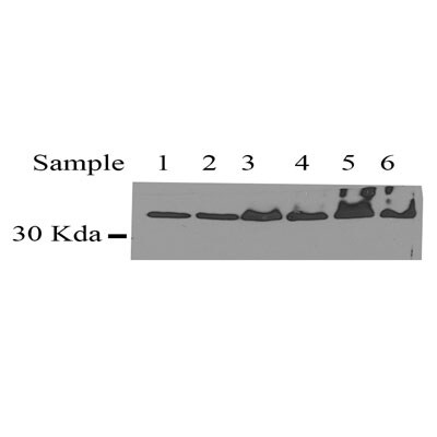
Western Blot: Rad51 Antibody (14B4) [NB100-148]
Western Blot: Rad51 Antibody (14B4) [NB100-148] - Various whole cell extracts (30 ug) were separated by 10% SDS-PAGE, and the membrane was blotted with Rad51 antibody [14B4] diluted at 1:500. The HRP-conjugated anti-mouset IgG antibody (NBP2-19382) was used to detect the primary antibody, and the signal was developed with Trident ECL plus-Enhanced.![Western Blot: Rad51 Antibody (14B4) [NB100-148]](https://resources.rndsystems.com/images/products/Rad51-Antibody-14B4-Western-Blot-NB100-148-img0011.jpg "Western Blot: Rad51 Antibody (14B4) [NB100-148]")
Western Blot: Rad51 Antibody (14B4) [NB100-148]
Western Blot: Rad51 Antibody (14B4) [NB100-148] - Various whole cell extracts (30 ug) were separated by 10% SDS-PAGE, and the membrane was blotted with Rad51 antibody [14B4] diluted at 1:500. The HRP-conjugated anti-mouset IgG antibody (NBP2-19382) was used to detect the primary antibody, and the signal was developed with Trident ECL plus-Enhanced.![Immunocytochemistry/ Immunofluorescence: Rad51 Antibody (14B4) [NB100-148]](https://resources.rndsystems.com/images/products/Rad51-Antibody-14B4-Immunocytochemistry-Immunofluorescence-NB100-148-img0004.jpg "Immunocytochemistry/ Immunofluorescence: Rad51 Antibody (14B4) [NB100-148]")
Immunocytochemistry/ Immunofluorescence: Rad51 Antibody (14B4) [NB100-148]
Immunocytochemistry/Immunofluorescence: Rad51 Antibody (14B4) [NB100-148] - Staining of RAD51 nuclear foci in U2OS cells using RAD51 14B4 antibody. Cells were pre-extracted with CSK buffer before fixation with 4% PFA. RAD51 14B4 was used at 1:1000 dultion. DAPI was used to counterstain the nucleus. Scale bar, 10 mciro-meter.![Immunoprecipitation: Rad51 Antibody (14B4) [NB100-148]](https://resources.rndsystems.com/images/products/Rad51-Antibody-14B4-Immunoprecipitation-NB100-148-img0005.jpg "Immunoprecipitation: Rad51 Antibody (14B4) [NB100-148]")
Immunoprecipitation: Rad51 Antibody (14B4) [NB100-148]
Immunoprecipitation: Rad51 Antibody (14B4) [NB100-148] - HeLa whole cell extract prepared with lysis 180 buffer (40 mM Tris-HCl pH8.0, 180 mM NaCl, 1 mMEDTA, 0.5% NP-40). Rabbit anti-RAD51 antibody was used for subsequent WB detection of immunoprecipated RAD51.![Knockdown Validated: Rad51 Antibody (14B4) [NB100-148]](https://resources.rndsystems.com/images/products/Rad51-Antibody-14B4-Western-Blot-NB100-148-img0006.jpg "Western Blot: Rad51 Antibody (14B4) [NB100-148]")
 [NB100-148] -")
Western Blot: Rad51 Antibody (14B4) [NB100-148] -
Western Blot: Rad51 Antibody (14B4) [NB100-148] - YFP-PALB2 & YFP-PALB2 146AAAA binds RAD51 equally.Lysates from HEK293T cells expressing the indicated constructs were subjected to GFP-Trap pulldown & immunoblotting against YFP or RAD51. YFP alone was used as a negative control. Image collected & cropped by CiteAb from the following publication (https://pubmed.ncbi.nlm.nih.gov/31017574), licensed under a CC-BY license. Not internally tested by Novus Biologicals. [NB100-148] -")
Immunocytochemistry/ Immunofluorescence: Rad51 Antibody (14B4) [NB100-148] -
Immunocytochemistry/ Immunofluorescence: Rad51 Antibody (14B4) [NB100-148] - Bractoppin Selectively Interrupts BRCA1-Dependent Steps in DNA Repair by Homologous Recombination(A) Confocal images depicting at high magnification the recruitment of the RPA32 protein into nuclear foci after the indicated treatments. Experiments were carried out as described as in Figure 5A. Staining in the upper row is for RPA32 (green), middle row, for mCherry-BRCA1 tBRCT (red); lower row, merged red & green staining, with DNA staining (DAPI) in blue. Scale bar represents 10 μm.(B) Recruitment of RAD51 protein into nuclear foci, measured & depicted as described in (A). Scale bar represents 10 μm.(C) Percentage of cells positive for radiation-induced nuclear RPA32 foci (mean ± SD; n = 5,300, 0 Gy; 3,200, 16 Gy; 3,600, BRCA1 tBRCT; 3,400, Bractoppin; 3,500, CCBT2047) enumerated by high-content imaging (see the STAR Methods). Statistical significance was performed using an unpaired two-tailed t test. ***p ≤ 0.001. Similar results were observed in three independent repeats.(D) Percentage of cells containing nuclear RAD51 foci enumerated & depicted as described in (B). ***p ≤ 0.001. Image collected & cropped by CiteAb from the following publication (https://pubmed.ncbi.nlm.nih.gov/29606576), licensed under a CC-BY license. Not internally tested by Novus Biologicals. [NB100-148] -")
Western Blot: Rad51 Antibody (14B4) [NB100-148] -
Western Blot: Rad51 Antibody (14B4) [NB100-148] - MiCas9’s beneficial effects are Brex27 & RAD51 dependent.a Indel rates by spCas9, miCas9, & miCas9-mutant at the on-target site for sg-AAVS1 & the off-target site 1 for sg-EMX1. b Indel rates estimated by T7E1 assay by spCas9 & miCas9 with or without the use of RAD51 inhibitor, B02. c Co-IP results by using anti-flag (left) or anti-RAD51 (right) antibodies followed western blot for RAD51 or Cas9. d ChIP assay results with anti-RAD51 antibody at the proximal region of the sg-AAVS1 target locus. Sp: spCas9, Mi: miCas9, Mi-mut: miCas9-mutant. #Reads: Average amplicon reads per sample. Three independent experiments were performed for each condition. Data are presented as mean ± standard error of means (SEM). Unpaired t-test (two tailed) was used to compare data using GraphPad Prism 8 software (GraphPad Software, Inc., San Diego, CA). Source data are available in the Source Data file. Image collected & cropped by CiteAb from the following publication (https://pubmed.ncbi.nlm.nih.gov/33247137), licensed under a CC-BY license. Not internally tested by Novus Biologicals. [NB100-148] -")
Western Blot: Rad51 Antibody (14B4) [NB100-148] -
Various whole cell extracts (30 ug) were separated by 10% SDS-PAGE, and the membrane was blotted with Rad51 antibody [14B4] (NB100-148) diluted at 1:500. The HRP-conjugated anti-rabbit IgG antibody was used to detect the primary antibody, and the signal was developed with Trident femto Western HRP Substrate.Applications for Rad51 Antibody (14B4) - Azide and BSA Free
Application
Recommended Usage
Immunocytochemistry/ Immunofluorescence
1:100 - 1:1000
Immunohistochemistry
1:100 - 1:1000
Immunohistochemistry-Paraffin
1:100 - 1:1000
Western Blot
1:500 - 1:3000
Application Notes
ChIP usage reported in scientific literature (PMID: 24023853). Rad51 antibody validated for IHC-Fr, ICC/IF from verified customer reviews. Use in In vitro assay reported in scientific literature (PMID: 27815389). PLA-Assay dependent.
Reviewed Applications
Read 5 reviews rated 4.8 using NB100-148 in the following applications:
Formulation, Preparation, and Storage
Purification
Antigen Affinity-purified
Formulation
PBS
Format
Azide and BSA Free
Preservative
No Preservative
Concentration
Concentrations vary lot to lot. See vial label for concentration. If unlisted please contact technical services.
Shipping
The product is shipped with polar packs. Upon receipt, store it immediately at the temperature recommended below.
Stability & Storage
Store at 4C short term. Aliquot and store at -20C long term. Avoid freeze-thaw cycles.
Background: Rad51
Alternate Names
BRCC5, hRAD51, HsRad51, HsT16930, RAD51 (S. cerevisiae) homolog (E coli RecA homolog), RAD51 homolog (RecA homolog, E. coli) (S. cerevisiae), RAD51 homolog A, RAD51ABRCA1/BRCA2-containing complex, subunit 5, RAD51L3, RecA, E. coli, homolog of, RECADNA repair protein RAD51 homolog 1, RecA-like protein, recombination protein A
Entrez Gene IDs
5888 (Human)
Gene Symbol
RAD51
UniProt
Additional Rad51 Products
Product Documents for Rad51 Antibody (14B4) - Azide and BSA Free
Certificate of Analysis
To download a Certificate of Analysis, please enter a lot or batch number in the search box below.
Product Specific Notices for Rad51 Antibody (14B4) - Azide and BSA Free
This product is for research use only and is not approved for use in humans or in clinical diagnosis. Primary Antibodies are guaranteed for 1 year from date of receipt.
Citations for Rad51 Antibody (14B4) - Azide and BSA Free
 Powered by Bioz
Customer Reviews for Rad51 Antibody (14B4) - Azide and BSA Free (5)
4.8 out of 5
5 Customer Ratings
Have you used Rad51 Antibody (14B4) - Azide and BSA Free?
Submit a review and receive an Amazon gift card!
$25/€18/£15/$25CAN/¥2500 Yen for a review with an image
$10/€7/£6/$10CAN/¥1110 Yen for a review without an image
Submit a review
Customer Images
Showing
1
-
5 of
5 reviews
Showing All
Filter By:
-
Application: ImmunocytochemistrySample Tested: human immortalized breast cancer cellsSpecies: HumanVerified Customer | Posted 06/14/2021HCC1937 cells treated with PARPi for 96 h. Nuclei are stained in blue (DAPI) and RAD51 in red.Cells were fixed with 4% PFA. RAD51 Ig was diluted 1:200.

-
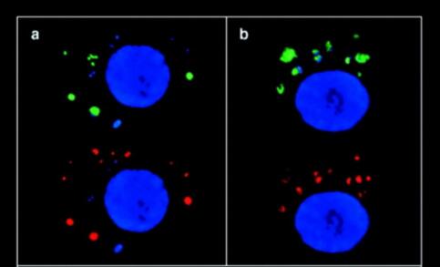
Application: ImmunocytochemistrySample Tested: testis cellsSpecies: MouseVerified Customer | Posted 10/09/2019We used NB100-148 antibody to detect Rad51 foci in germ cell, and colocalize with dmc1(red color).
-
Application: Immunohistochemistry-FrozenSample Tested: See PMID 23942865Species: OtherVerified Customer | Posted 12/12/2014
-
Application: Immunohistochemistry-FrozenSample Tested: whole C. elegans nematodeSpecies: OtherVerified Customer | Posted 11/07/2013
-
Application: Western BlotSample Tested: Hela whole cell lysateSpecies: HumanVerified Customer | Posted 06/20/2012
There are no reviews that match your criteria.
Protocols
Find general support by application which include: protocols, troubleshooting, illustrated assays, videos and webinars.
- Antigen Retrieval Protocol (PIER)
- Antigen Retrieval for Frozen Sections Protocol
- Appropriate Fixation of IHC/ICC Samples
- Cellular Response to Hypoxia Protocols
- ChIP Protocol Video
- Chromatin Immunoprecipitation (ChIP) Protocol
- Chromatin Immunoprecipitation Protocol
- Chromogenic IHC Staining of Formalin-Fixed Paraffin-Embedded (FFPE) Tissue Protocol
- Chromogenic Immunohistochemistry Staining of Frozen Tissue
- ClariTSA™ Fluorophore Kits
- Detection & Visualization of Antibody Binding
- Fluorescent IHC Staining of Frozen Tissue Protocol
- Graphic Protocol for Heat-induced Epitope Retrieval
- Graphic Protocol for the Preparation and Fluorescent IHC Staining of Frozen Tissue Sections
- Graphic Protocol for the Preparation and Fluorescent IHC Staining of Paraffin-embedded Tissue Sections
- Graphic Protocol for the Preparation of Gelatin-coated Slides for Histological Tissue Sections
- ICC Cell Smear Protocol for Suspension Cells
- ICC Immunocytochemistry Protocol Videos
- ICC for Adherent Cells
- IHC Sample Preparation (Frozen sections vs Paraffin)
- Immunocytochemistry (ICC) Protocol
- Immunocytochemistry Troubleshooting
- Immunofluorescence of Organoids Embedded in Cultrex Basement Membrane Extract
- Immunofluorescent IHC Staining of Formalin-Fixed Paraffin-Embedded (FFPE) Tissue Protocol
- Immunohistochemistry (IHC) and Immunocytochemistry (ICC) Protocols
- Immunohistochemistry Frozen Troubleshooting
- Immunohistochemistry Paraffin Troubleshooting
- Immunoprecipitation Protocol
- Preparing Samples for IHC/ICC Experiments
- Preventing Non-Specific Staining (Non-Specific Binding)
- Primary Antibody Selection & Optimization
- Protocol for Heat-Induced Epitope Retrieval (HIER)
- Protocol for Making a 4% Formaldehyde Solution in PBS
- Protocol for VisUCyte™ HRP Polymer Detection Reagent
- Protocol for the Fluorescent ICC Staining of Cell Smears - Graphic
- Protocol for the Fluorescent ICC Staining of Cultured Cells on Coverslips - Graphic
- Protocol for the Preparation & Fixation of Cells on Coverslips
- Protocol for the Preparation and Chromogenic IHC Staining of Frozen Tissue Sections
- Protocol for the Preparation and Chromogenic IHC Staining of Frozen Tissue Sections - Graphic
- Protocol for the Preparation and Chromogenic IHC Staining of Paraffin-embedded Tissue Sections
- Protocol for the Preparation and Chromogenic IHC Staining of Paraffin-embedded Tissue Sections - Graphic
- Protocol for the Preparation and Fluorescent ICC Staining of Cells on Coverslips
- Protocol for the Preparation and Fluorescent ICC Staining of Non-adherent Cells
- Protocol for the Preparation and Fluorescent ICC Staining of Stem Cells on Coverslips
- Protocol for the Preparation and Fluorescent IHC Staining of Frozen Tissue Sections
- Protocol for the Preparation and Fluorescent IHC Staining of Paraffin-embedded Tissue Sections
- Protocol for the Preparation of Gelatin-coated Slides for Histological Tissue Sections
- Protocol for the Preparation of a Cell Smear for Non-adherent Cell ICC - Graphic
- R&D Systems Quality Control Western Blot Protocol
- TUNEL and Active Caspase-3 Detection by IHC/ICC Protocol
- The Importance of IHC/ICC Controls
- Troubleshooting Guide: Immunohistochemistry
- Troubleshooting Guide: Western Blot Figures
- Western Blot Conditions
- Western Blot Protocol
- Western Blot Protocol for Cell Lysates
- Western Blot Troubleshooting
- Western Blot Troubleshooting Guide
- View all Protocols, Troubleshooting, Illustrated assays and Webinars
Loading...