RBPMS Antibody - Azide and BSA Free
Novus Biologicals | Catalog # NBP2-20112
![Immunohistochemistry-Paraffin: RBPMS Antibody [NBP2-20112]](https://resources.rndsystems.com/images/products/RBPMS-Antibody-Immunohistochemistry-Paraffin-NBP2-20112-img0012.jpg "Immunohistochemistry-Paraffin: RBPMS Antibody [NBP2-20112]")
Loading...
Key Product Details
Validated by
Knockout/Knockdown
Species Reactivity
Validated:
Human, Mouse, Rat, Monkey
Cited:
Human, Mouse
Predicted:
Bovine (98%). Backed by our 100% Guarantee.
Applications
Validated:
Immunohistochemistry, Immunohistochemistry-Paraffin, Immunohistochemistry Whole-Mount, Western Blot, Flow Cytometry, Immunocytochemistry/ Immunofluorescence
Cited:
Immunohistochemistry, Immunohistochemistry-Paraffin, Immunohistochemistry Whole-Mount, Immunofluorescence, IF/ICC, IF/IHC
Label
Unconjugated
Antibody Source
Polyclonal Rabbit IgG
Format
Azide and BSA Free
Loading...
Product Specifications
Immunogen
Recombinant protein encompassing a sequence within the center region of human RBPMS. The exact sequence is proprietary.
Clonality
Polyclonal
Host
Rabbit
Isotype
IgG
Theoretical MW
22 kDa.
Disclaimer note: The observed molecular weight of the protein may vary from the listed predicted molecular weight due to post translational modifications, post translation cleavages, relative charges, and other experimental factors.
Disclaimer note: The observed molecular weight of the protein may vary from the listed predicted molecular weight due to post translational modifications, post translation cleavages, relative charges, and other experimental factors.
Scientific Data Images for RBPMS Antibody - Azide and BSA Free
Immunohistochemistry-Paraffin: RBPMS Antibody [NBP2-20112]
Immunohistochemistry-Paraffin: RBPMS Antibody [NBP2-20112] - Mouse intestine. RBPMS stained by RBPMS antibody diluted at 1:2000.Antigen Retrieval: Citrate buffer, pH 6.0, 15 min.
Western Blot: RBPMS Antibody [NBP2-20112] -
Western Blot: RBPMS Antibody [NBP2-20112] - Various whole cell extracts (30 ug) were separated by 12% SDS-PAGE, and the membrane was blotted with RBPMS antibody (NBP2-20112) diluted at 1:500. The HRP-conjugated anti-rabbit IgG antibody was used to detect the primary antibody.
Immunohistochemistry-Paraffin: RBPMS Antibody [NBP2-20112] -
Immunohistochemistry-Paraffin: RBPMS Antibody [NBP2-20112] - RBPMS antibody detects RBPMS protein at cytoplasm and nucleus by immunohistochemical analysis.Sample: Paraffin-embedded mouse lymph node.
RBPMS stained by RBPMS antibody (NBP2-20112) diluted at 1:500.
Antigen Retrieval: Citrate buffer, pH 6.0, 15 min

Immunohistochemistry-Paraffin: RBPMS Antibody [NBP2-20112] -
Immunohistochemistry-Paraffin: RBPMS Antibody [NBP2-20112] - RBPMS antibody detects RBPMS protein at cytoplasm and nucleus by immunohistochemical analysis.Sample: Paraffin-embedded rat colon.
RBPMS stained by RBPMS antibody (NBP2-20112) diluted at 1:500.
Antigen Retrieval: Citrate buffer, pH 6.0, 15 min

Immunocytochemistry/ Immunofluorescence: RBPMS Antibody [NBP2-20112] -
Immunocytochemistry/ Immunofluorescence: RBPMS Antibody [NBP2-20112] - RBPMS antibody detects RBPMS protein at cytoplasm and nucleus by immunofluorescent analysis.Sample: A549 cells were fixed in 4% paraformaldehyde at RT for 15 min.
Green: RBPMS stained by RBPMS antibody (NBP2-20112) diluted at 1:500.
Red: alpha Tubulin, a cytoskeleton marker, stained by alpha Tubulin antibody [GT114] diluted at 1:1000.

Immunohistochemistry-Paraffin: RBPMS Antibody [NBP2-20112] -
RBPMS antibody detects RBPMS protein by immunohistochemical analysis.Sample: Paraffin-embedded rat kidney.
RBPMS stained by RBPMS antibody (NBP2-20112) diluted at 1:500.
Antigen Retrieval: Citrate buffer, pH 6.0, 15 min

Western Blot: RBPMS Antibody [NBP2-20112] -
Non-transfected (-) and transfected (+) HCT-116 whole cell extract were separated by 12% SDS-PAGE, and the membrane was blotted with RBPMS antibody (NBP2-20112) diluted at 1:500. The HRP-conjugated anti-rabbit IgG antibody was used to detect the primary antibody.
Immunocytochemistry/ Immunofluorescence: RBPMS Antibody [NBP2-20112] -
RBPMS antibody detects RBPMS protein at cytoplasm and nucleus by immunofluorescent analysis.Sample: A431 cells were fixed in 4% paraformaldehyde at RT for 15 min.
Green: RBPMS stained by RBPMS antibody (NBP2-20112) diluted at 1:500.
Red: alpha Tubulin, a cytoskeleton marker, stained by alpha Tubulin antibody [GT114] diluted at 1:1000.
Blue: Fluoroshield with DAPI.

Immunohistochemistry-Paraffin: RBPMS Antibody [NBP2-20112] -
RBPMS antibody detects RBPMS protein by immunohistochemical analysis.Samples: Paraffin-embedded mouse retina.
Green: RBPMS protein stained by RBPMS antibody (NBP2-20112) diluted at 1:250.
Red: beta Tubulin 3/ Tuj1, a marker, stained by beta Tubulin 3/ Tuj1 antibody [GT1338] diluted at 1:500.
Blue: Fluoroshield with DAPI.
br>Antigen Retrieval: Citrate buffer, pH 6.0, 15 min

Western Blot: RBPMS Antibody [NBP2-20112] -
Various tissue extracts (50 ug) were separated by 12% SDS-PAGE, and the membrane was blotted with RBPMS antibody (NBP2-20112) diluted at 1:3000. The HRP-conjugated anti-rabbit IgG antibody was used to detect the primary antibody.
Western Blot: RBPMS Antibody [NBP2-20112] -
Various whole cell extracts (30 ug) were separated by 12% SDS-PAGE, and the membrane was blotted with RBPMS antibody (NBP2-20112) diluted at 1:500. The HRP-conjugated anti-rabbit IgG antibody was used to detect the primary antibody.
Western Blot: RBPMS Antibody [NBP2-20112] -
Non-transfected (-) and transfected (+) 293T whole cell extracts (30 ug) were separated by 12% SDS-PAGE, and the membrane was blotted with RBPMS antibody (NBP2-20112) diluted at 1:1000. The HRP-conjugated anti-rabbit IgG antibody was used to detect the primary antibody.
Immunohistochemistry-Paraffin: RBPMS Antibody [NBP2-20112] -
RBPMS antibody detects RBPMS protein at cytoplasm and nucleus by immunohistochemical analysis.Sample: Paraffin-embedded rat lymph node.
RBPMS stained by RBPMS antibody (NBP2-20112) diluted at 1:2000.
Antigen Retrieval: Citrate buffer, pH 6.0, 15 min

Immunocytochemistry/ Immunofluorescence: RBPMS Antibody - Azide and BSA Free [NBP2-20112] -
Nicotinamide provides a robust, long-term retinal ganglion cell neuroprotection following intravitreal MPTP administration. A Retinal ganglion cells were labelled in whole-mounted retina with anti-RBPMS (magenta) in Thy1-CFP mice. B There was no significant difference in RBPMS+ retinal ganglion cell density between the vehicle control group and the groups treated with NAM prior to injection of 5 mg/mL MPTP or 50 mg/mL MPTP, supporting the protection of retinal ganglion cells by NAM. Scale bar = 20 um in A Image collected and cropped by CiteAb from the following open publication (https://pubmed.ncbi.nlm.nih.gov/38773545), licensed under a CC-BY license. Not internally tested by Novus Biologicals.Applications for RBPMS Antibody - Azide and BSA Free
Application
Recommended Usage
Flow Cytometry
Validated from a verified customer review.
Immunocytochemistry/ Immunofluorescence
1:100-1:1000
Immunohistochemistry
1:100-1:1000
Immunohistochemistry Whole-Mount
Assay dependent
Immunohistochemistry-Paraffin
1:100-1:1000
Western Blot
1:500-1:3000
Reviewed Applications
Read 1 review rated 4 using NBP2-20112 in the following applications:
Flow Cytometry Panel Builder
Bio-Techne Knows Flow Cytometry
Save time and reduce costly mistakes by quickly finding compatible reagents using the Panel Builder Tool.
Advanced Features
- Spectra Viewer - Custom analysis of spectra from multiple fluorochromes
- Spillover Popups - Visualize the spectra of individual fluorochromes
- Antigen Density Selector - Match fluorochrome brightness with antigen density

Formulation, Preparation, and Storage
Purification
Antigen Affinity-purified
Formulation
PBS, 20% Glycerol
Format
Azide and BSA Free
Preservative
0.025% Proclin 300
Concentration
Concentrations vary lot to lot. See vial label for concentration. If unlisted please contact technical services.
Shipping
The product is shipped with polar packs. Upon receipt, store it immediately at the temperature recommended below.
Stability & Storage
Aliquot and store at -20C or -80C. Avoid freeze-thaw cycles.
Background: RBPMS
Long Name
RNA binding protein, mRNA processing factor
Alternate Names
HERMES, RBPMS
Gene Symbol
RBPMS
Additional RBPMS Products
Product Documents for RBPMS Antibody - Azide and BSA Free
Certificate of Analysis
To download a Certificate of Analysis, please enter a lot or batch number in the search box below.
Product Specific Notices for RBPMS Antibody - Azide and BSA Free
This product is for research use only and is not approved for use in humans or in clinical diagnosis. Primary Antibodies are guaranteed for 1 year from date of receipt.
Citations for RBPMS Antibody - Azide and BSA Free
 Powered by Bioz
Customer Reviews for RBPMS Antibody - Azide and BSA Free (1)
4 out of 5
1 Customer Rating
Have you used RBPMS Antibody - Azide and BSA Free?
Submit a review and receive an Amazon gift card!
$25/€18/£15/$25CAN/¥2500 Yen for a review with an image
$10/€7/£6/$10CAN/¥1110 Yen for a review without an image
Submit a review
Customer Images
Showing
1
-
1 of
1 review
Showing All
Filter By:
-
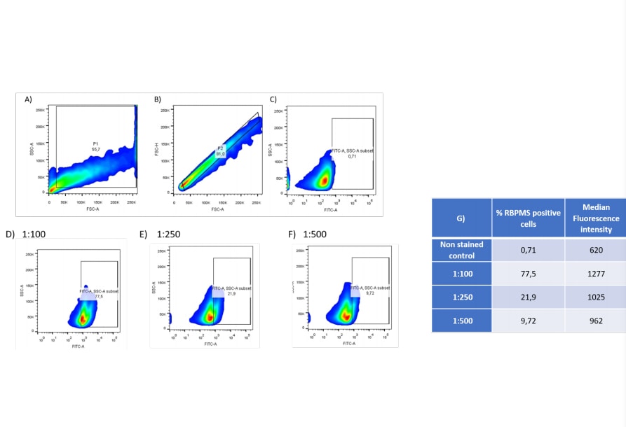
Application: Flow CytometrySample Tested: mouse retinaSpecies: MouseVerified Customer | Posted 04/09/2021RBPMS FACS assay of retina cells. A.;Gate strategy and B.; duplets exclusion.C.; FITC fluorescence of unstained cells. D – F 1; FITC fluorescence of RBPMS of cells stained at 1:100, 1:250 and 1:500 respectively.G.; % of RBPMS positive cells and MFI.

There are no reviews that match your criteria.
Protocols
Find general support by application which include: protocols, troubleshooting, illustrated assays, videos and webinars.
- 7-Amino Actinomycin D (7-AAD) Cell Viability Flow Cytometry Protocol
- Antigen Retrieval Protocol (PIER)
- Antigen Retrieval for Frozen Sections Protocol
- Appropriate Fixation of IHC/ICC Samples
- Cellular Response to Hypoxia Protocols
- Chromogenic IHC Staining of Formalin-Fixed Paraffin-Embedded (FFPE) Tissue Protocol
- Chromogenic Immunohistochemistry Staining of Frozen Tissue
- ClariTSA™ Fluorophore Kits
- Detection & Visualization of Antibody Binding
- Extracellular Membrane Flow Cytometry Protocol
- Flow Cytometry Protocol for Cell Surface Markers
- Flow Cytometry Protocol for Staining Membrane Associated Proteins
- Flow Cytometry Staining Protocols
- Flow Cytometry Troubleshooting Guide
- Fluorescent IHC Staining of Frozen Tissue Protocol
- Graphic Protocol for Heat-induced Epitope Retrieval
- Graphic Protocol for the Preparation and Fluorescent IHC Staining of Frozen Tissue Sections
- Graphic Protocol for the Preparation and Fluorescent IHC Staining of Paraffin-embedded Tissue Sections
- Graphic Protocol for the Preparation of Gelatin-coated Slides for Histological Tissue Sections
- ICC Cell Smear Protocol for Suspension Cells
- ICC Immunocytochemistry Protocol Videos
- ICC for Adherent Cells
- IHC Sample Preparation (Frozen sections vs Paraffin)
- Immunocytochemistry (ICC) Protocol
- Immunocytochemistry Troubleshooting
- Immunofluorescence of Organoids Embedded in Cultrex Basement Membrane Extract
- Immunofluorescent IHC Staining of Formalin-Fixed Paraffin-Embedded (FFPE) Tissue Protocol
- Immunohistochemistry (IHC) and Immunocytochemistry (ICC) Protocols
- Immunohistochemistry Frozen Troubleshooting
- Immunohistochemistry Paraffin Troubleshooting
- Intracellular Flow Cytometry Protocol Using Alcohol (Methanol)
- Intracellular Flow Cytometry Protocol Using Detergents
- Intracellular Nuclear Staining Flow Cytometry Protocol Using Detergents
- Intracellular Staining Flow Cytometry Protocol Using Alcohol Permeabilization
- Intracellular Staining Flow Cytometry Protocol Using Detergents to Permeabilize Cells
- Preparing Samples for IHC/ICC Experiments
- Preventing Non-Specific Staining (Non-Specific Binding)
- Primary Antibody Selection & Optimization
- Propidium Iodide Cell Viability Flow Cytometry Protocol
- Protocol for Heat-Induced Epitope Retrieval (HIER)
- Protocol for Liperfluo
- Protocol for Making a 4% Formaldehyde Solution in PBS
- Protocol for VisUCyte™ HRP Polymer Detection Reagent
- Protocol for the Characterization of Human Th22 Cells
- Protocol for the Characterization of Human Th9 Cells
- Protocol for the Fluorescent ICC Staining of Cell Smears - Graphic
- Protocol for the Fluorescent ICC Staining of Cultured Cells on Coverslips - Graphic
- Protocol for the Preparation & Fixation of Cells on Coverslips
- Protocol for the Preparation and Chromogenic IHC Staining of Frozen Tissue Sections
- Protocol for the Preparation and Chromogenic IHC Staining of Frozen Tissue Sections - Graphic
- Protocol for the Preparation and Chromogenic IHC Staining of Paraffin-embedded Tissue Sections
- Protocol for the Preparation and Chromogenic IHC Staining of Paraffin-embedded Tissue Sections - Graphic
- Protocol for the Preparation and Fluorescent ICC Staining of Cells on Coverslips
- Protocol for the Preparation and Fluorescent ICC Staining of Non-adherent Cells
- Protocol for the Preparation and Fluorescent ICC Staining of Stem Cells on Coverslips
- Protocol for the Preparation and Fluorescent IHC Staining of Frozen Tissue Sections
- Protocol for the Preparation and Fluorescent IHC Staining of Paraffin-embedded Tissue Sections
- Protocol for the Preparation of Gelatin-coated Slides for Histological Tissue Sections
- Protocol for the Preparation of a Cell Smear for Non-adherent Cell ICC - Graphic
- Protocol: Annexin V and PI Staining by Flow Cytometry
- Protocol: Annexin V and PI Staining for Apoptosis by Flow Cytometry
- R&D Systems Quality Control Western Blot Protocol
- TUNEL and Active Caspase-3 Detection by IHC/ICC Protocol
- The Importance of IHC/ICC Controls
- Troubleshooting Guide: Fluorokine Flow Cytometry Kits
- Troubleshooting Guide: Immunohistochemistry
- Troubleshooting Guide: Western Blot Figures
- Western Blot Conditions
- Western Blot Protocol
- Western Blot Protocol for Cell Lysates
- Western Blot Troubleshooting
- Western Blot Troubleshooting Guide
- View all Protocols, Troubleshooting, Illustrated assays and Webinars
Loading...