PD-1 protein is type I transmembrane receptor belonging to the CD28 family of immune regulatory receptors (1). Other members of this family include CD28, CTLA‑4, ICOS, and BTLA (2-5). Mature human PD-1 consists of an extracellular region (ECD) with one immunoglobulin-like V‑type domain, a transmembrane domain, and a cytoplasmic region. The mature ECD of human PD-1 shares 61% amino acid sequence identity with mouse PD-1 ECD. PD-1 protein acts as a monomeric receptor and interacts in a 1:1 stoichiometric ratio with its ligands PD-L1 (B7-H1) and PD-L2 (B7-DC) (6, 7). PD‑1 is expressed on activated T cells, B cells, monocytes, and dendritic cells while PD-L1 expression is constitutive on the same cells and also on nonhematopoietic cells such as lung endothelial cells and hepatocytes (8, 9). Ligation of PD-L1 with PD-1 induces co-inhibitory signals on T cells promoting their apoptosis, anergy, and functional exhaustion (10). Thus, the PD-1:PD-L1 interaction is a key regulator of the threshold of immune response and peripheral immune tolerance (11).
Best Seller
Recombinant Human PD-1 Fc Chimera Protein, CF
R&D Systems | Catalog # 1086-PD
Analyzed by SEC-MALS

Loading...
Key Product Details
- Recombinant Human PD-1 Protein supports immune checkpoint drug development by enabling ligand binding and inhibition assays.
- R&D Systems NS0-derived Recombinant Human PD-1 Fc Chimera Protein (1086-PD)
- Quality control testing to verify active proteins with lot specific assays by in-house scientists
- All R&D Systems proteins are covered with a 100% guarantee
Source
NS0
Accession Number
Structure / Form
Disulfide-linked homodimer
Applications
Binding Activity
Loading...
Why choose R&D Systems PD-1 Protein, Fc Chimera?
- Guaranteed Bioactivity and High Purity: Bioactivity tested by functional ELISA and purity determined by SDS-PAGE to be greater than 95%.
- Lot-to-Lot Consistency: Stringent QC testing performed on each lot to ensure consistent activity and purity.
- Bulk Quantities Available: Bulk up and save with large mass quantities to meet your research needs. Supply agreements available, partner with us. Please contact us.
- Most Respected, Most Cited Brand in Proteins: With over 35 years of providing the best recombinant proteins to the scientific community, R&D Systems continues to lead the industry in quality, activity, and purity.
Find the non-Fc, His-tagged PD-1 protein here PD-1 Protein His
Visit Bio-Techne to explore other immune checkpoint proteins Immune Checkpoint Proteins
Product Specifications
Source
Mouse myeloma cell line, NS0-derived human PD-1 protein
| Human PD-1 (Leu25-Gln167) Accession # Q15116.3 |
IEGRMD | Human IgG1 (Pro100-Lys330) |
| N-terminus | C-terminus |
Purity
>95%, by SDS-PAGE visualized with Silver Staining and quantitative densitometry by Coomassie® Blue Staining.
Endotoxin Level
<0.01 EU per 1 μg of the protein by the LAL method.
N-terminal Sequence Analysis
Leu25
Predicted Molecular Mass
42.6 kDa (monomer)
SDS-PAGE
60 - 70 kDa, under reducing conditions.
Activity
Measured by its binding ability in a functional ELISA.
When Recombinant Human PD-1 Fc Chimera is immobilized at 0.1 µg/mL (100 µL/well), Recombinant Human B7-H1/PD-L1 Fc Chimera (Catalog # 156-B7) binds with a typical ED50 of 0.15-0.75 μg/mL.
When Recombinant Human PD-1 Fc Chimera is immobilized at 0.1 µg/mL (100 µL/well), Recombinant Human B7-H1/PD-L1 Fc Chimera (Catalog # 156-B7) binds with a typical ED50 of 0.15-0.75 μg/mL.
Reviewed Applications
Read 10 reviews rated 4.7 using 1086-PD in the following applications:
Scientific Data Images for Recombinant Human PD-1 Fc Chimera Protein, CF
Recombinant Human PD‑1 Fc Chimera Protein SEC-MALS.
Recombinant human PD-1/Fc (Catalog # 1086-PD) has a molecular weight (MW) of 125.1 kDa as analyzed by SEC-MALS, suggesting that this protein is a homodimer. MW may differ from predicted MW due to post-translational modifications (PTMs) present (i.e. Glycosylation).
Bioactivity of Human PD-1 Protein
When Recombinant Human PD-1 Fc Chimera (Catalog # 1086-PD) is coated at 0.1 µg/mL, Recombinant Human B7-H1/PD-L1 Fc Chimera (156-B7) binds with a typical ED50 of 0.15-0.75 µg/mL.Formulation, Preparation, and Storage
1086-PD
| Formulation | Lyophilized from a 0.2 μm filtered solution in PBS. |
| Reconstitution | Reconstitute at 0.5 mg/mL in sterile PBS.
Loading...
|
| Shipping | The product is shipped at ambient temperature. Upon receipt, store it immediately at the temperature recommended below. |
| Stability & Storage | Use a manual defrost freezer and avoid repeated freeze-thaw cycles.
|
Calculators
Background: PD-1
References
- Ishida, Y. et al. (1992) EMBO J. 11:3887.
- Sharpe, A.H. and G.J. Freeman (2002) Nat. Rev. Immunol. 2:116.
- Coyle, A. and J. Gutierrez-Ramos (2001) Nat. Immunol. 2:203.
- Nishimura, H. and T. Honjo (2001) Trends Immunol. 22:265.
- Watanabe, N. et al. (2003) Nat. Immunol. 4:670.
- Zhang, X. et al. (2004) Immunity 20:337.
- Lázár-Molnár, E. et al. (2008) Proc. Natl. Acad. Sci. USA 105:10483.
- Nishimura, H. et al. (1996) Int. Immunol. 8:773.
- Keir, M.E. et al. (2008) Annu. Rev. Immunol. 26:677.
- Butte, M.J. et al. (2007) Immunity 27:111.
- Okazaki, T. et al. (2013) Nat. Immunol. 14:1212.
- Iwai, Y. et al. (2002) Proc. Natl. Acad. Sci. USA 99:12293.
- Nogrady, B. (2014) Nature 513:S10.
Long Name
Programmed Death-1
Alternate Names
CD279, PD1, PDCD1, SLEB2
Entrez Gene IDs
Gene Symbol
PDCD1
UniProt
Additional PD-1 Products
Product Documents for Recombinant Human PD-1 Fc Chimera Protein, CF
Certificate of Analysis
To download a Certificate of Analysis, please enter a lot or batch number in the search box below.
Note: Certificate of Analysis not available for kit components.
Product Specific Notices for Recombinant Human PD-1 Fc Chimera Protein, CF
For research use only
Citations for Recombinant Human PD-1 Fc Chimera Protein, CF
 Powered by Bioz
Customer Reviews for Recombinant Human PD-1 Fc Chimera Protein, CF (10)
4.7 out of 5
10 Customer Ratings
Have you used Recombinant Human PD-1 Fc Chimera Protein, CF?
Submit a review and receive an Amazon gift card!
$25/€18/£15/$25CAN/¥2500 Yen for a review with an image
$10/€7/£6/$10CAN/¥1110 Yen for a review without an image
Submit a review
Customer Images
Showing
1
-
5 of
10 reviews
Showing All
Filter By:
-
Application: Cell migration/motilityVerified Customer | Posted 07/13/2024

-
Application: In vitro bioactivity in cell cultureVerified Customer | Posted 06/26/2020
-
Application: Binding assay/Protein-protein interactionVerified Customer | Posted 01/15/2020
-
Application: Binding assay/Protein-protein interactionVerified Customer | Posted 01/12/2020
-
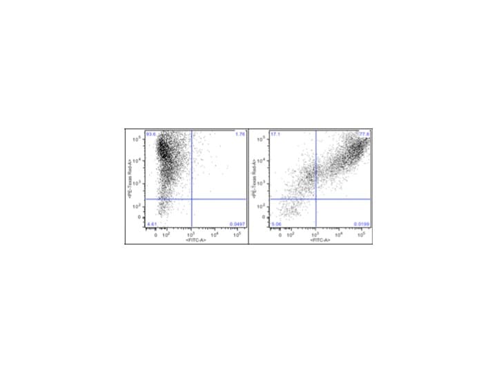
Application: Binding assay/Protein-protein interactionVerified Customer | Posted 11/22/2019Cells expressing a negative control (left) or hPD-L1 mCherry (right) were treated with 0.25 ug of hPD-1 hIgG1. Cells were analyzed by flow cytometry with binding detected using an anti-human 488 secondary antibody.
-
Application: Binding assay/Protein-protein interactionVerified Customer | Posted 08/20/2018
-
Application: Binding assay/Protein-protein interactionVerified Customer | Posted 12/08/2017
-
Application: Binding assay/Protein-protein interactionVerified Customer | Posted 11/03/2017
-
Verified Customer | Posted 04/21/2017
-
Application: Binding assay/Protein-protein interactionVerified Customer | Posted 07/08/2016Sorry that I just rate the catalog based on my alike recombinant PD1 peptide. I have one question for the peptide. Owing the Ig like peptide with Fc fragment. So when we detect it binding to PDL1 on cell assay, it is difficult to get definite conclusion if it is PDL1 binding or others. My protocol is as below: -Cells with PDL1 high level was treated by PD1 like peptide, -4degree 1hr -collected and lysis -WB by anti-IgG to Fc recognization. Result: weak signal. you don't know if it is non-specific bindingBio-Techne ResponseThank you for reviewing our product. We are sorry to hear that this antibody did not perform as expected. We have been in touch with the customer to resolve this issue according to our Product Guarantee and to the customer’s satisfaction.
There are no reviews that match your criteria.
Loading...