Rev-erb A alpha/NR1D1 Antibody (4F6) - Azide and BSA Free
Novus Biologicals | Catalog # H00009572-M02
![Western Blot: Rev-erb A alpha/NR1D1 Antibody (4F6) [H00009572-M02]](https://resources.rndsystems.com/images/products/Rev-erb-A-alpha-NR1D1-Antibody-4F6-Western-Blot-H00009572-M02-img0022.jpg "Western Blot: Rev-erb A alpha/NR1D1 Antibody (4F6) [H00009572-M02]")
Loading...
Key Product Details
Species Reactivity
Validated:
Human, Mouse
Cited:
Human, Mouse
Applications
Validated:
Immunohistochemistry, Immunohistochemistry-Paraffin, Western Blot, ELISA, Immunocytochemistry/ Immunofluorescence, Chromatin Immunoprecipitation (ChIP), Knockdown Validated
Cited:
Immunohistochemistry-Paraffin, Western Blot, IF/IHC
Label
Unconjugated
Antibody Source
Monoclonal Mouse IgG1 kappa Clone # 4F6
Format
Azide and BSA Free
Loading...
Product Specifications
Immunogen
NR1D1 (AAH56148, 1 a.a. ~ 614 a.a) full-length recombinant protein with GST tag. MW of the GST tag alone is 26 KDa. MTTLDSNNNTGGVITYIGSSGSSPSRTSPESLYSDNSNGSFQSLTQGCPTYFPPSPTGSLTQDPARSFGSIPPSLSDDGSPSSSSSSSSSSSSFYNGSPPGSLQVAMEDSSRVSPSKSTSNITKLNGMVLLCKVCGDVASGFHYGVHACEGCKGFFRRSIQQNIQYKRCLKNENCSIVRINRNRCQQCRFKKCLSVGMSRDAVRFGRIPKREKQRMLAEMQSAMNLANNQLSSQCPLETSPTQHPTPGPMGPSPPPAPVPSPLVGFSQFPQQLTPPRSPSPEPTVEDVISQVARAHREIFTYAHDKLGSSPGNFNANHASGSPPATTPHRWENQGCPPAPNDNNTLAAQRHNEALNGLRQAPSSYPPTWPPGPAHHSCHQSNSNGHRLCPTHVYAAPEGKAPANSPRQGNSKNVLLACPMNMYPHGRSGRTVQEIWEDFSMSFTPAVREVVEFAKHIPGFRDLSQHDQVTLLKAGTFEVLMVRFASLFNVKDQTVMFLSRTTYSLQELGAMGMGDLLSAMFDFSEKLNSLALTEEELGLFTAVVLVSADRSGMENSASVEQLQETLLRALRALVLKNRPLETSRFTKLLLKLPDLRTLNNMHSEKLLSFRVDAQ
Specificity
NR1D1 (4F6)
Clonality
Monoclonal
Host
Mouse
Isotype
IgG1 kappa
Scientific Data Images for Rev-erb A alpha/NR1D1 Antibody (4F6) - Azide and BSA Free
Western Blot: Rev-erb A alpha/NR1D1 Antibody (4F6) [H00009572-M02]
Western Blot: Rev-erb A alpha/NR1D1 Antibody (4F6) [H00009572-M02] - NR1D1 monoclonal antibody (M02), clone 4F6. Analysis of NR1D1 expression in NIH/3T3.![Immunocytochemistry/ Immunofluorescence: Rev-erb A alpha/NR1D1 Antibody (4F6) [H00009572-M02]](https://resources.rndsystems.com/images/products/Rev-erb-A-alpha-NR1D1-Antibody-4F6-Immunocytochemistry-Immunofluorescence-H00009572-M02-img0016.jpg "Immunocytochemistry/ Immunofluorescence: Rev-erb A alpha/NR1D1 Antibody (4F6) [H00009572-M02]")
Immunocytochemistry/ Immunofluorescence: Rev-erb A alpha/NR1D1 Antibody (4F6) [H00009572-M02]
Immunocytochemistry/Immunofluorescence: Rev-erb A alpha/NR1D1 Antibody (4F6) [H00009572-M02] - Analysis of monoclonal antibody to NR1D1 on HeLa cell. Antibody concentration 10 ug/ml.![Immunohistochemistry-Paraffin: Rev-erb A alpha/NR1D1 Antibody (4F6) [H00009572-M02]](https://resources.rndsystems.com/images/products/Rev-erb-A-alpha-NR1D1-Antibody-4F6-Immunohistochemistry-Paraffin-H00009572-M02-img0024.jpg "Immunohistochemistry-Paraffin: Rev-erb A alpha/NR1D1 Antibody (4F6) [H00009572-M02]")
Immunohistochemistry-Paraffin: Rev-erb A alpha/NR1D1 Antibody (4F6) [H00009572-M02]
Rev-erb-A-alpha-NR1D1-Antibody-4F6-Immunohistochemistry-Paraffin-H00009572-M02-img0024.jpg![Western Blot: Rev-erb A alpha/NR1D1 Antibody (4F6) [H00009572-M02]](https://resources.rndsystems.com/images/products/Rev-erb-A-alpha-NR1D1-Antibody-4F6-Western-Blot-H00009572-M02-img0002.jpg "Western Blot: Rev-erb A alpha/NR1D1 Antibody (4F6) [H00009572-M02]")
Western Blot: Rev-erb A alpha/NR1D1 Antibody (4F6) [H00009572-M02]
Western Blot: Rev-erb A alpha/NR1D1 Antibody (4F6) [H00009572-M02] - Analysis of NR1D1 expression in transfected 293T cell line by NR1D1 monoclonal antibody (M02), clone 4F6. Lane 1: NR1D1 transfected lysate (66.8 KDa). Lane 2: Non-transfected lysate.![Western Blot: Rev-erb A alpha/NR1D1 Antibody (4F6) [H00009572-M02]](https://resources.rndsystems.com/images/products/Rev-erb-A-alpha-NR1D1-Antibody-4F6-Western-Blot-H00009572-M02-img0019.jpg "Western Blot: Rev-erb A alpha/NR1D1 Antibody (4F6) [H00009572-M02]")
Western Blot: Rev-erb A alpha/NR1D1 Antibody (4F6) [H00009572-M02]
Western Blot: Rev-erb A alpha/NR1D1 Antibody (4F6) [H00009572-M02] - Analysis of NR1D1 over-expressed 293 cell line, cotransfected with NR1D1 Validated Chimera RNAi ( Cat # H00009572-R01V ) (Lane 2) or non-transfected control (Lane 1). Blot probed with NR1D1 monoclonal antibody (M02), clone 4F6 (Cat # H00009572-M02 ). GAPDH ( 36.1 kDa ) used as specificity and loading control.![Western Blot: Rev-erb A alpha/NR1D1 Antibody (4F6) [H00009572-M02]](https://resources.rndsystems.com/images/products/Rev-erb-A-alpha-NR1D1-Antibody-4F6-Western-Blot-H00009572-M02-img0021.jpg "Western Blot: Rev-erb A alpha/NR1D1 Antibody (4F6) [H00009572-M02]")
Western Blot: Rev-erb A alpha/NR1D1 Antibody (4F6) [H00009572-M02]
Western Blot: Rev-erb A alpha/NR1D1 Antibody (4F6) [H00009572-M02] - NR1D1 monoclonal antibody (M02), clone 4F6 Analysis of NR1D1 expression in Hela S3 NE.![Immunohistochemistry-Paraffin: Rev-erb A alpha/NR1D1 Antibody (4F6) [H00009572-M02]](https://resources.rndsystems.com/images/products/Rev-erb-A-alpha-NR1D1-Antibody-4F6-Immunohistochemistry-Paraffin-H00009572-M02-img0011.jpg "Immunohistochemistry-Paraffin: Rev-erb A alpha/NR1D1 Antibody (4F6) [H00009572-M02]")
Immunohistochemistry-Paraffin: Rev-erb A alpha/NR1D1 Antibody (4F6) [H00009572-M02]
Immunohistochemistry-Paraffin: Rev-erb A alpha/NR1D1 Antibody (4F6) [H00009572-M02] - analysis of NR1D1 in paraffin embedded mouse lung tissue using anti-NR1D1 antibody. Image from verified customer review.![Immunohistochemistry-Paraffin: Rev-erb A alpha/NR1D1 Antibody (4F6) [H00009572-M02]](https://resources.rndsystems.com/images/products/Rev-erb-A-alpha-NR1D1-Antibody-4F6-Immunohistochemistry-Paraffin-H00009572-M02-img0012.jpg "Immunohistochemistry-Paraffin: Rev-erb A alpha/NR1D1 Antibody (4F6) [H00009572-M02]")
Immunohistochemistry-Paraffin: Rev-erb A alpha/NR1D1 Antibody (4F6) [H00009572-M02]
Immunohistochemistry-Paraffin: Rev-erb A alpha/NR1D1 Antibody (4F6) [H00009572-M02] - analysis of NR1D1 in paraffin embedded human lung cancer tissue using anti-NR1D1 antibody. Image from verified customer review.![Immunohistochemistry-Paraffin: Rev-erb A alpha/NR1D1 Antibody (4F6) [H00009572-M02]](https://resources.rndsystems.com/images/products/Rev-erb-A-alpha-NR1D1-Antibody-4F6-Immunohistochemistry-Paraffin-H00009572-M02-img0014.jpg "Immunohistochemistry-Paraffin: Rev-erb A alpha/NR1D1 Antibody (4F6) [H00009572-M02]")
Immunohistochemistry-Paraffin: Rev-erb A alpha/NR1D1 Antibody (4F6) [H00009572-M02]
Immunohistochemistry-Paraffin: Rev-erb A alpha/NR1D1 Antibody (4F6) [H00009572-M02] - analysis of Rev-erb A alpha in paraffin embedded mouse lung sections using anti-Rev-erb A alpha antibody. Image from verified customer review.![Immunohistochemistry-Paraffin: Rev-erb A alpha/NR1D1 Antibody (4F6) [H00009572-M02]](https://resources.rndsystems.com/images/products/Rev-erb-A-alpha-NR1D1-Antibody-4F6-Immunohistochemistry-Paraffin-H00009572-M02-img0008.jpg "Immunohistochemistry-Paraffin: Rev-erb A alpha/NR1D1 Antibody (4F6) [H00009572-M02]")
Immunohistochemistry-Paraffin: Rev-erb A alpha/NR1D1 Antibody (4F6) [H00009572-M02]
Immunohistochemistry-Paraffin: Rev-erb A alpha/NR1D1 Antibody (4F6) [H00009572-M02] - Staining on human breast cancer tissue. Image from verified customer review.![Immunohistochemistry-Paraffin: Rev-erb A alpha/NR1D1 Antibody (4F6) [H00009572-M02]](https://resources.rndsystems.com/images/products/Rev-erb-A-alpha-NR1D1-Antibody-4F6-Immunohistochemistry-Paraffin-H00009572-M02-img0018.jpg "Immunohistochemistry-Paraffin: Rev-erb A alpha/NR1D1 Antibody (4F6) [H00009572-M02]")
Immunohistochemistry-Paraffin: Rev-erb A alpha/NR1D1 Antibody (4F6) [H00009572-M02]
Immunohistochemistry-Paraffin: Rev-erb A alpha/NR1D1 Antibody (4F6) [H00009572-M02] - Analysis of monoclonal antibody to NR1D1 on formalin-fixed paraffin-embedded human kidney. Antibody concentration 3 ug/ml.![ELISA: Rev-erb A alpha/NR1D1 Antibody (4F6) [H00009572-M02]](https://resources.rndsystems.com/images/products/Rev-erb-A-alpha-NR1D1-Antibody-4F6-ELISA-H00009572-M02-img0023.jpg "ELISA: Rev-erb A alpha/NR1D1 Antibody (4F6) [H00009572-M02]")
ELISA: Rev-erb A alpha/NR1D1 Antibody (4F6) [H00009572-M02]
ELISA: Rev-erb A alpha/NR1D1 Antibody (4F6) [H00009572-M02] - Detection limit for recombinant GST tagged NR1D1 is approximately 1ng/ml as a capture antibody. [H00009572-M02] -")
Immunohistochemistry: Rev-erb A alpha/NR1D1 Antibody (4F6) [H00009572-M02] -
Representative tissue microarray specimens stained for NR1D1. Immunoreactivity of representative tumor specimens is shown. Low and high NR1D1 expression groups are defined in methods. The low NR1D1 expression ranges from immunoreactive scores 2 to 6 (left (P2xI1), middle (P3xI1), and right (P3xI2)), and the high NR1D1 expression shows immunoreactive score 12 (all, P4xI3). P, percentage of stained tumor cells; I, intensity of staining. Scale bar, 50 μm [H00009572-M02] -")
Immunohistochemistry: Rev-erb A alpha/NR1D1 Antibody (4F6) [H00009572-M02] -
Immunohistochemistry: Rev-erb A alpha/NR1D1 Antibody (4F6) [H00009572-M02] - Representative tissue microarray specimens stained for NR1D1. Immunoreactivity of representative tumor specimens is shown. Low & high NR1D1 expression groups are defined in methods. The low NR1D1 expression ranges from immunoreactive scores 2 to 6 (left (P2xI1), middle (P3xI1), & right (P3xI2)), & the high NR1D1 expression shows immunoreactive score 12 (all, P4xI3). P, percentage of stained tumor cells; I, intensity of staining. Scale bar, 50 μm Image collected & cropped by CiteAb from the following publication (https://pubmed.ncbi.nlm.nih.gov/31779659), licensed under a CC-BY license. Not internally tested by Novus Biologicals.Applications for Rev-erb A alpha/NR1D1 Antibody (4F6) - Azide and BSA Free
Application
Recommended Usage
ELISA
1:100-1:2000
Immunocytochemistry/ Immunofluorescence
1:10-1:500
Immunohistochemistry
1:10-1:500
Immunohistochemistry-Paraffin
1:10-1:500
Western Blot
1:500
Application Notes
Antibody reactive against recominant protein and cell lysate for Western Blot. Has also been used for immunofluoresence, immunohistochemistry (paraffin), RNAi validation and ELISA.
Reviewed Applications
Read 4 reviews rated 4.3 using H00009572-M02 in the following applications:
Formulation, Preparation, and Storage
Purification
Protein A purified
Formulation
In 1x PBS, pH 7.4
Format
Azide and BSA Free
Preservative
No Preservative
Concentration
Concentrations vary lot to lot. See vial label for concentration. If unlisted please contact technical services.
Shipping
The product is shipped with polar packs. Upon receipt, store it immediately at the temperature recommended below.
Stability & Storage
Aliquot and store at -20C or -80C. Avoid freeze-thaw cycles.
Background: Rev-erb A alpha/NR1D1
Alternate Names
Ear-1, NR1D1, Reverb A alpha, THRA1, THRAL
Entrez Gene IDs
9572 (Human)
Gene Symbol
NR1D1
OMIM
602408 (Human)
UniProt
Additional Rev-erb A alpha/NR1D1 Products
Product Documents for Rev-erb A alpha/NR1D1 Antibody (4F6) - Azide and BSA Free
Certificate of Analysis
To download a Certificate of Analysis, please enter a lot or batch number in the search box below.
Product Specific Notices for Rev-erb A alpha/NR1D1 Antibody (4F6) - Azide and BSA Free
This product is produced by and distributed for Abnova, a company based in Taiwan.
This product is for research use only and is not approved for use in humans or in clinical diagnosis. Primary Antibodies are guaranteed for 1 year from date of receipt.
Related Research Areas
Citations for Rev-erb A alpha/NR1D1 Antibody (4F6) - Azide and BSA Free
 Powered by Bioz
Customer Reviews for Rev-erb A alpha/NR1D1 Antibody (4F6) - Azide and BSA Free (4)
4.3 out of 5
4 Customer Ratings
Have you used Rev-erb A alpha/NR1D1 Antibody (4F6) - Azide and BSA Free?
Submit a review and receive an Amazon gift card!
$25/€18/£15/$25CAN/¥2500 Yen for a review with an image
$10/€7/£6/$10CAN/¥1110 Yen for a review without an image
Submit a review
Customer Images
-(01-mg)_H00009572-M02_10486.jpg)
Showing
1
-
4 of
4 reviews
Showing All
Filter By:
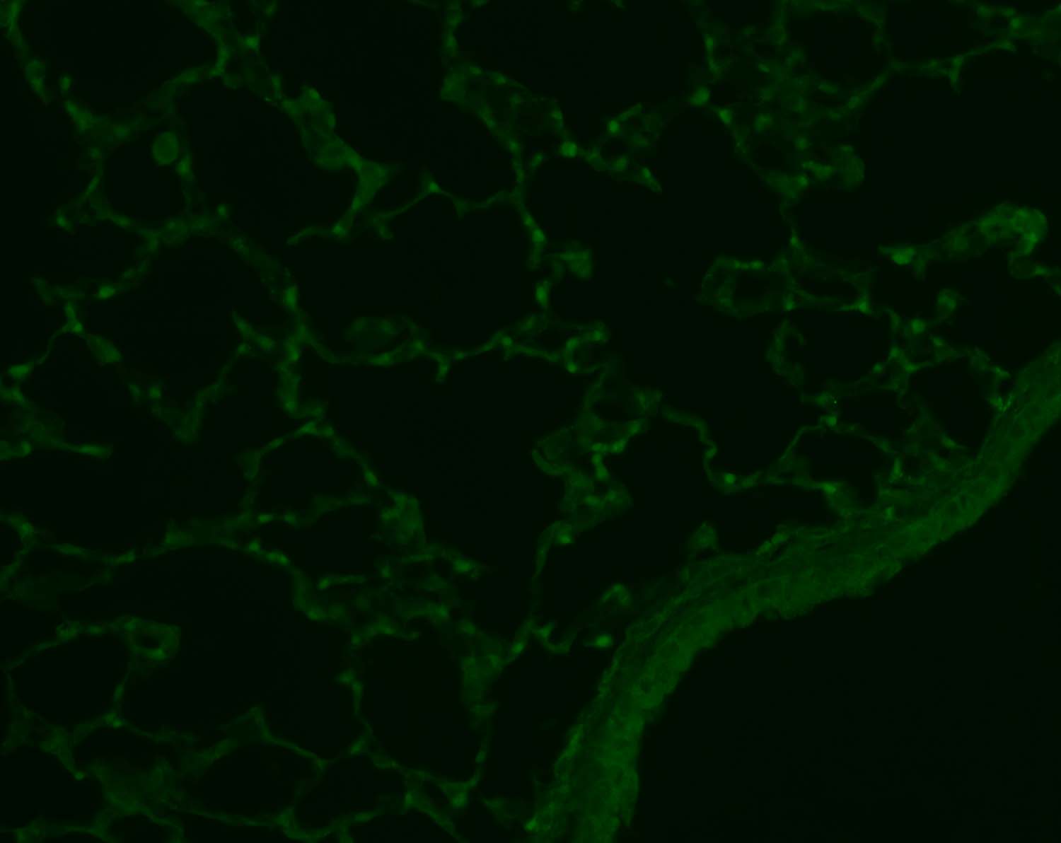
-
Application: ImmunofluorescenceSample Tested: Mouse lung paraffin embedded sectionsSpecies: MouseVerified Customer | Posted 02/26/2015Nr1d1 expression in mouse lungs

-
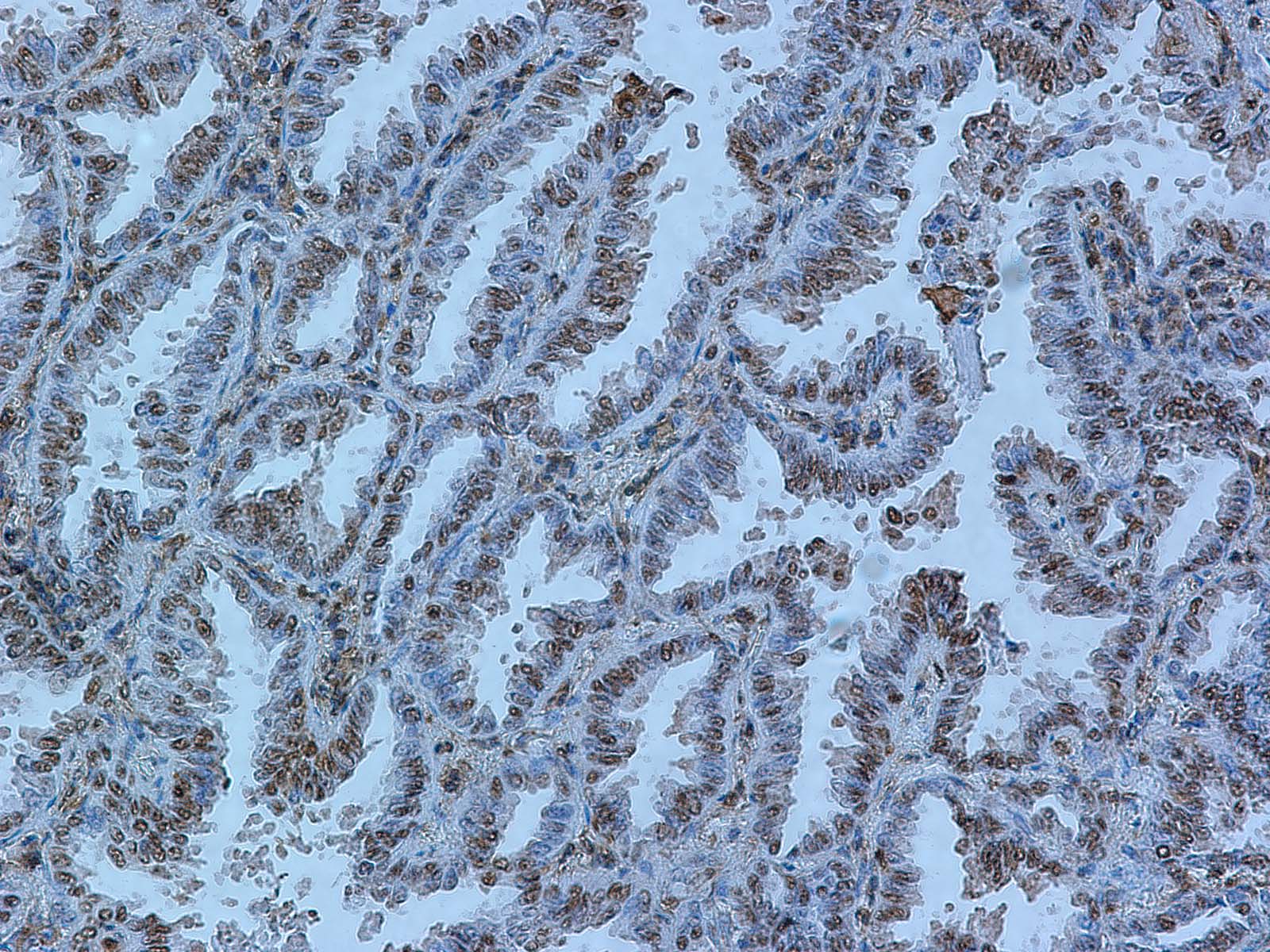
Application: Immunohistochemistry-ParaffinSample Tested: Human lung cancer tissueSpecies: HumanVerified Customer | Posted 02/22/2015Human lung cancer slides
-
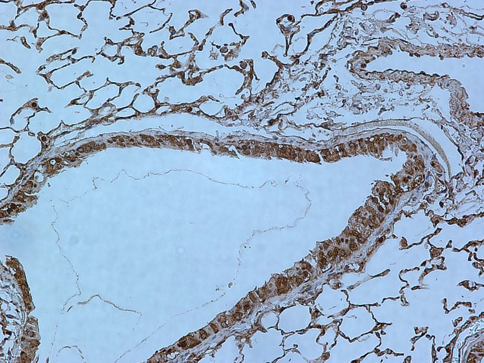
Application: Immunohistochemistry-ParaffinSample Tested: Mouse lung paraffin embedded sectionsSpecies: MouseVerified Customer | Posted 02/22/2015Mouse lung stained for Rev-erb alpha (Nr1d1)
-
Application: Immunohistochemistry-ParaffinSample Tested: Human Breast Cancer TissueSpecies: HumanVerified Customer | Posted 09/26/2014breast cancer tissue
There are no reviews that match your criteria.
Protocols
Find general support by application which include: protocols, troubleshooting, illustrated assays, videos and webinars.
- Antigen Retrieval Protocol (PIER)
- Antigen Retrieval for Frozen Sections Protocol
- Appropriate Fixation of IHC/ICC Samples
- Cellular Response to Hypoxia Protocols
- ChIP Protocol Video
- Chromatin Immunoprecipitation (ChIP) Protocol
- Chromatin Immunoprecipitation Protocol
- Chromogenic IHC Staining of Formalin-Fixed Paraffin-Embedded (FFPE) Tissue Protocol
- Chromogenic Immunohistochemistry Staining of Frozen Tissue
- ClariTSA™ Fluorophore Kits
- Detection & Visualization of Antibody Binding
- ELISA Sample Preparation & Collection Guide
- ELISA Troubleshooting Guide
- Fluorescent IHC Staining of Frozen Tissue Protocol
- Graphic Protocol for Heat-induced Epitope Retrieval
- Graphic Protocol for the Preparation and Fluorescent IHC Staining of Frozen Tissue Sections
- Graphic Protocol for the Preparation and Fluorescent IHC Staining of Paraffin-embedded Tissue Sections
- Graphic Protocol for the Preparation of Gelatin-coated Slides for Histological Tissue Sections
- How to Run an R&D Systems DuoSet ELISA
- How to Run an R&D Systems Quantikine ELISA
- How to Run an R&D Systems Quantikine™ QuicKit™ ELISA
- ICC Cell Smear Protocol for Suspension Cells
- ICC Immunocytochemistry Protocol Videos
- ICC for Adherent Cells
- IHC Sample Preparation (Frozen sections vs Paraffin)
- Immunocytochemistry (ICC) Protocol
- Immunocytochemistry Troubleshooting
- Immunofluorescence of Organoids Embedded in Cultrex Basement Membrane Extract
- Immunofluorescent IHC Staining of Formalin-Fixed Paraffin-Embedded (FFPE) Tissue Protocol
- Immunohistochemistry (IHC) and Immunocytochemistry (ICC) Protocols
- Immunohistochemistry Frozen Troubleshooting
- Immunohistochemistry Paraffin Troubleshooting
- Preparing Samples for IHC/ICC Experiments
- Preventing Non-Specific Staining (Non-Specific Binding)
- Primary Antibody Selection & Optimization
- Protocol for Heat-Induced Epitope Retrieval (HIER)
- Protocol for Making a 4% Formaldehyde Solution in PBS
- Protocol for VisUCyte™ HRP Polymer Detection Reagent
- Protocol for the Fluorescent ICC Staining of Cell Smears - Graphic
- Protocol for the Fluorescent ICC Staining of Cultured Cells on Coverslips - Graphic
- Protocol for the Preparation & Fixation of Cells on Coverslips
- Protocol for the Preparation and Chromogenic IHC Staining of Frozen Tissue Sections
- Protocol for the Preparation and Chromogenic IHC Staining of Frozen Tissue Sections - Graphic
- Protocol for the Preparation and Chromogenic IHC Staining of Paraffin-embedded Tissue Sections
- Protocol for the Preparation and Chromogenic IHC Staining of Paraffin-embedded Tissue Sections - Graphic
- Protocol for the Preparation and Fluorescent ICC Staining of Cells on Coverslips
- Protocol for the Preparation and Fluorescent ICC Staining of Non-adherent Cells
- Protocol for the Preparation and Fluorescent ICC Staining of Stem Cells on Coverslips
- Protocol for the Preparation and Fluorescent IHC Staining of Frozen Tissue Sections
- Protocol for the Preparation and Fluorescent IHC Staining of Paraffin-embedded Tissue Sections
- Protocol for the Preparation of Gelatin-coated Slides for Histological Tissue Sections
- Protocol for the Preparation of a Cell Smear for Non-adherent Cell ICC - Graphic
- Quantikine HS ELISA Kit Assay Principle, Alkaline Phosphatase
- Quantikine HS ELISA Kit Principle, Streptavidin-HRP Polymer
- R&D Systems Quality Control Western Blot Protocol
- Sandwich ELISA (Colorimetric) – Biotin/Streptavidin Detection Protocol
- Sandwich ELISA (Colorimetric) – Direct Detection Protocol
- TUNEL and Active Caspase-3 Detection by IHC/ICC Protocol
- The Importance of IHC/ICC Controls
- Troubleshooting Guide: ELISA
- Troubleshooting Guide: Immunohistochemistry
- Troubleshooting Guide: Western Blot Figures
- Western Blot Conditions
- Western Blot Protocol
- Western Blot Protocol for Cell Lysates
- Western Blot Troubleshooting
- Western Blot Troubleshooting Guide
- View all Protocols, Troubleshooting, Illustrated assays and Webinars
Loading...