Rhodopsin Antibody (4D2) - BSA Free
Novus Biologicals | Catalog # NBP2-59690
![Western Blot: Rhodopsin Antibody (4D2) [NBP2-59690]](https://resources.rndsystems.com/images/products/Rhodopsin-Antibody-4D2-Western-Blot-NBP2-59690-img0005.jpg "Western Blot: Rhodopsin Antibody (4D2) [NBP2-59690]")
Key Product Details
Species Reactivity
Validated:
Amphibian, Avian, Fish, Mammal, Zebrafish
Cited:
Human, Fish - Danio rerio (Zebrafish)
Applications
Validated:
Immunohistochemistry, Immunohistochemistry-Frozen, Western Blot, ELISA, Immunocytochemistry/ Immunofluorescence, Immunoprecipitation
Cited:
Immunohistochemistry-Frozen, ELISA, IF/IHC
Label
Unconjugated
Antibody Source
Monoclonal Mouse IgG1 Clone # 4D2
Format
BSA Free
Loading...
Product Specifications
Immunogen
Bovine Rhodopsin
Reactivity Notes
Extinct Spiny Shark (Acanthodes bridgei). Zebrafish reactivity reported from a verified customer review.
Localization
Membrane
Specificity
Binds specifically to the N-terminus of Rhodopsin. Does not detect Rhodopsin in invertebrates.
Clonality
Monoclonal
Host
Mouse
Isotype
IgG1
Theoretical MW
40 kDa.
Disclaimer note: The observed molecular weight of the protein may vary from the listed predicted molecular weight due to post translational modifications, post translation cleavages, relative charges, and other experimental factors.
Disclaimer note: The observed molecular weight of the protein may vary from the listed predicted molecular weight due to post translational modifications, post translation cleavages, relative charges, and other experimental factors.
Scientific Data Images for Rhodopsin Antibody (4D2) - BSA Free
Western Blot: Rhodopsin Antibody (4D2) [NBP2-59690]
Western Blot: Rhodopsin Antibody (4D2) [NBP2-59690] - Western Blot analysis of Human A549 cells showing detection of ~38.9kDa Rhodopsin protein using Mouse Anti-Rhodopsin Monoclonal Antibody, Clone 4D2 (NBP2-59690). Lane 1: MW ladder. Lane 2: Human A549 Cells 15 ug). Load: 15 ug. Block: 5% Skim Milk Powder in TBST. Primary Antibody: Mouse Anti-Rhodopsin Monoclonal Antibody (NBP2-59690) at 1:1000 for 2.5 hours at RT with shaking. Secondary Antibody: Goat anti-mouse IgG:HRP at 1:1000 for 1 hour at RT with shaking. Color Development: Chemiluminescent for HRP (Moss) for 5 min in RT. Predicted/Observed Size: ~38.9kDa. Other Band(s): Band appears at ~75 kDa indicating detection of the Rhodopsin dimer.![Immunohistochemistry: Rhodopsin Antibody (4D2) [NBP2-59690]](https://resources.rndsystems.com/images/products/Rhodopsin-Antibody-4D2-Immunohistochemistry-NBP2-59690-img0006.jpg "Immunohistochemistry: Rhodopsin Antibody (4D2) [NBP2-59690]")
Immunohistochemistry: Rhodopsin Antibody (4D2) [NBP2-59690]
Immunohistochemistry: Rhodopsin Antibody (4D2) [NBP2-59690] - Immunohistochemistry analysis using Mouse Anti-Rhodopsin Monoclonal Antibody, Clone 4D2 (NBP2-59690). Tissue: retina. Species: Mouse. Primary Antibody: Mouse Anti-Rhodopsin Monoclonal Antibody (NBP2-59690) at 1:1000. Secondary Antibody: FITC Goat Anti-Mouse (green). Counterstain: DAPI (blue) nuclear stain. Localization: Staining of photoreceptor outer segment (OS). Other layers of the retina: IS inner segment; ONL outer nuclear layer; OPL outer plexiform layer; INL inner nuclear layer; IPL inner plexiform layer; GCL ganglion cell layer..![Western Blot: Rhodopsin Antibody (4D2) [NBP2-59690]](https://resources.rndsystems.com/images/products/Rhodopsin-Antibody-4D2-Western-Blot-NBP2-59690-img0004.jpg "Western Blot: Rhodopsin Antibody (4D2) [NBP2-59690]")
Western Blot: Rhodopsin Antibody (4D2) [NBP2-59690]
Western Blot: Rhodopsin Antibody (4D2) [NBP2-59690] - Western Blot analysis of Bovine photoreceptor membranes showing detection of Rhodopsin protein using Mouse Anti-Rhodopsin Monoclonal Antibody, Clone 4D2 (NBP2-59690). Lane 1: MW ladder. Lane 2: 10 ug. Lane 3: 5 ug. Lane 4: 2.5 ug.. Primary Antibody: Mouse Anti-Rhodopsin Monoclonal Antibody (NBP2-59690) at 1:1000.![Immunohistochemistry-Frozen: Rhodopsin Antibody (4D2) [NBP2-59690]](https://resources.rndsystems.com/images/products/Rhodopsin-Antibody-4D2-Immunohistochemistry-Frozen-NBP2-59690-img0002.jpg "Immunohistochemistry-Frozen: Rhodopsin Antibody (4D2) [NBP2-59690]")
Immunohistochemistry-Frozen: Rhodopsin Antibody (4D2) [NBP2-59690]
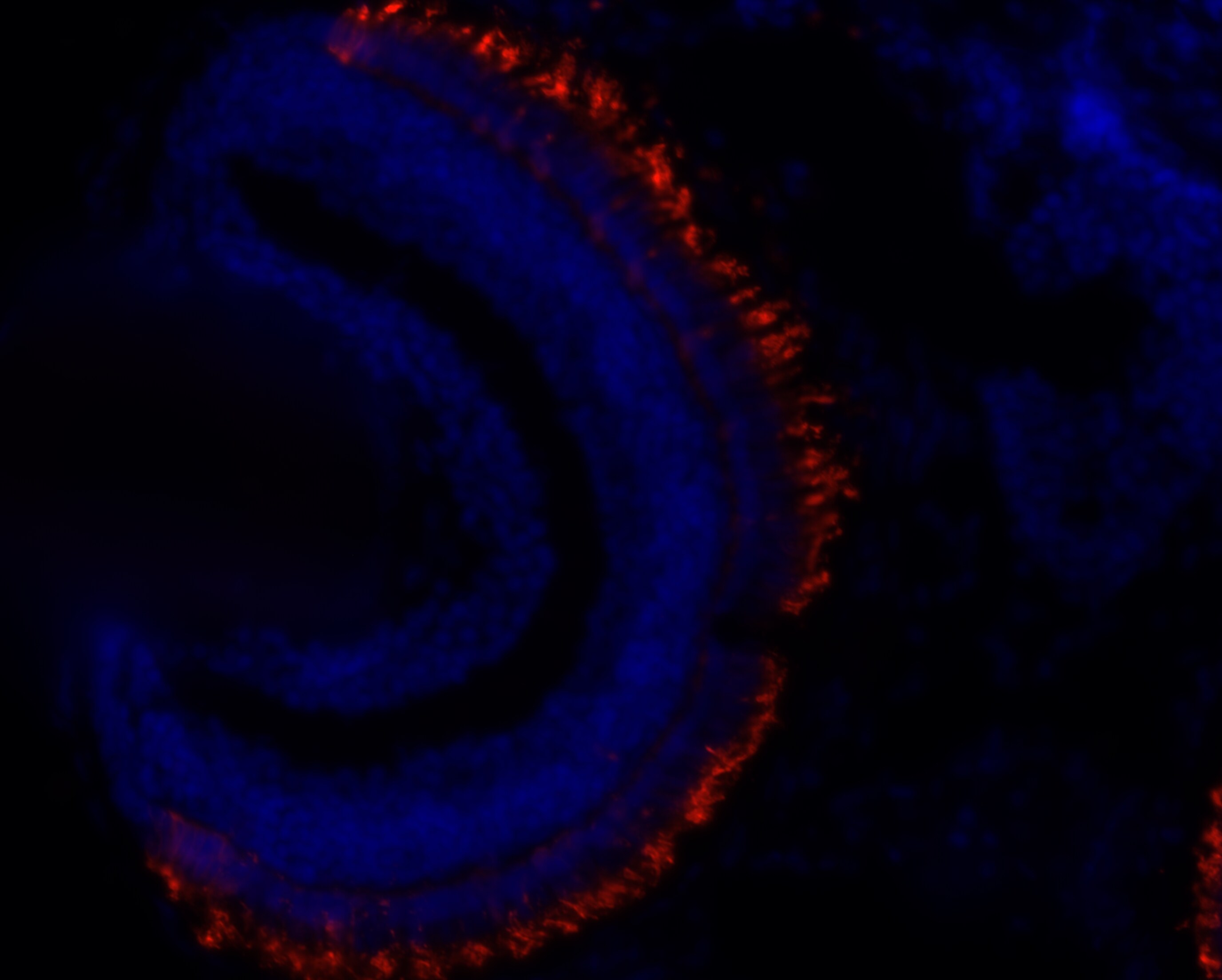
Immunohistochemistry-Frozen: Rhodopsin Antibody (4D2) [NBP2-59690] - Zebrafish retina (outer nuclear layer). PFA and methanol fixed 7 um sections. Rhodopsin antibody O/N incubation at 1:500. IHC-Fr image submitted by a verified customer review.Applications for Rhodopsin Antibody (4D2) - BSA Free
Application
Recommended Usage
Immunohistochemistry
1:1000
Western Blot
1:1000
Application Notes
1 ug/mL of this antibody was sufficient for detection of rhodopsin in 10 ug of rat eye lysate by colorimetric immunoblot analysis using Goat anti-mouse IgG:HRP as the secondary antibody. Rhodopsin antibody validated for IHC-Fr from a verified customer review.
Reviewed Applications
Read 1 review rated 5 using NBP2-59690 in the following applications:
Formulation, Preparation, and Storage
Purification
Protein G purified
Formulation
PBS (pH 7.4), 50% Glycerol
Format
BSA Free
Preservative
0.09% Sodium Azide
Concentration
1 mg/ml
Shipping
The product is shipped with polar packs. Upon receipt, store it immediately at the temperature recommended below.
Stability & Storage
Store at 4C short term. Aliquot and store at -20C long term. Avoid freeze-thaw cycles.
Background: Rhodopsin
Long Name
Rhodopsin
Alternate Names
OPN2, Opsin-2, RHO
Gene Symbol
RHO
Additional Rhodopsin Products
Product Documents for Rhodopsin Antibody (4D2) - BSA Free
Certificate of Analysis
To download a Certificate of Analysis, please enter a lot or batch number in the search box below.
Product Specific Notices for Rhodopsin Antibody (4D2) - BSA Free
This product is for research use only and is not approved for use in humans or in clinical diagnosis. Primary Antibodies are guaranteed for 1 year from date of receipt.
Citations for Rhodopsin Antibody (4D2) - BSA Free
 Powered by Bioz
Customer Reviews for Rhodopsin Antibody (4D2) - BSA Free (1)
5 out of 5
1 Customer Rating
Have you used Rhodopsin Antibody (4D2) - BSA Free?
Submit a review and receive an Amazon gift card!
$25/€18/£15/$25CAN/¥2500 Yen for a review with an image
$10/€7/£6/$10CAN/¥1110 Yen for a review without an image
Submit a review
Customer Images
Showing
1
-
1 of
1 review
Showing All
Filter By:
-
Application: Immunohistochemistry-FrozenSample Tested: Retina (outer nuclear layer)Species: ZebrafishVerified Customer | Posted 08/29/2019Zebrafish eye 5dpfo/n PFA and Methanol fixed 7um sections Put slides in 0.1% PBS-Tween 20 1-2 minutes In 10mM sodium citrate pH 8.5 autoclave (1 min at 121°C, door open at 99⁰C ) Put back in 0.1% PBS-Tween20 Wash 3 x 5min 0.1% PBS-Tween20 1 hour block solution (10% NFDM in 0.1% PBS-Tween20) Overnight with Primary antibody 1:500- 1:5000 in block solution Wash 3 x 10 min 0.1% PBS-Tween20 2 hour incubation with Secondary antibody (1:500) + DAPI (1:10000) in block solution Wash 3 X 10 min 0.1% PBS-Tween20 Enclose with prolong gold antifade Store in dark at 4°C.
 Bio-Techne ResponseThis review was submitted through the legacy Novus Innovators Program, reflecting a new species or application tested on a primary antibody.
There are no reviews that match your criteria.
Protocols
Find general support by application which include: protocols, troubleshooting, illustrated assays, videos and webinars.
- Antigen Retrieval Protocol (PIER)
- Antigen Retrieval for Frozen Sections Protocol
- Appropriate Fixation of IHC/ICC Samples
- Cellular Response to Hypoxia Protocols
- Chromogenic IHC Staining of Formalin-Fixed Paraffin-Embedded (FFPE) Tissue Protocol
- Chromogenic Immunohistochemistry Staining of Frozen Tissue
- ClariTSA™ Fluorophore Kits
- Detection & Visualization of Antibody Binding
- ELISA Sample Preparation & Collection Guide
- ELISA Troubleshooting Guide
- Fluorescent IHC Staining of Frozen Tissue Protocol
- Graphic Protocol for Heat-induced Epitope Retrieval
- Graphic Protocol for the Preparation and Fluorescent IHC Staining of Frozen Tissue Sections
- Graphic Protocol for the Preparation and Fluorescent IHC Staining of Paraffin-embedded Tissue Sections
- Graphic Protocol for the Preparation of Gelatin-coated Slides for Histological Tissue Sections
- How to Run an R&D Systems DuoSet ELISA
- How to Run an R&D Systems Quantikine ELISA
- How to Run an R&D Systems Quantikine™ QuicKit™ ELISA
- ICC Cell Smear Protocol for Suspension Cells
- ICC Immunocytochemistry Protocol Videos
- ICC for Adherent Cells
- IHC Sample Preparation (Frozen sections vs Paraffin)
- Immunocytochemistry (ICC) Protocol
- Immunocytochemistry Troubleshooting
- Immunofluorescence of Organoids Embedded in Cultrex Basement Membrane Extract
- Immunofluorescent IHC Staining of Formalin-Fixed Paraffin-Embedded (FFPE) Tissue Protocol
- Immunohistochemistry (IHC) and Immunocytochemistry (ICC) Protocols
- Immunohistochemistry Frozen Troubleshooting
- Immunohistochemistry Paraffin Troubleshooting
- Immunoprecipitation Protocol
- Preparing Samples for IHC/ICC Experiments
- Preventing Non-Specific Staining (Non-Specific Binding)
- Primary Antibody Selection & Optimization
- Protocol for Heat-Induced Epitope Retrieval (HIER)
- Protocol for Making a 4% Formaldehyde Solution in PBS
- Protocol for VisUCyte™ HRP Polymer Detection Reagent
- Protocol for the Fluorescent ICC Staining of Cell Smears - Graphic
- Protocol for the Fluorescent ICC Staining of Cultured Cells on Coverslips - Graphic
- Protocol for the Preparation & Fixation of Cells on Coverslips
- Protocol for the Preparation and Chromogenic IHC Staining of Frozen Tissue Sections
- Protocol for the Preparation and Chromogenic IHC Staining of Frozen Tissue Sections - Graphic
- Protocol for the Preparation and Chromogenic IHC Staining of Paraffin-embedded Tissue Sections
- Protocol for the Preparation and Chromogenic IHC Staining of Paraffin-embedded Tissue Sections - Graphic
- Protocol for the Preparation and Fluorescent ICC Staining of Cells on Coverslips
- Protocol for the Preparation and Fluorescent ICC Staining of Non-adherent Cells
- Protocol for the Preparation and Fluorescent ICC Staining of Stem Cells on Coverslips
- Protocol for the Preparation and Fluorescent IHC Staining of Frozen Tissue Sections
- Protocol for the Preparation and Fluorescent IHC Staining of Paraffin-embedded Tissue Sections
- Protocol for the Preparation of Gelatin-coated Slides for Histological Tissue Sections
- Protocol for the Preparation of a Cell Smear for Non-adherent Cell ICC - Graphic
- Quantikine HS ELISA Kit Assay Principle, Alkaline Phosphatase
- Quantikine HS ELISA Kit Principle, Streptavidin-HRP Polymer
- R&D Systems Quality Control Western Blot Protocol
- Sandwich ELISA (Colorimetric) – Biotin/Streptavidin Detection Protocol
- Sandwich ELISA (Colorimetric) – Direct Detection Protocol
- TUNEL and Active Caspase-3 Detection by IHC/ICC Protocol
- The Importance of IHC/ICC Controls
- Troubleshooting Guide: ELISA
- Troubleshooting Guide: Immunohistochemistry
- Troubleshooting Guide: Western Blot Figures
- Western Blot Conditions
- Western Blot Protocol
- Western Blot Protocol for Cell Lysates
- Western Blot Troubleshooting
- Western Blot Troubleshooting Guide
- View all Protocols, Troubleshooting, Illustrated assays and Webinars
Loading...