RUNX2/CBFA1 Antibody (1D8) - Azide and BSA Free
Novus Biologicals | Catalog # H00000860-M01
![Western Blot: RUNX2/CBFA1 Antibody (1D8) [H00000860-M01]](https://resources.rndsystems.com/images/products/RUNX2-CBFA1-Antibody-1D8-Western-Blot-H00000860-M01-img0013.jpg "Western Blot: RUNX2/CBFA1 Antibody (1D8) [H00000860-M01]")
Loading...
Key Product Details
Species Reactivity
Validated:
Human, Mouse, Rat
Cited:
Human
Applications
Validated:
Immunohistochemistry, Immunohistochemistry-Paraffin, Western Blot, ELISA, Sandwich ELISA, Immunocytochemistry/ Immunofluorescence
Cited:
Immunohistochemistry-Paraffin
Label
Unconjugated
Antibody Source
Monoclonal Mouse IgG2B Clone # 1D8
Format
Azide and BSA Free
Loading...
Product Specifications
Immunogen
RUNX2/CBFA1 (NP_004339, 251 a.a. ~ 350 a.a) partial recombinant protein with GST tag. MW of the GST tag alone is 26 KDa. NPRPSLNSAPSPFNPQGQSQITDPRQAQSSPPWSYDQSYPSYLSQMTSPSIHSTTPLSSTRGTGLPAITDVPRRISDDDTATSDFCLWPSTLSKKSQAGA
Clonality
Monoclonal
Host
Mouse
Isotype
IgG2B
Theoretical MW
56.6 kDa.
Disclaimer note: The observed molecular weight of the protein may vary from the listed predicted molecular weight due to post translational modifications, post translation cleavages, relative charges, and other experimental factors.
Disclaimer note: The observed molecular weight of the protein may vary from the listed predicted molecular weight due to post translational modifications, post translation cleavages, relative charges, and other experimental factors.
Description
Quality control test: Antibody Reactive Against Recombinant Protein.
Scientific Data Images for RUNX2/CBFA1 Antibody (1D8) - Azide and BSA Free
Western Blot: RUNX2/CBFA1 Antibody (1D8) [H00000860-M01]
Western Blot: RUNX2/CBFA1 Antibody (1D8) [H00000860-M01] - Analysis of RUNX2 expression in transfected 293T cell line by RUNX2 monoclonal antibody (M01), clone 1D8.Lane 1: RUNX2 transfected lysate (Predicted MW: 60.28 KDa).Lane 2: Non-transfected lysate.![Immunocytochemistry/ Immunofluorescence: RUNX2/CBFA1 Antibody (1D8) [H00000860-M01]](https://resources.rndsystems.com/images/products/RUNX2-CBFA1-Antibody-1D8-Immunocytochemistry-Immunofluorescence-H00000860-M01-img0008.jpg "Immunocytochemistry/ Immunofluorescence: RUNX2/CBFA1 Antibody (1D8) [H00000860-M01]")
Immunocytochemistry/ Immunofluorescence: RUNX2/CBFA1 Antibody (1D8) [H00000860-M01]
Immunocytochemistry/Immunofluorescence: RUNX2/CBFA1 Antibody (1D8) [H00000860-M01] - Analysis of monoclonal antibody to RUNX2 on U-2 OS cell. Antibody concentration 10 ug/ml.![Immunohistochemistry-Paraffin: RUNX2/CBFA1 Antibody (1D8) [H00000860-M01]](https://resources.rndsystems.com/images/products/RUNX2-CBFA1-Antibody-1D8-Immunohistochemistry-Paraffin-H00000860-M01-img0010.jpg "Immunohistochemistry-Paraffin: RUNX2/CBFA1 Antibody (1D8) [H00000860-M01]")
Immunohistochemistry-Paraffin: RUNX2/CBFA1 Antibody (1D8) [H00000860-M01]
Immunohistochemistry-Paraffin: RUNX2/CBFA1 Antibody (1D8) [H00000860-M01] - Analysis of monoclonal antibody to RUNX2 on formalin-fixed paraffin-embedded human placenta. Antibody concentration 3 ug/ml.![Western Blot: RUNX2/CBFA1 Antibody (1D8) [H00000860-M01]](https://resources.rndsystems.com/images/products/RUNX2-CBFA1-Antibody-1D8-Western-Blot-H00000860-M01-img0001.jpg "Western Blot: RUNX2/CBFA1 Antibody (1D8) [H00000860-M01]")
Western Blot: RUNX2/CBFA1 Antibody (1D8) [H00000860-M01]
Western Blot: RUNX2/CBFA1 Antibody (1D8) [H00000860-M01] - Western Blot analysis of RUNX2 expression in SJCRH30 ( Cat # L027V1 ).![Western Blot: RUNX2/CBFA1 Antibody (1D8) [H00000860-M01]](https://resources.rndsystems.com/images/products/RUNX2-CBFA1-Antibody-1D8-Western-Blot-H00000860-M01-img0003.jpg "Western Blot: RUNX2/CBFA1 Antibody (1D8) [H00000860-M01]")
Western Blot: RUNX2/CBFA1 Antibody (1D8) [H00000860-M01]
Western Blot: RUNX2/CBFA1 Antibody (1D8) [H00000860-M01] - Western Blot analysis of RUNX2 expression in K-562 ( Cat # L009V1 ).![Western Blot: RUNX2/CBFA1 Antibody (1D8) [H00000860-M01]](https://resources.rndsystems.com/images/products/RUNX2-CBFA1-Antibody-1D8-Western-Blot-H00000860-M01-img0012.jpg "Western Blot: RUNX2/CBFA1 Antibody (1D8) [H00000860-M01]")
Western Blot: RUNX2/CBFA1 Antibody (1D8) [H00000860-M01]
Western Blot: RUNX2/CBFA1 Antibody (1D8) [H00000860-M01] - RUNX2 monoclonal antibody (M01), clone 1D8. Analysis of RUNX2 expression in Jurkat.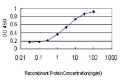
Sandwich ELISA: RUNX2/CBFA1 Antibody (1D8) [H00000860-M01] - Detection limit for recombinant GST tagged RUNX2 is approximately 0.03ng/ml as a capture antibody.
Applications for RUNX2/CBFA1 Antibody (1D8) - Azide and BSA Free
Application
Recommended Usage
Western Blot
1:500
Application Notes
Antibody reactivity against cell lysate and recombinant protein for WB. It has also been used for IF, IHC-P and ELISA.
Formulation, Preparation, and Storage
Purification
IgG purified
Formulation
In 1x PBS, pH 7.4
Format
Azide and BSA Free
Preservative
No Preservative
Concentration
Concentrations vary lot to lot. See vial label for concentration. If unlisted please contact technical services.
Shipping
The product is shipped with polar packs. Upon receipt, store it immediately at the temperature recommended below.
Stability & Storage
Aliquot and store at -20C or -80C. Avoid freeze-thaw cycles.
Background: RUNX2/CBFA1
Functionally, RUNX2 promotes the expression of osteoblast-specific genes vital for the osteoblast differentiation and proliferation process including type I collagen, osteocalcin (OCN), and alkaline phosphatase (APC) (1, 3). Further evidence for the role of RUNX2 is highlighted by a study of Runx2-/-mice which completely lack osteoblasts (4). Additionally, RUNX2 is also required for chondrocyte maturation, which are the cells responsible for cartilage formation (1, 3, 5). Given the role of RUNX2 in bone and cartilage maturation and formation, it is clear that defects or mutations in RUNX2 cause various bone and bone-related diseases (3, 6, 7). For instance, cleidocranial dysplasia (CCD), which presents with delayed cranial suture closure phenotypes, hypoplastic clavicles, extra teeth, and short stature, is caused by haploinsufficiency in RUNX2 (2, 3, 6). Furthermore, metaphyseal dysplasia with maxillary hypoplasia and brachydactyly (MDMHB) is a bone dysplasia disorder with a phenotype of abnormalities in the long bones, an underdeveloped jawbone, and short fingers that is caused by a duplication in RUNX2 (6). Finally, RUNX2 has been shown to be upregulated in mouse models of the joint disorder osteoarthritis (OA) and may be a potential molecular target for disease treatment (7).
Alternative names for RUNX2 include Acute myeloid leukemia 3 protein CBFA1, CBF-alpha-1, CCD1, CCDAML3, CLCD, Core-binding factor subunit alpha-1, MGC120023, ML3, oncogene AML-3, OSF2, osteoblast-specific transcription factor 2, PEA2aA, PEA2-alpha A, PEBP2A, polyomavirus enhancer-binding protein 2 alpha A subunit, runt related transcription factor 2, SL3/AKV core-binding factor alpha A subunit, and SL3-3 enhancer factor 1 alpha A subunit.
References
1. Ferreira, L. B., Gimba, E., Vinagre, J., Sobrinho-Simoes, M., & Soares, P. (2020). Molecular Aspects of Thyroid Calcification. International journal of molecular sciences. https://doi.org/10.3390/ijms21207718
2. Kim, W. J., Shin, H. L., Kim, B. S., Kim, H. J., & Ryoo, H. M. (2020). RUNX2-modifying enzymes: therapeutic targets for bone diseases. Experimental & molecular medicine. https://doi.org/10.1038/s12276-020-0471-4
3. Vimalraj, S., Arumugam, B., Miranda, P. J., & Selvamurugan, N. (2015). Runx2: Structure, function, and phosphorylation in osteoblast differentiation. International journal of biological macromolecules. https://doi.org/10.1016/j.ijbiomac.2015.04.008
4. Uniprot (Q13950)
5. Komori T. (2017). Roles of Runx2 in Skeletal Development. Advances in experimental medicine and biology. https://doi.org/10.1007/978-981-10-3233-2_6
6. Moffatt, P., Ben Amor, M., Glorieux, F. H., Roschger, P., Klaushofer, K., Schwartzentruber, J. A., Paterson, A. D., Hu, P., Marshall, C., FORGE Canada Consortium, Fahiminiya, S., Majewski, J., Beaulieu, C. L., Boycott, K. M., & Rauch, F. (2013). Metaphyseal dysplasia with maxillary hypoplasia and brachydactyly is caused by a duplication in RUNX2. American journal of human genetics. https://doi.org/10.1016/j.ajhg.2012.12.001
7. Chen, D., Kim, D. J., Shen, J., Zou, Z., & O'Keefe, R. J. (2019). Runx2 plays a central role in Osteoarthritis development. Journal of orthopaedic translation. https://doi.org/10.1016/j.jot.2019.11.008
Long Name
Runt-related Transcription Factor 2
Alternate Names
CBFA1
Entrez Gene IDs
860 (Human)
Gene Symbol
RUNX2
OMIM
119600 (Human)
UniProt
Additional RUNX2/CBFA1 Products
Product Documents for RUNX2/CBFA1 Antibody (1D8) - Azide and BSA Free
Certificate of Analysis
To download a Certificate of Analysis, please enter a lot or batch number in the search box below.
Product Specific Notices for RUNX2/CBFA1 Antibody (1D8) - Azide and BSA Free
This product is produced by and distributed for Abnova, a company based in Taiwan.
This product is for research use only and is not approved for use in humans or in clinical diagnosis. Primary Antibodies are guaranteed for 1 year from date of receipt.
Citations for RUNX2/CBFA1 Antibody (1D8) - Azide and BSA Free
 Powered by Bioz
Customer Reviews for RUNX2/CBFA1 Antibody (1D8) - Azide and BSA Free
There are currently no reviews for this product. Be the first to review RUNX2/CBFA1 Antibody (1D8) - Azide and BSA Free and earn rewards!
Have you used RUNX2/CBFA1 Antibody (1D8) - Azide and BSA Free?
Submit a review and receive an Amazon gift card!
$25/€18/£15/$25CAN/¥2500 Yen for a review with an image
$10/€7/£6/$10CAN/¥1110 Yen for a review without an image
Submit a review
Protocols
Find general support by application which include: protocols, troubleshooting, illustrated assays, videos and webinars.
- Antigen Retrieval Protocol (PIER)
- Antigen Retrieval for Frozen Sections Protocol
- Appropriate Fixation of IHC/ICC Samples
- Cellular Response to Hypoxia Protocols
- Chromogenic IHC Staining of Formalin-Fixed Paraffin-Embedded (FFPE) Tissue Protocol
- Chromogenic Immunohistochemistry Staining of Frozen Tissue
- ClariTSA™ Fluorophore Kits
- Detection & Visualization of Antibody Binding
- ELISA Sample Preparation & Collection Guide
- ELISA Troubleshooting Guide
- Fluorescent IHC Staining of Frozen Tissue Protocol
- Graphic Protocol for Heat-induced Epitope Retrieval
- Graphic Protocol for the Preparation and Fluorescent IHC Staining of Frozen Tissue Sections
- Graphic Protocol for the Preparation and Fluorescent IHC Staining of Paraffin-embedded Tissue Sections
- Graphic Protocol for the Preparation of Gelatin-coated Slides for Histological Tissue Sections
- How to Run an R&D Systems DuoSet ELISA
- How to Run an R&D Systems Quantikine ELISA
- How to Run an R&D Systems Quantikine™ QuicKit™ ELISA
- ICC Cell Smear Protocol for Suspension Cells
- ICC Immunocytochemistry Protocol Videos
- ICC for Adherent Cells
- IHC Sample Preparation (Frozen sections vs Paraffin)
- Immunocytochemistry (ICC) Protocol
- Immunocytochemistry Troubleshooting
- Immunofluorescence of Organoids Embedded in Cultrex Basement Membrane Extract
- Immunofluorescent IHC Staining of Formalin-Fixed Paraffin-Embedded (FFPE) Tissue Protocol
- Immunohistochemistry (IHC) and Immunocytochemistry (ICC) Protocols
- Immunohistochemistry Frozen Troubleshooting
- Immunohistochemistry Paraffin Troubleshooting
- Preparing Samples for IHC/ICC Experiments
- Preventing Non-Specific Staining (Non-Specific Binding)
- Primary Antibody Selection & Optimization
- Protocol for Heat-Induced Epitope Retrieval (HIER)
- Protocol for Making a 4% Formaldehyde Solution in PBS
- Protocol for VisUCyte™ HRP Polymer Detection Reagent
- Protocol for the Fluorescent ICC Staining of Cell Smears - Graphic
- Protocol for the Fluorescent ICC Staining of Cultured Cells on Coverslips - Graphic
- Protocol for the Preparation & Fixation of Cells on Coverslips
- Protocol for the Preparation and Chromogenic IHC Staining of Frozen Tissue Sections
- Protocol for the Preparation and Chromogenic IHC Staining of Frozen Tissue Sections - Graphic
- Protocol for the Preparation and Chromogenic IHC Staining of Paraffin-embedded Tissue Sections
- Protocol for the Preparation and Chromogenic IHC Staining of Paraffin-embedded Tissue Sections - Graphic
- Protocol for the Preparation and Fluorescent ICC Staining of Cells on Coverslips
- Protocol for the Preparation and Fluorescent ICC Staining of Non-adherent Cells
- Protocol for the Preparation and Fluorescent ICC Staining of Stem Cells on Coverslips
- Protocol for the Preparation and Fluorescent IHC Staining of Frozen Tissue Sections
- Protocol for the Preparation and Fluorescent IHC Staining of Paraffin-embedded Tissue Sections
- Protocol for the Preparation of Gelatin-coated Slides for Histological Tissue Sections
- Protocol for the Preparation of a Cell Smear for Non-adherent Cell ICC - Graphic
- Quantikine HS ELISA Kit Assay Principle, Alkaline Phosphatase
- Quantikine HS ELISA Kit Principle, Streptavidin-HRP Polymer
- R&D Systems Quality Control Western Blot Protocol
- Sandwich ELISA (Colorimetric) – Biotin/Streptavidin Detection Protocol
- Sandwich ELISA (Colorimetric) – Direct Detection Protocol
- TUNEL and Active Caspase-3 Detection by IHC/ICC Protocol
- The Importance of IHC/ICC Controls
- Troubleshooting Guide: ELISA
- Troubleshooting Guide: Immunohistochemistry
- Troubleshooting Guide: Western Blot Figures
- Western Blot Conditions
- Western Blot Protocol
- Western Blot Protocol for Cell Lysates
- Western Blot Troubleshooting
- Western Blot Troubleshooting Guide
- View all Protocols, Troubleshooting, Illustrated assays and Webinars
FAQs for RUNX2/CBFA1 Antibody (1D8) - Azide and BSA Free
Showing
1
-
1 of
1 FAQ
Showing All
-
Q: We would like an anti-RUNX2 for IHC-P which share cross reactivity with Rat, but not with Human.
A: We don't have any data for our RUNX2 antibodies that confirms they will NOT detect the human protein. When we can confirm that an antibody will not react with a certain species, we display a (-) sign on the datasheet. Otherwise, if the species is not listed it means that it has not been tested.


