![Immunocytochemistry/ Immunofluorescence: RUVBL1 Antibody [NBP2-20245]](https://resources.rndsystems.com/images/products/RUVBL1-Antibody-Immunocytochemistry-Immunofluorescence-NBP2-20245-img0012.jpg "Immunocytochemistry/ Immunofluorescence: RUVBL1 Antibody [NBP2-20245]")
Loading...
Key Product Details
Validated by
Knockout/Knockdown
Species Reactivity
Validated:
Human, Mouse, Rat
Cited:
Human
Predicted:
Bovine (99%), Chicken (99%), Chimpanzee (100%), Rhesus Macaque (100%), Xenopus (97%), Zebrafish (97%). Backed by our 100% Guarantee.
Applications
Validated:
Immunohistochemistry, Immunohistochemistry-Paraffin, Western Blot, Immunocytochemistry/ Immunofluorescence, Immunoprecipitation, Knockdown Validated
Cited:
Western Blot, Immunocytochemistry/ Immunofluorescence, Knockdown Validated
Label
Unconjugated
Antibody Source
Polyclonal Rabbit IgG
Loading...
Product Specifications
Immunogen
Recombinant protein encompassing a sequence within the center region of human RUVBL1. The exact sequence is proprietary.
Reactivity Notes
Xenopus laevis (97%).
Localization
Nucleus matrix; Nucleus; nucleoplasm; Cytoplasm; Membrane
Clonality
Polyclonal
Host
Rabbit
Isotype
IgG
Theoretical MW
50 kDa.
Disclaimer note: The observed molecular weight of the protein may vary from the listed predicted molecular weight due to post translational modifications, post translation cleavages, relative charges, and other experimental factors.
Disclaimer note: The observed molecular weight of the protein may vary from the listed predicted molecular weight due to post translational modifications, post translation cleavages, relative charges, and other experimental factors.
Scientific Data Images for RUVBL1 Antibody
Immunocytochemistry/ Immunofluorescence: RUVBL1 Antibody [NBP2-20245]
Immunocytochemistry/Immunofluorescence: RUVBL1 Antibody [NBP2-20245] - HeLa cells were fixed in 4% paraformaldehyde at RT for 15 min. Green: RUVBL1 protein stained by RUVBL1 antibody diluted at 1:500. Blue: Hoechst 33342 staining. Scale bar = 10 um.![Immunohistochemistry-Paraffin: RUVBL1 Antibody [NBP2-20245]](https://resources.rndsystems.com/images/products/RUVBL1-Antibody-Immunohistochemistry-Paraffin-NBP2-20245-img0010.jpg "Immunohistochemistry-Paraffin: RUVBL1 Antibody [NBP2-20245]")
Immunohistochemistry-Paraffin: RUVBL1 Antibody [NBP2-20245]
Immunohistochemistry-Paraffin: RUVBL1 Antibody [NBP2-20245] - Mouse testis. RUVBL1 antibody diluted at 1:500. Antigen Retrieval: Citrate buffer, pH 6.0, 15 min.![Immunohistochemistry-Paraffin: RUVBL1 Antibody [NBP2-20245]](https://resources.rndsystems.com/images/products/RUVBL1-Antibody-Immunohistochemistry-Paraffin-NBP2-20245-img0002.jpg "Immunohistochemistry-Paraffin: RUVBL1 Antibody [NBP2-20245]")
Immunohistochemistry-Paraffin: RUVBL1 Antibody [NBP2-20245]
Immunohistochemistry-Paraffin: RUVBL1 Antibody [NBP2-20245] - Human colon carcinoma, using RUVBL1antibody at 1:500 dilution. Antigen Retrieval: Trilogy™ (EDTA based, pH 8.0) buffer, 15min.![Immunohistochemistry-Paraffin: RUVBL1 Antibody [NBP2-20245]](https://resources.rndsystems.com/images/products/RUVBL1-Antibody-Immunohistochemistry-Paraffin-NBP2-20245-img0007.jpg "Immunohistochemistry-Paraffin: RUVBL1 Antibody [NBP2-20245]")
Immunohistochemistry-Paraffin: RUVBL1 Antibody [NBP2-20245]
Immunohistochemistry-Paraffin: RUVBL1 Antibody [NBP2-20245] - Rat heart. RUVBL1 antibody diluted at 1:500. Antigen Retrieval: Citrate buffer, pH 6.0, 15 min.![Immunohistochemistry-Paraffin: RUVBL1 Antibody [NBP2-20245]](https://resources.rndsystems.com/images/products/RUVBL1-Antibody-Immunohistochemistry-Paraffin-NBP2-20245-img0008.jpg "Immunohistochemistry-Paraffin: RUVBL1 Antibody [NBP2-20245]")
Immunohistochemistry-Paraffin: RUVBL1 Antibody [NBP2-20245]
Immunohistochemistry-Paraffin: RUVBL1 Antibody [NBP2-20245] - Mouse colon. RUVBL1 antibody diluted at 1:500. Antigen Retrieval: Citrate buffer, pH 6.0, 15 min.![Immunohistochemistry-Paraffin: RUVBL1 Antibody [NBP2-20245]](https://resources.rndsystems.com/images/products/RUVBL1-Antibody-Immunohistochemistry-Paraffin-NBP2-20245-img0009.jpg "Immunohistochemistry-Paraffin: RUVBL1 Antibody [NBP2-20245]")
Immunohistochemistry-Paraffin: RUVBL1 Antibody [NBP2-20245]
Immunohistochemistry-Paraffin: RUVBL1 Antibody [NBP2-20245] - Rat testis. RUVBL1 antibody diluted at 1:500. Antigen Retrieval: Citrate buffer, pH 6.0, 15 min.![Immunoprecipitation: RUVBL1 Antibody [NBP2-20245]](https://resources.rndsystems.com/images/products/RUVBL1-Antibody-Immunoprecipitation-NBP2-20245-img0006.jpg "Immunoprecipitation: RUVBL1 Antibody [NBP2-20245]")
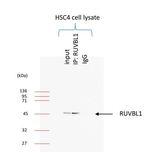
Immunoprecipitation: RUVBL1 Antibody [NBP2-20245]
Immunoprecipitation: RUVBL1 Antibody [NBP2-20245] - Immunoprecipitation of RUVBL protein from Jurkat whole cell extracts using 5 ug of RUVBL1 antibody. Western blot analysis was performed using RUVBL1 antibody. EasyBlot anti-Rabbit IgG was used as a secondary reagent.
Western Blot: RUVBL1 Antibody [NBP2-20245] -
RUVBL1/2 do not effect EBOV minigenome activity. (A) Schematic diagram of the EBOV minigenome system. The EBOV minigenome system consists of six plasmids: Four support plasmids encode replication complex components NP, L, VP35, & VP30. The EBOV minigenome plasmid encodes a firefly luciferase reporter gene flanked by the leader & trailer sequences of EBOV, & the plasmid that encodes Renilla luciferase is used for normalization. (B) Minigenome activity upon the knockdown of either RUVBL1, RUVBL2, or in combination. Below are protein levels confirmed by immunoblot. HeLa cells were transfected with 80 nM scrambled siRNA, 30 nM siRNA targeting RUVBL1, or 50 nM siRNA targeting RUVBL2. Twenty-four h after siRNA addition, the minigenome components were transfected. Forty-eight h later, minigenome reporter activity was measured. (C) Overexpression of FLAG-RUVBL1 & HA-RUVBL2 in the EBOV minigenome. HeLa cells were left untransfected, or transfected with vector control (VC), or increasing amounts of FLAG-RUVBL1 (125, 250, & 500 ng) or HA-RUVBL2 (125, 250, & 500 ng). Twenty-four h after exogenous transfection, the minigenome components were transfected. Forty-eight h later, minigenome reporter activity was measured. Data represent mean ± SEM from one representative experiment (n = 3) of at least three experiments (* p < 0.05). Image collected & cropped by CiteAb from the following publication (https://pubmed.ncbi.nlm.nih.gov/31018511), licensed under a CC-BY license. Not internally tested by Novus Biologicals.
Immunocytochemistry/ Immunofluorescence: RUVBL1 Antibody [NBP2-20245] -
Immunocytochemistry/ Immunofluorescence: RUVBL1 Antibody [NBP2-20245] - Endogenous RUVBL1 & RUVBL2 colocalize with HA-NP. HeLa cells were transfected with vector control, HA-NP, or HA-VP35. Twenty-four h later, the cells were fixed & processed for immunofluorescence detection of endogenous RUVBL1 or RUVBL2 in the presence of vector control, HA-NP, or HA-VP35. Representative images of (A) endogenous RUVBL1 localization pattern with control vector (top panels), HA-NP (middle panels), or HA-VP35 (bottom panels) & (B) endogenous RUVBL2 localization pattern with control vector (top panels), HA-NP (middle panels), or HA-VP35 (bottom panels) are shown. HA-NP or HA-VP35 (green), RUVBL1/2 (red), & Hoechst 33342 nuclear stain (blue) were visualized by confocal microscopy. Scale bars = 20 µM. Image collected & cropped by CiteAb from the following publication (https://pubmed.ncbi.nlm.nih.gov/31018511), licensed under a CC-BY license. Not internally tested by Novus Biologicals.
Western Blot: RUVBL1 Antibody [NBP2-20245] -
Western Blot: RUVBL1 Antibody [NBP2-20245] - Various whole cell extracts (30 ug) were separated by 10% SDS-PAGE, and the membrane was blotted with RUVBL1 antibody (NBP2-20245) diluted at 1:1000.
Western Blot: RUVBL1 Antibody [NBP2-20245] -
Mouse tissue extract (50 ug) was separated by 10% SDS-PAGE, and the membrane was blotted with RUVBL1 antibody (NBP2-20245) diluted at 1:1000. The HRP-conjugated anti-rabbit IgG antibody was used to detect the primary antibody.
Western Blot: RUVBL1 Antibody [NBP2-20245] -
Non-transfected (-) and transfected (+) 293T whole cell extracts (30 ug) were separated by 10% SDS-PAGE, and the membrane was blotted with RUVBL1 antibody (NBP2-20245) diluted at 1:5000. The HRP-conjugated anti-rabbit IgG antibody was used to detect the primary antibody.Applications for RUVBL1 Antibody
Application
Recommended Usage
Immunocytochemistry/ Immunofluorescence
1:100-1:1000
Immunohistochemistry
1:100-1:1000
Immunohistochemistry-Paraffin
1:100-1:1000
Immunoprecipitation
1:100-1:500
Knockdown Validated
Reported in scientific literature (PMID: 31018511).
Western Blot
1:500-1:3000
Reviewed Applications
Read 2 reviews rated 4.5 using NBP2-20245 in the following applications:
Formulation, Preparation, and Storage
Purification
Antigen Affinity-purified
Formulation
PBS, 20% Glycerol
Preservative
0.025% Proclin 300
Concentration
Concentrations vary lot to lot. See vial label for concentration. If unlisted please contact technical services.
Shipping
The product is shipped with polar packs. Upon receipt, store it immediately at the temperature recommended below.
Stability & Storage
Aliquot and store at -20C or -80C. Avoid freeze-thaw cycles.
Background: RUVBL1
Alternate Names
49 kDa TATA box-binding protein-interacting protein, 54 kDa erythrocyte cytosolic protein, EC 3.6.1, EC 3.6.4.12,49 kDa TBP-interacting protein, ECP54, INO80 complex subunit H, INO80HTIP49A, NMP 238, NMP238ECP-54, Nuclear matrix protein 238, PONTIN, Pontin 52, Pontin52, RuvB (E coli homolog)-like 1, ruvB-like 1, RuvB-like 1 (E. coli), RVB1, TATA binding protein interacting protein 49 kDa, TIH1, TIP49a, TIP49TAP54-alpha, TIP60-associated protein 54-alpha
Gene Symbol
RUVBL1
UniProt
Additional RUVBL1 Products
Product Documents for RUVBL1 Antibody
Certificate of Analysis
To download a Certificate of Analysis, please enter a lot or batch number in the search box below.
Product Specific Notices for RUVBL1 Antibody
This product is for research use only and is not approved for use in humans or in clinical diagnosis. Primary Antibodies are guaranteed for 1 year from date of receipt.
Citations for RUVBL1 Antibody
 Powered by Bioz
Customer Reviews for RUVBL1 Antibody (2)
4.5 out of 5
2 Customer Ratings
Have you used RUVBL1 Antibody?
Submit a review and receive an Amazon gift card!
$25/€18/£15/$25CAN/¥2500 Yen for a review with an image
$10/€7/£6/$10CAN/¥1110 Yen for a review without an image
Submit a review
Customer Images
Showing
1
-
2 of
2 reviews
Showing All
Filter By:
-
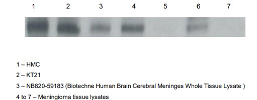
Application: Western BlotSample Tested: Human Brain Whole Tissue Lysate (Adult Normal), Novus NB820-59177Species: HumanVerified Customer | Posted 09/30/2019These have been attached. The protein content of all samples were quantified prior to loading, and standardized at concentrations of 40ug.

-
Application: ImmunoprecipitationSample Tested: Cancer Cell lysateSpecies: HumanVerified Customer | Posted 11/30/2017
There are no reviews that match your criteria.
Protocols
Find general support by application which include: protocols, troubleshooting, illustrated assays, videos and webinars.
- Antigen Retrieval Protocol (PIER)
- Antigen Retrieval for Frozen Sections Protocol
- Appropriate Fixation of IHC/ICC Samples
- Cellular Response to Hypoxia Protocols
- Chromogenic IHC Staining of Formalin-Fixed Paraffin-Embedded (FFPE) Tissue Protocol
- Chromogenic Immunohistochemistry Staining of Frozen Tissue
- ClariTSA™ Fluorophore Kits
- Detection & Visualization of Antibody Binding
- Fluorescent IHC Staining of Frozen Tissue Protocol
- Graphic Protocol for Heat-induced Epitope Retrieval
- Graphic Protocol for the Preparation and Fluorescent IHC Staining of Frozen Tissue Sections
- Graphic Protocol for the Preparation and Fluorescent IHC Staining of Paraffin-embedded Tissue Sections
- Graphic Protocol for the Preparation of Gelatin-coated Slides for Histological Tissue Sections
- ICC Cell Smear Protocol for Suspension Cells
- ICC Immunocytochemistry Protocol Videos
- ICC for Adherent Cells
- IHC Sample Preparation (Frozen sections vs Paraffin)
- Immunocytochemistry (ICC) Protocol
- Immunocytochemistry Troubleshooting
- Immunofluorescence of Organoids Embedded in Cultrex Basement Membrane Extract
- Immunofluorescent IHC Staining of Formalin-Fixed Paraffin-Embedded (FFPE) Tissue Protocol
- Immunohistochemistry (IHC) and Immunocytochemistry (ICC) Protocols
- Immunohistochemistry Frozen Troubleshooting
- Immunohistochemistry Paraffin Troubleshooting
- Immunoprecipitation Protocol
- Preparing Samples for IHC/ICC Experiments
- Preventing Non-Specific Staining (Non-Specific Binding)
- Primary Antibody Selection & Optimization
- Protocol for Heat-Induced Epitope Retrieval (HIER)
- Protocol for Making a 4% Formaldehyde Solution in PBS
- Protocol for VisUCyte™ HRP Polymer Detection Reagent
- Protocol for the Fluorescent ICC Staining of Cell Smears - Graphic
- Protocol for the Fluorescent ICC Staining of Cultured Cells on Coverslips - Graphic
- Protocol for the Preparation & Fixation of Cells on Coverslips
- Protocol for the Preparation and Chromogenic IHC Staining of Frozen Tissue Sections
- Protocol for the Preparation and Chromogenic IHC Staining of Frozen Tissue Sections - Graphic
- Protocol for the Preparation and Chromogenic IHC Staining of Paraffin-embedded Tissue Sections
- Protocol for the Preparation and Chromogenic IHC Staining of Paraffin-embedded Tissue Sections - Graphic
- Protocol for the Preparation and Fluorescent ICC Staining of Cells on Coverslips
- Protocol for the Preparation and Fluorescent ICC Staining of Non-adherent Cells
- Protocol for the Preparation and Fluorescent ICC Staining of Stem Cells on Coverslips
- Protocol for the Preparation and Fluorescent IHC Staining of Frozen Tissue Sections
- Protocol for the Preparation and Fluorescent IHC Staining of Paraffin-embedded Tissue Sections
- Protocol for the Preparation of Gelatin-coated Slides for Histological Tissue Sections
- Protocol for the Preparation of a Cell Smear for Non-adherent Cell ICC - Graphic
- R&D Systems Quality Control Western Blot Protocol
- TUNEL and Active Caspase-3 Detection by IHC/ICC Protocol
- The Importance of IHC/ICC Controls
- Troubleshooting Guide: Immunohistochemistry
- Troubleshooting Guide: Western Blot Figures
- Western Blot Conditions
- Western Blot Protocol
- Western Blot Protocol for Cell Lysates
- Western Blot Troubleshooting
- Western Blot Troubleshooting Guide
- View all Protocols, Troubleshooting, Illustrated assays and Webinars
Loading...