SARS Nucleocapsid Protein Antibody - BSA Free
Novus Biologicals | Catalog # NB100-56576
![Immunohistochemistry: SARS Nucleocapsid Protein Antibody - BSA Free [NB100-56576]](https://resources.rndsystems.com/images/products/SARS-Nucleocapsid-Protein-Antibody-Immunohistochemistry-NB100-56576-img0005.jpg "Immunohistochemistry: SARS Nucleocapsid Protein Antibody - BSA Free [NB100-56576]")
Key Product Details
Validated by
Species Reactivity
Validated:
Cited:
Applications
Validated:
Cited:
Label
Antibody Source
Format
Product Specifications
Immunogen
Reactivity Notes
Specificity
Clonality
Host
Isotype
Scientific Data Images for SARS Nucleocapsid Protein Antibody - BSA Free
Immunohistochemistry: SARS Nucleocapsid Protein Antibody - BSA Free [NB100-56576]
Immunohistochemistry: SARS Nucleocapsid Protein Antibody [NB100-56576] - Immunostaining of severe acute respiratory syndrome coronavirus 2 in pulmonary tissues from fatal coronavirus disease cases. A) P5 (Patient 5): scattered immunostaining of tracheal epithelial cells. B) P5: higher magnification shows immunostaining of ciliated cells. C) P8: immunostaining of desquamated type I pneumocyte in an alveolar lumen. D) P4: colocalization of SARS-CoV-2 viral antigen (red) with type II pneumocyte stained by surfactant (brown; arrow). E) P4: colocalization of SARS-CoV-2 viral antigen (red) with macrophages stained by CD163 (brown; arrows); virus immunostaining within type II pneumocytes is also seen (arrowheads). F) P4: extensive immunostaining of hyaline membranes in a region of exudative DAD. G) P3: scattered immunostaining within macrophage in hilar lymph node; anthracosis is also present. Emerg Infect Dis. 2020 May 21;26(9) 10.3201/eid2609.202095, PMID: 32437316![Immunohistochemistry: SARS Nucleocapsid Protein Antibody - BSA Free [NB100-56576]](https://resources.rndsystems.com/images/products/SARS-Nucleocapsid-Protein-Antibody-Immunohistochemistry-NB100-56576-img0004.jpg "Immunohistochemistry: SARS Nucleocapsid Protein Antibody - BSA Free [NB100-56576]")
Immunohistochemistry: SARS Nucleocapsid Protein Antibody - BSA Free [NB100-56576]
Immunohistochemistry: SARS Nucleocapsid Protein Antibody [NB100-56576] - Pathological changes in rhesus macaques infected with SARS-CoV-2. (g) SARSCoV-2 antigen is detected by immunohistochemistry in type I pneumocytes. Magnification 400x. (j) SARS-CoV-2 antigen is detected by immunohistochemistry in type I pneumocytes (asterisk) and type II pneumocytes (arrow) as well as alveolar macrophages (arrowheads). Magnification 400x. (k) SARS-CoV-2 antigen is detected by immunohistochemistry in mediastinal lymph node. Magnification 400x. (l) SARSCoV-2 antigen is detected by immunohistochemistry in macrophages and lymphocytes in the lamina propria of the cecum. Magnification 400x. bioRxiv March 21, 2020 https://doi.org/10.1101/2020.03.21.001628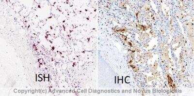
![Immunohistochemistry: SARS Nucleocapsid Protein Antibody - BSA Free [NB100-56576]](https://resources.rndsystems.com/images/products/SARS-Nucleocapsid-Protein-Antibody-Immunohistochemistry-NB100-56576-img0003.jpg "Immunohistochemistry: SARS Nucleocapsid Protein Antibody - BSA Free [NB100-56576]")
Immunohistochemistry: SARS Nucleocapsid Protein Antibody - BSA Free [NB100-56576]
SARS-Nucleocapsid-Protein-Antibody-Immunohistochemistry-NB100-56576-img0003.jpg![Western Blot: SARS Nucleocapsid Protein AntibodyBSA Free [NB100-56576]](https://resources.rndsystems.com/images/products/SARS-Nucleocapsid-Protein-Antibody-Western-Blot-NB100-56576-img0007.jpg "Western Blot: SARS Nucleocapsid Protein AntibodyBSA Free [NB100-56576]")
Western Blot: SARS Nucleocapsid Protein AntibodyBSA Free [NB100-56576]
Western Blot: SARS Nucleocapsid Protein Antibody [NB100-56576] - Western blot shows recombinant SARS-CoV-2 Nucleocapsid protein. PVDF membrane was probed with 1 ug/mL of Rabbit Anti-SARS-CoV-2 Nucleocapsid Polyclonal Antibody (Catalog # NB100-56576) followed by HRP-conjugated Anti-Rabbit IgG Secondary Antibody (HAF008). A specific band was detected for SARS-CoV-2 Nucleocapsid at approximately 55 kDa (as indicated). This experiment was conducted under reducing conditions and using Western Blot Buffer Group 1![Western Blot: SARS Nucleocapsid Protein AntibodyBSA Free [NB100-56576]](https://resources.rndsystems.com/images/products/SARS-Nucleocapsid-Protein-Antibody-Western-Blot-NB100-56576-img0002.jpg "Western Blot: SARS Nucleocapsid Protein AntibodyBSA Free [NB100-56576]")
Western Blot: SARS Nucleocapsid Protein AntibodyBSA Free [NB100-56576]
Western Blot: SARS Nucleocapsid Protein Antibody [NB100-56576] - analysis of SARS Nucleocapsid in (A) untransfected mouse melanoma cell lysate and (B) transfected cell lysate using this antibody.
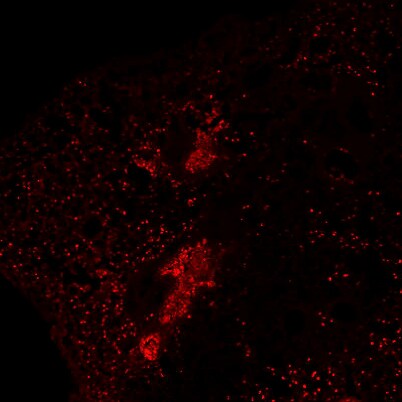
Immunohistochemistry-Frozen: Rabbit Polyclonal SARS Nucleocapsid Protein Antibody [NB100-56576]
Immunohistochemistry-Frozen: Rabbit Polyclonal SARS Nucleocapsid Protein Antibody [NB100-56576] - Immunostaining of mouse adult lung infected with SARS-CoV-2. Image from a verified customer review.Applications for SARS Nucleocapsid Protein Antibody - BSA Free
Immunocytochemistry/ Immunofluorescence
Immunohistochemistry
Immunohistochemistry-Frozen
Immunohistochemistry-Paraffin
SDS-PAGE
Western Blot
Reviewed Applications
Read 1 review rated 4 using NB100-56576 in the following applications:
Formulation, Preparation, and Storage
Purification
Formulation
Format
Preservative
Concentration
Shipping
Stability & Storage
Background: SARS Nucleocapsid Protein
Alternate Names
Gene Symbol
Additional SARS Nucleocapsid Protein Products
Product Documents for SARS Nucleocapsid Protein Antibody - BSA Free
Certificate of Analysis
To download a Certificate of Analysis, please enter a lot or batch number in the search box below.
Product Specific Notices for SARS Nucleocapsid Protein Antibody - BSA Free
This product is for research use only and is not approved for use in humans or in clinical diagnosis. Primary Antibodies are guaranteed for 1 year from date of receipt.
Citations for SARS Nucleocapsid Protein Antibody - BSA Free
 Powered by Bioz
Customer Reviews for SARS Nucleocapsid Protein Antibody - BSA Free (1)
Have you used SARS Nucleocapsid Protein Antibody - BSA Free?
Submit a review and receive an Amazon gift card!
$25/€18/£15/$25CAN/¥2500 Yen for a review with an image
$10/€7/£6/$10CAN/¥1110 Yen for a review without an image
Submit a review
Customer Images
-
Application: Immunohistochemistry-FrozenSample Tested: Adult lungSpecies: MouseVerified Customer | Posted 04/09/2025Immunostaining of Lung infected with SARS-CoV-2

There are no reviews that match your criteria.
Protocols
Find general support by application which include: protocols, troubleshooting, illustrated assays, videos and webinars.
- Antigen Retrieval Protocol (PIER)
- Antigen Retrieval for Frozen Sections Protocol
- Appropriate Fixation of IHC/ICC Samples
- Cellular Response to Hypoxia Protocols
- Chromogenic IHC Staining of Formalin-Fixed Paraffin-Embedded (FFPE) Tissue Protocol
- Chromogenic Immunohistochemistry Staining of Frozen Tissue
- ClariTSA™ Fluorophore Kits
- Detection & Visualization of Antibody Binding
- Fluorescent IHC Staining of Frozen Tissue Protocol
- Graphic Protocol for Heat-induced Epitope Retrieval
- Graphic Protocol for the Preparation and Fluorescent IHC Staining of Frozen Tissue Sections
- Graphic Protocol for the Preparation and Fluorescent IHC Staining of Paraffin-embedded Tissue Sections
- Graphic Protocol for the Preparation of Gelatin-coated Slides for Histological Tissue Sections
- ICC Cell Smear Protocol for Suspension Cells
- ICC Immunocytochemistry Protocol Videos
- ICC for Adherent Cells
- IHC Sample Preparation (Frozen sections vs Paraffin)
- ISH-IHC Protocol for Chromogenic Detection on Formalin Fixed Paraffin Embedded (FFPE) Tissue
- Immunocytochemistry (ICC) Protocol
- Immunocytochemistry Troubleshooting
- Immunofluorescence of Organoids Embedded in Cultrex Basement Membrane Extract
- Immunofluorescent IHC Staining of Formalin-Fixed Paraffin-Embedded (FFPE) Tissue Protocol
- Immunohistochemistry (IHC) and Immunocytochemistry (ICC) Protocols
- Immunohistochemistry Frozen Troubleshooting
- Immunohistochemistry Paraffin Troubleshooting
- Preparing Samples for IHC/ICC Experiments
- Preventing Non-Specific Staining (Non-Specific Binding)
- Primary Antibody Selection & Optimization
- Protocol for Heat-Induced Epitope Retrieval (HIER)
- Protocol for Making a 4% Formaldehyde Solution in PBS
- Protocol for VisUCyte™ HRP Polymer Detection Reagent
- Protocol for the Fluorescent ICC Staining of Cell Smears - Graphic
- Protocol for the Fluorescent ICC Staining of Cultured Cells on Coverslips - Graphic
- Protocol for the Preparation & Fixation of Cells on Coverslips
- Protocol for the Preparation and Chromogenic IHC Staining of Frozen Tissue Sections
- Protocol for the Preparation and Chromogenic IHC Staining of Frozen Tissue Sections - Graphic
- Protocol for the Preparation and Chromogenic IHC Staining of Paraffin-embedded Tissue Sections
- Protocol for the Preparation and Chromogenic IHC Staining of Paraffin-embedded Tissue Sections - Graphic
- Protocol for the Preparation and Fluorescent ICC Staining of Cells on Coverslips
- Protocol for the Preparation and Fluorescent ICC Staining of Non-adherent Cells
- Protocol for the Preparation and Fluorescent ICC Staining of Stem Cells on Coverslips
- Protocol for the Preparation and Fluorescent IHC Staining of Frozen Tissue Sections
- Protocol for the Preparation and Fluorescent IHC Staining of Paraffin-embedded Tissue Sections
- Protocol for the Preparation of Gelatin-coated Slides for Histological Tissue Sections
- Protocol for the Preparation of a Cell Smear for Non-adherent Cell ICC - Graphic
- R&D Systems Quality Control Western Blot Protocol
- TUNEL and Active Caspase-3 Detection by IHC/ICC Protocol
- The Importance of IHC/ICC Controls
- Troubleshooting Guide: Immunohistochemistry
- Troubleshooting Guide: Western Blot Figures
- Western Blot Conditions
- Western Blot Protocol
- Western Blot Protocol for Cell Lysates
- Western Blot Troubleshooting
- Western Blot Troubleshooting Guide
- View all Protocols, Troubleshooting, Illustrated assays and Webinars
FAQs for SARS Nucleocapsid Protein Antibody - BSA Free
-
Q: does your antibody NB100-56576 recognizes SARS-CoV-2 or only SARS-Cov? (Image 1 indicates Cov2 in the legend, but the data sheet only states SARS coronavirus
A:
This antibody was originally developed against SARS, but due to identity to SARS-CoV-2 was predicted to cross-react. the image in question (#1) is from a recent publication that used NB100-56576 to detect SARS-CoV-2 nucleocapsid. Here is the link to the publication if you would like to take a look https://www.biorxiv.org/content/10.1101/2020.03.21.001628v1