SCP1 Antibody - BSA Free
Novus Biologicals | Catalog # NB300-229
![Simple Western: SCP1 AntibodyBSA Free [NB300-229]](https://resources.rndsystems.com/images/products/SCP1-Antibody-Simple-Western-NB300-229-img0010.jpg "Simple Western: SCP1 AntibodyBSA Free [NB300-229]")
Key Product Details
Species Reactivity
Validated:
Mouse, Rat, Chicken, Human (Negative), Mammal, Monkey, Parasite
Cited:
Human, Mouse, Rat, Mammal, Parasite, Primate - Macaca mulatta (Rhesus Macaque)
Applications
Validated:
Immunohistochemistry, Immunohistochemistry-Paraffin, Immunohistochemistry-Frozen, Western Blot, Immunocytochemistry/ Immunofluorescence, Simple Western, Immunoprecipitation, Chromatin Immunoprecipitation, Chromatin Immunoprecipitation (ChIP)
Cited:
Immunohistochemistry, Immunohistochemistry-Paraffin, Western Blot, Immunocytochemistry/ Immunofluorescence, Immunoprecipitation, Chemotaxis, IF/IHC
Label
Unconjugated
Antibody Source
Polyclonal Rabbit IgG
Format
BSA Free
Loading...
Product Specifications
Immunogen
A synthetic peptide made to the C-terminus of the mouse SCP1 protein sequence. [UniProt# Q62209]
Reactivity Notes
Mammal reactivity reported in scientific literature (PMID: 25981592). Parasite reactivity reported in scientific literature (PMID: 27084479). Chicken reactivity reported in scientific literature (PMID: 28174243). Use in Monkey reported in scientific literature (PMID: 31907447). This antibody has not been shown to have human reactivity.
Localization
Nuclear
Clonality
Polyclonal
Host
Rabbit
Isotype
IgG
Scientific Data Images for SCP1 Antibody - BSA Free
Simple Western: SCP1 AntibodyBSA Free [NB300-229]
Simple Western: SCP1 Antibody [NB300-229] - Simple Western lane view shows a specific band for SCP1 in 0.5 mg/ml of Human Testis (left) and Mouse Testis (right) lysate. This experiment was performed under reducing conditions using the 12-230 kDa separation system.![Western Blot: SCP1 AntibodyBSA Free [NB300-229]](https://resources.rndsystems.com/images/products/SCP1-Antibody-Western-Blot-NB300-229-img0014.jpg "Western Blot: SCP1 AntibodyBSA Free [NB300-229]")
Western Blot: SCP1 AntibodyBSA Free [NB300-229]
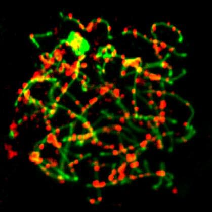
Western Blot: SCP1 Antibody [NB300-229] - Meiotic chromosome spreads from wild type and Fance mutant (MT) primary spermatocytes isolated from the testes at 20 dpp. Each chromatin spread was staged by analyzing SYCP3 (red) and SYCP1 (green), which are axial element and central region components of the synaptonemal complex respectively. Most of the Fance mutant spermatocytes displayed normal synapsis (middle panels); however a sub-set displayed synapsis abnormalities including an association between non-homologous chromosome ends (arrow head) and abnormal SYCP3 structures (arrows). Dearth and Delayed Maturation of Testicular Germ Cells in Fanconi Anemia E Mutant Male Mice. PLoS One (2016)![Immunocytochemistry/ Immunofluorescence: SCP1 Antibody - BSA Free [NB300-229]](https://resources.rndsystems.com/images/products/SCP1-Antibody---BSA-Free-Immunocytochemistry-Immunofluorescence-NB300-229-img0017.jpg "Immunocytochemistry/ Immunofluorescence: SCP1 Antibody - BSA Free [NB300-229]")
Immunocytochemistry/ Immunofluorescence: SCP1 Antibody - BSA Free [NB300-229]
SCP1-Antibody---BSA-Free-Immunocytochemistry-Immunofluorescence-NB300-229-img0017.jpg![Immunohistochemistry-Paraffin: SCP1 Antibody - BSA Free [NB300-229]](https://resources.rndsystems.com/images/products/SCP1-Antibody-Immunohistochemistry-Paraffin-NB300-229-img0008.jpg "Immunohistochemistry-Paraffin: SCP1 Antibody - BSA Free [NB300-229]")
Immunohistochemistry-Paraffin: SCP1 Antibody - BSA Free [NB300-229]
Immunohistochemistry-Paraffin: SCP1 Antibody [NB300-229] - Punctate staining of murine SCP1 in mouse ovary using NB300-229.![Immunocytochemistry/ Immunofluorescence: SCP1 Antibody - BSA Free [NB300-229]](https://resources.rndsystems.com/images/products/SCP1-Antibody-Immunocytochemistry-Immunofluorescence-NB300-229-img0007.jpg "Immunocytochemistry/ Immunofluorescence: SCP1 Antibody - BSA Free [NB300-229]")
Immunocytochemistry/ Immunofluorescence: SCP1 Antibody - BSA Free [NB300-229]
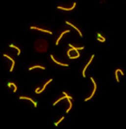
Immunocytochemistry/Immunofluorescence: SCP1 Antibody [NB300-229] - SCP1 labeled in mouse pachytene preparation (red), using NB300-229. CDK2 staining, near teleomeres, is also present (green).![Immunocytochemistry/ Immunofluorescence: SCP1 Antibody - BSA Free [NB300-229]](https://resources.rndsystems.com/images/products/SCP1-Antibody-Immunocytochemistry-Immunofluorescence-NB300-229-img0012.jpg "Immunocytochemistry/ Immunofluorescence: SCP1 Antibody - BSA Free [NB300-229]")
Immunocytochemistry/ Immunofluorescence: SCP1 Antibody - BSA Free [NB300-229]
Immunocytochemistry/Immunofluorescence: SCP1 Antibody [NB300-229] - Spermatocytes cells fixed in PFA. Detected with anti-mouse 594. ICC/IF image submitted by a verified customer review.![Immunocytochemistry/ Immunofluorescence: SCP1 Antibody - BSA Free [NB300-229]](https://resources.rndsystems.com/images/products/SCP1-Antibody-Immunocytochemistry-Immunofluorescence-NB300-229-img0013.jpg "Immunocytochemistry/ Immunofluorescence: SCP1 Antibody - BSA Free [NB300-229]")
Immunocytochemistry/ Immunofluorescence: SCP1 Antibody - BSA Free [NB300-229]
Immunocytochemistry/Immunofluorescence: SCP1 Antibody [NB300-229] - Mouse spermatozoa. Green: SCP1 staining. ICC/IF image submitted by a verified customer review.![Immunocytochemistry/ Immunofluorescence: SCP1 Antibody - BSA Free [NB300-229]](https://resources.rndsystems.com/images/products/SCP1-Antibody-Immunocytochemistry-Immunofluorescence-NB300-229-img0015.jpg "Immunocytochemistry/ Immunofluorescence: SCP1 Antibody - BSA Free [NB300-229]")
Immunocytochemistry/ Immunofluorescence: SCP1 Antibody - BSA Free [NB300-229]
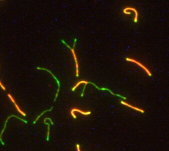
Immunocytochemistry/Immunofluorescence: SCP1 Antibody [NB300-229] - Mouse spermatocyte. Red: scp1 colocalized with scp3 (green). ICC/IF image submitted by a verified customer review.![Immunocytochemistry/ Immunofluorescence: SCP1 Antibody - BSA Free [NB300-229]](https://resources.rndsystems.com/images/products/SCP1-Antibody---BSA-Free-Immunocytochemistry-Immunofluorescence-NB300-229-img0016.jpg "Immunocytochemistry/ Immunofluorescence: SCP1 Antibody - BSA Free [NB300-229]")
Immunocytochemistry/ Immunofluorescence: SCP1 Antibody - BSA Free [NB300-229]
SCP1-Antibody---BSA-Free-Immunocytochemistry-Immunofluorescence-NB300-229-img0016.jpg
Chromatin Immunoprecipitation: SCP1 Antibody - BSA Free [NB300-229] -
Chromatin Immunoprecipitation: SCP1 Antibody - BSA Free [NB300-229] - Meiotic spreads.Meiotic chromosome spreads from wild type & Fance mutant (MT) primary spermatocytes isolated from the testes at 20 dpp. Each chromatin spread was staged by analyzing SYCP3 (red) & SYCP1 (green), which are axial element & central region components of the synaptonemal complex respectively. Most of the Fance mutant spermatocytes displayed normal synapsis (middle panels); however a sub-set displayed synapsis abnormalities including an association between non-homologous chromosome ends (arrow head) & abnormal SYCP3 structures (arrows). Image collected & cropped by CiteAb from the following publication (https://dx.plos.org/10.1371/journal.pone.0159800), licensed under a CC-BY license. Not internally tested by Novus Biologicals.
Immunocytochemistry/ Immunofluorescence: SCP1 Antibody - BSA Free [NB300-229] -
Immunocytochemistry/ Immunofluorescence: SCP1 Antibody - BSA Free [NB300-229] - Meiotic progression & chromosome paring in XX, XO & XY oocytes.(A) Meiotic progression in XX, XO & XY oocytes. The graphs show the percentages of the meiotic stage at the day of culture indicated. L, leptotene; Z, zygotene; P, pachytene; D, diplotene. (B) Pairing of homologous chromosomes in XX, XO & XY oocytes. Images show the immunofluorescence analysis of SYCP3 (white) & SYCP1 (red), & FISH analysis of the X chromosome (green) & Y chromosome (purple). The dashed squares in the merged images are shown at high magnification (right). The numbers of samples showing the phenotype are shown with the total number tested (left). Arrowheads indicate asynapsed bivalents at the end of the chromosomes. Scale bars, 10 μm. (C) Pairing rates of autosomes & sex chromosomes. Each value was calculated from three independent experiments (see also Materials & Methods). P values were calculated by Tukey’s HSD test. ***P<0.001, **P<0.01, *P<0.05; NS, not significant. Image collected & cropped by CiteAb from the following publication (https://pubmed.ncbi.nlm.nih.gov/32214314), licensed under a CC-BY license. Not internally tested by Novus Biologicals.
Immunocytochemistry/ Immunofluorescence: SCP1 Antibody - BSA Free [NB300-229] -
Immunocytochemistry/ Immunofluorescence: SCP1 Antibody - BSA Free [NB300-229] - Meiotic prophase I progression & apoptosis in spermatocytes in ex vivo culture at different temperatures.a Representative images of chromosomal spreads at different stages of meiotic prophase I, prepared from in vivo-developed testes of 5-week-old Acr-GFP mice & from testis explants cultured at 34, 37, or 38 °C for 5 weeks as indicated. Samples were stained for SCP3, SCP1, & DNA (Hoechst 33342). Scale bars, 10 μm. b Proportions of spermatocytes in the leptotene (lep), zygotene (zygo), pachytene (pachy), & diplotene (diplo) stages, found in explants cultured for 5 weeks at the indicated temperatures classified visually on the chromosome spreads after immunofluorescence-staining for SCP1 & SCP3 as (a), according to the criteria described in the text. Values obtained from chromosome spreads prepared from pooled testicular cells of two in vivo-developed testes from different individuals & those from 6 to 7 ex vivo-grown explants are summarized. The total number of spermatocyte nuclei counted is indicated at the upper right of each panel. Percentages of spermatocytes nuclei in which all the autosomes have completed synapsis (i.e., the sum of pachytene & diplotene spermatocytes) were 63, 52, 16, & 0.5% for in vivo & ex vivo samples at 34, 37, & 38 °C, respectively. c Detection of cleaved Caspase-3 (red) & SCP3 (green) in testis explants following the temperature shift from 34 to 38 °C. Double-stained images overlaid with DNA staining (gray) & the signals for cleaved Caspase-3 alone are shown in the upper & lower panels, respectively. Enlarged images at positions indicated by rectangles are also shown below. Yellow arrowheads, Caspase-3+/SCP3+ double-positive cells (the dying spermatocytes). Scale bars, 40 μm. Image collected & cropped by CiteAb from the following publication (https://pubmed.ncbi.nlm.nih.gov/35618762), licensed under a CC-BY license. Not internally tested by Novus Biologicals.
Immunocytochemistry/ Immunofluorescence: SCP1 Antibody - BSA Free [NB300-229] -
Immunocytochemistry/ Immunofluorescence: SCP1 Antibody - BSA Free [NB300-229] - No major meiotic DSB repair or chromosome synapsis defects are observed in Cxxc1 CKO testis.(A) Immunostaining of SYCP3 & gamma H2AX on adult Cxxc1 het & CKO chromosome spreads. Green, SYCP3; magenta, gamma H2AX. Scale bar, 10 μm. (B) Spermatocyte stage proportion in adult Cxxc1 het (n = 1,062 from two individuals) & CKO (n = 1,105 from two individuals) spermatocytes based on SYCP3/SYCP1/ gamma H2AX staining. p = 0.7 by Chi-square test. (C) Immunostaining of SYCP3 & SYCP1 on adult Cxxc1 het & CKO chromosome spreads. Green, SYCP3; orange, SYCP1. Scale bar, 10 μm. (D) Crossover number measured by MLH1 staining on chromosome spreads of adult Cxxc1 het & CKO spermatocytes. Left, magenta, SYCP3; green, MLH1. Scale bar, 10 μm. Right, number of MLH1 foci per late pachynema in Cxxc1 het (n = 32 from two individuals) & CKO (n = 33 from two individuals). Bars represent mean ± SD. p = 0.4 by Student’s t-test. Image collected & cropped by CiteAb from the following publication (https://pubmed.ncbi.nlm.nih.gov/30365547), licensed under a CC-BY license. Not internally tested by Novus Biologicals.
Immunocytochemistry/ Immunofluorescence: SCP1 Antibody - BSA Free [NB300-229] -
Immunocytochemistry/ Immunofluorescence: SCP1 Antibody - BSA Free [NB300-229] - UHRF1 deletion disrupted the meiotic progression & synaptonemal complex assembly.a Relative amounts of four spermatocyte populations (leptotene stage, zygotene stage, pachytene stage, & diplotene stage) during the prophase I in testes based on analyzing >600 spermatocytes in each stage. b, c The immunostaining of SYCP3 in the testicular sections (b) & surface-spread chromatin preparations of Uhrf1 deletion & control mice (c); d the percentage of spermatocytes with abnormal SYCP3 location. e Double immunofluorescence of testicular spread preparations of the adult mice, SYCP3 (green) & SYCP1 (red). f The percentage of spermatocytes with abnormal SYCP1 location. Lep leptotene, Zyg zygotene, Pac pachytene, Dip diplotene. Data are presented as mean ± SEM of three mice. ***p ≤ 0.001. Scale bar, 25 μm in b, 5 μm in c, e. Image collected & cropped by CiteAb from the following publication (https://pubmed.ncbi.nlm.nih.gov/32081844), licensed under a CC-BY license. Not internally tested by Novus Biologicals.Applications for SCP1 Antibody - BSA Free
Application
Recommended Usage
Chromatin Immunoprecipitation
1:10 - 1:500. Use reported in scientific literature (PMID 27486799)
Chromatin Immunoprecipitation (ChIP)
1:10-1:500
Immunocytochemistry/ Immunofluorescence
1:100 - 1:750
Immunohistochemistry
1:750
Immunohistochemistry-Frozen
1:750
Immunohistochemistry-Paraffin
1:750
Simple Western
1:100
Application Notes
In Simple Western only 10 - 15 uL of the recommended dilution is used per data point.
See Simple Western Antibody Database for Simple Western validation: Tested in Human Testis and Mouse Testis lysate 0.5 mg/mL, separated by Size, antibody dilution of 1:100. Separated by Size-Wes, Sally Sue/Peggy Sue.
See Simple Western Antibody Database for Simple Western validation: Tested in Human Testis and Mouse Testis lysate 0.5 mg/mL, separated by Size, antibody dilution of 1:100. Separated by Size-Wes, Sally Sue/Peggy Sue.
Reviewed Applications
Read 3 reviews rated 5 using NB300-229 in the following applications:
Formulation, Preparation, and Storage
Purification
Immunogen affinity purified
Formulation
PBS
Format
BSA Free
Preservative
0.02% Sodium Azide
Concentration
1.0 mg/ml
Shipping
The product is shipped with polar packs. Upon receipt, store it immediately at the temperature recommended below.
Stability & Storage
Store at 4C short term. Aliquot and store at -20C long term. Avoid freeze-thaw cycles.
Background: SCP1
Alternate Names
Cancer/testis antigen 8, CT8MGC104417, HOM-TES-14, SCP1SCP-1, synaptonemal complex protein 1
Gene Symbol
SYCP1
Additional SCP1 Products
Product Documents for SCP1 Antibody - BSA Free
Certificate of Analysis
To download a Certificate of Analysis, please enter a lot or batch number in the search box below.
Product Specific Notices for SCP1 Antibody - BSA Free
This product is for research use only and is not approved for use in humans or in clinical diagnosis. Primary Antibodies are guaranteed for 1 year from date of receipt.
Citations for SCP1 Antibody - BSA Free
 Powered by Bioz
Customer Reviews for SCP1 Antibody - BSA Free (3)
5 out of 5
3 Customer Ratings
Have you used SCP1 Antibody - BSA Free?
Submit a review and receive an Amazon gift card!
$25/€18/£15/$25CAN/¥2500 Yen for a review with an image
$10/€7/£6/$10CAN/¥1110 Yen for a review without an image
Submit a review
Customer Images
Showing
1
-
3 of
3 reviews
Showing All
Filter By:
-
Application: ImmunocytochemistrySample Tested: spermatocyteSpecies: MouseVerified Customer | Posted 02/17/2021Red colour is scp1 colocalize with scp3(green)

-
Application: ImmunocytochemistrySample Tested: Mouse spermatozoaSpecies: MouseVerified Customer | Posted 09/12/2019green colour is scp1 staining. It's good for IF.
-
Application: ImmunocytochemistrySample Tested: SpermatocytesSpecies: MouseVerified Customer | Posted 04/11/2017Spermatocytes cells fixed in PFA. Detected with anti-mouse 594
There are no reviews that match your criteria.
Protocols
View specific protocols for SCP1 Antibody - BSA Free (NB300-229):
Immunofluorescence Procedure
1. Freshly prepared slides are soaked in 1X ADB for 75 minutes.
2. Primary antibodies are added concurrently (SCP1 and CDK2).
3. The primary antibodies are incubated overnight in a hudid chamber (37 degrees Celcius).
4. The slides are washed for 40 minutes in 1X ADB.
5. The slides are detected with the appropriate secondary antibodies (RDAR for SCP1 and FDAM for CDK2).
6. The slides are incubated for 4 hours in a humid chamber (37 degrees Celcius).
7. The slides are washed for 20 minutes in 1X ADB, followed by 3 washes, 10 minutes each, in 1X PBS.
8. The slides are counterstained with DAPI.
9. Images are captured after allowing the slides to remain in the dark overnight at RT.
Find general support by application which include: protocols, troubleshooting, illustrated assays, videos and webinars.
- Antigen Retrieval Protocol (PIER)
- Antigen Retrieval for Frozen Sections Protocol
- Appropriate Fixation of IHC/ICC Samples
- Cellular Response to Hypoxia Protocols
- ChIP Protocol Video
- Chromatin Immunoprecipitation (ChIP) Protocol
- Chromatin Immunoprecipitation Protocol
- Chromogenic IHC Staining of Formalin-Fixed Paraffin-Embedded (FFPE) Tissue Protocol
- Chromogenic Immunohistochemistry Staining of Frozen Tissue
- ClariTSA™ Fluorophore Kits
- Detection & Visualization of Antibody Binding
- Fluorescent IHC Staining of Frozen Tissue Protocol
- Graphic Protocol for Heat-induced Epitope Retrieval
- Graphic Protocol for the Preparation and Fluorescent IHC Staining of Frozen Tissue Sections
- Graphic Protocol for the Preparation and Fluorescent IHC Staining of Paraffin-embedded Tissue Sections
- Graphic Protocol for the Preparation of Gelatin-coated Slides for Histological Tissue Sections
- ICC Cell Smear Protocol for Suspension Cells
- ICC Immunocytochemistry Protocol Videos
- ICC for Adherent Cells
- IHC Sample Preparation (Frozen sections vs Paraffin)
- Immunocytochemistry (ICC) Protocol
- Immunocytochemistry Troubleshooting
- Immunofluorescence of Organoids Embedded in Cultrex Basement Membrane Extract
- Immunofluorescent IHC Staining of Formalin-Fixed Paraffin-Embedded (FFPE) Tissue Protocol
- Immunohistochemistry (IHC) and Immunocytochemistry (ICC) Protocols
- Immunohistochemistry Frozen Troubleshooting
- Immunohistochemistry Paraffin Troubleshooting
- Immunoprecipitation Protocol
- Preparing Samples for IHC/ICC Experiments
- Preventing Non-Specific Staining (Non-Specific Binding)
- Primary Antibody Selection & Optimization
- Protocol for Heat-Induced Epitope Retrieval (HIER)
- Protocol for Making a 4% Formaldehyde Solution in PBS
- Protocol for VisUCyte™ HRP Polymer Detection Reagent
- Protocol for the Fluorescent ICC Staining of Cell Smears - Graphic
- Protocol for the Fluorescent ICC Staining of Cultured Cells on Coverslips - Graphic
- Protocol for the Preparation & Fixation of Cells on Coverslips
- Protocol for the Preparation and Chromogenic IHC Staining of Frozen Tissue Sections
- Protocol for the Preparation and Chromogenic IHC Staining of Frozen Tissue Sections - Graphic
- Protocol for the Preparation and Chromogenic IHC Staining of Paraffin-embedded Tissue Sections
- Protocol for the Preparation and Chromogenic IHC Staining of Paraffin-embedded Tissue Sections - Graphic
- Protocol for the Preparation and Fluorescent ICC Staining of Cells on Coverslips
- Protocol for the Preparation and Fluorescent ICC Staining of Non-adherent Cells
- Protocol for the Preparation and Fluorescent ICC Staining of Stem Cells on Coverslips
- Protocol for the Preparation and Fluorescent IHC Staining of Frozen Tissue Sections
- Protocol for the Preparation and Fluorescent IHC Staining of Paraffin-embedded Tissue Sections
- Protocol for the Preparation of Gelatin-coated Slides for Histological Tissue Sections
- Protocol for the Preparation of a Cell Smear for Non-adherent Cell ICC - Graphic
- R&D Systems Quality Control Western Blot Protocol
- TUNEL and Active Caspase-3 Detection by IHC/ICC Protocol
- The Importance of IHC/ICC Controls
- Troubleshooting Guide: Immunohistochemistry
- Troubleshooting Guide: Western Blot Figures
- Western Blot Conditions
- Western Blot Protocol
- Western Blot Protocol for Cell Lysates
- Western Blot Troubleshooting
- Western Blot Troubleshooting Guide
- View all Protocols, Troubleshooting, Illustrated assays and Webinars
Loading...