SCP1 Antibody [Biotin]
Novus Biologicals | Catalog # NB300-229B
Key Product Details
Species Reactivity
Validated:
Mouse, Rat, Chicken, Human (Negative), Mammal, Monkey, Parasite
Cited:
Mouse
Applications
Validated:
Immunohistochemistry, Immunohistochemistry-Paraffin, Immunohistochemistry-Frozen, Western Blot, Immunocytochemistry/ Immunofluorescence, Immunoprecipitation, Chromatin Immunoprecipitation, Chromatin Immunoprecipitation (ChIP)
Cited:
Immunocytochemistry/ Immunofluorescence
Label
Biotin
Antibody Source
Polyclonal Rabbit IgG
Loading...
Product Specifications
Immunogen
A synthetic peptide made to the C-terminus of the mouse SCP1 protein sequence. [UniProt# Q62209]
Reactivity Notes
Mammal reactivity reported in scientific literature (PMID: 25981592). Parasite reactivity reported in scientific literature (PMID: 27084479). Chicken reactivity reported in scientific literature (PMID: 28174243). Use in Monkey reported in scientific literature (PMID: 31907447). This antibody has not been shown to have human reactivity.
Localization
Nuclear
Clonality
Polyclonal
Host
Rabbit
Isotype
IgG
Scientific Data Images for SCP1 Antibody [Biotin]

SCP1-Antibody-[Biotin]-Immunocytochemistry-Immunofluorescence-NB300-229B-img0002.jpg
SCP1-Antibody-[Biotin]-Immunocytochemistry-Immunofluorescence-NB300-229B-img0001.jpg
![SCP1 Antibody [Biotin]](https://resources.rndsystems.com/images/products/nb300-229b_rabbit-polyclonal-scp1-antibody-biotin-31020241534383.jpg "Immunocytochemistry/ Immunofluorescence: SCP1 Antibody [Biotin] [NB300-229B] -")
Immunocytochemistry/ Immunofluorescence: SCP1 Antibody [Biotin] [NB300-229B] -
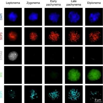
Immunocytochemistry/ Immunofluorescence: SCP1 Antibody [Biotin] [NB300-229B] - Immunofluorescence staining of spermatocyte nuclei. Immunofluorescence images & signal quantification of stage-specific spermatocyte nuclei through meiosis prophase I. Details for signal quantification are described in Methods. Microscopic images are selected from two independent experiments in which two different combinations of primary antibodies are used; one using SCP3, H1t & SCP1, another one using SCP3 & STRA8. *Early & late pachytene nuclei cannot be unambiguously differentiated in the absence of H1t staining, & are therefore merged for counting & signal quantification. Source data are provided as a Source Data file Image collected & cropped by CiteAb from the following publication (https://pubmed.ncbi.nlm.nih.gov/31444359), licensed under a CC-BY license. Not internally tested by Novus Biologicals.Applications for SCP1 Antibody [Biotin]
Application
Recommended Usage
Chromatin Immunoprecipitation
Optimal dilutions of this antibody should be experimentally determined.
Chromatin Immunoprecipitation (ChIP)
Optimal dilutions of this antibody should be experimentally determined.
Immunocytochemistry/ Immunofluorescence
Optimal dilutions of this antibody should be experimentally determined.
Immunohistochemistry
Optimal dilutions of this antibody should be experimentally determined.
Immunohistochemistry-Frozen
Optimal dilutions of this antibody should be experimentally determined.
Immunohistochemistry-Paraffin
Optimal dilutions of this antibody should be experimentally determined.
Immunoprecipitation
Optimal dilutions of this antibody should be experimentally determined.
Western Blot
Optimal dilutions of this antibody should be experimentally determined.
Application Notes
Optimal dilution of this antibody should be experimentally determined.
Formulation, Preparation, and Storage
Purification
Immunogen affinity purified
Formulation
PBS
Preservative
0.05% Sodium Azide
Concentration
Please see the vial label for concentration. If unlisted please contact technical services.
Shipping
The product is shipped with polar packs. Upon receipt, store it immediately at the temperature recommended below.
Stability & Storage
Store at 4C in the dark.
Background: SCP1
Alternate Names
Cancer/testis antigen 8, CT8MGC104417, HOM-TES-14, SCP1SCP-1, synaptonemal complex protein 1
Gene Symbol
SYCP1
Additional SCP1 Products
Product Documents for SCP1 Antibody [Biotin]
Certificate of Analysis
To download a Certificate of Analysis, please enter a lot or batch number in the search box below.
Product Specific Notices for SCP1 Antibody [Biotin]
This product is for research use only and is not approved for use in humans or in clinical diagnosis. Primary Antibodies are guaranteed for 1 year from date of receipt.
Citations for SCP1 Antibody [Biotin]
 Powered by Bioz
Customer Reviews for SCP1 Antibody [Biotin]
There are currently no reviews for this product. Be the first to review SCP1 Antibody [Biotin] and earn rewards!
Have you used SCP1 Antibody [Biotin]?
Submit a review and receive an Amazon gift card!
$25/€18/£15/$25CAN/¥2500 Yen for a review with an image
$10/€7/£6/$10CAN/¥1110 Yen for a review without an image
Submit a review
Protocols
Find general support by application which include: protocols, troubleshooting, illustrated assays, videos and webinars.
- Antigen Retrieval Protocol (PIER)
- Antigen Retrieval for Frozen Sections Protocol
- Appropriate Fixation of IHC/ICC Samples
- Cellular Response to Hypoxia Protocols
- ChIP Protocol Video
- Chromatin Immunoprecipitation (ChIP) Protocol
- Chromatin Immunoprecipitation Protocol
- Chromogenic IHC Staining of Formalin-Fixed Paraffin-Embedded (FFPE) Tissue Protocol
- Chromogenic Immunohistochemistry Staining of Frozen Tissue
- ClariTSA™ Fluorophore Kits
- Detection & Visualization of Antibody Binding
- Fluorescent IHC Staining of Frozen Tissue Protocol
- Graphic Protocol for Heat-induced Epitope Retrieval
- Graphic Protocol for the Preparation and Fluorescent IHC Staining of Frozen Tissue Sections
- Graphic Protocol for the Preparation and Fluorescent IHC Staining of Paraffin-embedded Tissue Sections
- Graphic Protocol for the Preparation of Gelatin-coated Slides for Histological Tissue Sections
- ICC Cell Smear Protocol for Suspension Cells
- ICC Immunocytochemistry Protocol Videos
- ICC for Adherent Cells
- IHC Sample Preparation (Frozen sections vs Paraffin)
- Immunocytochemistry (ICC) Protocol
- Immunocytochemistry Troubleshooting
- Immunofluorescence of Organoids Embedded in Cultrex Basement Membrane Extract
- Immunofluorescent IHC Staining of Formalin-Fixed Paraffin-Embedded (FFPE) Tissue Protocol
- Immunohistochemistry (IHC) and Immunocytochemistry (ICC) Protocols
- Immunohistochemistry Frozen Troubleshooting
- Immunohistochemistry Paraffin Troubleshooting
- Immunoprecipitation Protocol
- Preparing Samples for IHC/ICC Experiments
- Preventing Non-Specific Staining (Non-Specific Binding)
- Primary Antibody Selection & Optimization
- Protocol for Heat-Induced Epitope Retrieval (HIER)
- Protocol for Making a 4% Formaldehyde Solution in PBS
- Protocol for VisUCyte™ HRP Polymer Detection Reagent
- Protocol for the Fluorescent ICC Staining of Cell Smears - Graphic
- Protocol for the Fluorescent ICC Staining of Cultured Cells on Coverslips - Graphic
- Protocol for the Preparation & Fixation of Cells on Coverslips
- Protocol for the Preparation and Chromogenic IHC Staining of Frozen Tissue Sections
- Protocol for the Preparation and Chromogenic IHC Staining of Frozen Tissue Sections - Graphic
- Protocol for the Preparation and Chromogenic IHC Staining of Paraffin-embedded Tissue Sections
- Protocol for the Preparation and Chromogenic IHC Staining of Paraffin-embedded Tissue Sections - Graphic
- Protocol for the Preparation and Fluorescent ICC Staining of Cells on Coverslips
- Protocol for the Preparation and Fluorescent ICC Staining of Non-adherent Cells
- Protocol for the Preparation and Fluorescent ICC Staining of Stem Cells on Coverslips
- Protocol for the Preparation and Fluorescent IHC Staining of Frozen Tissue Sections
- Protocol for the Preparation and Fluorescent IHC Staining of Paraffin-embedded Tissue Sections
- Protocol for the Preparation of Gelatin-coated Slides for Histological Tissue Sections
- Protocol for the Preparation of a Cell Smear for Non-adherent Cell ICC - Graphic
- R&D Systems Quality Control Western Blot Protocol
- TUNEL and Active Caspase-3 Detection by IHC/ICC Protocol
- The Importance of IHC/ICC Controls
- Troubleshooting Guide: Immunohistochemistry
- Troubleshooting Guide: Western Blot Figures
- Western Blot Conditions
- Western Blot Protocol
- Western Blot Protocol for Cell Lysates
- Western Blot Troubleshooting
- Western Blot Troubleshooting Guide
- View all Protocols, Troubleshooting, Illustrated assays and Webinars
Loading...