Septin-2 Antibody - BSA Free
Novus Biologicals | Catalog # NBP1-85212
![Western Blot: Septin-2 Antibody [NBP1-85212]](https://resources.rndsystems.com/images/products/Septin-2-Antibody-Western-Blot-NBP1-85212-img0019.jpg "Western Blot: Septin-2 Antibody [NBP1-85212]")
![Western Blot: Septin-2 Antibody [NBP1-85212]](https://resources.rndsystems.com/images/products/Septin-2-Antibody-Western-Blot-NBP1-85212-img0014.jpg "Western Blot: Septin-2 Antibody [NBP1-85212]")
Loading...
Key Product Details
Validated by
Knockout/Knockdown
Species Reactivity
Validated:
Human, Mouse, Rat
Cited:
Human
Applications
Validated:
Immunohistochemistry-Paraffin, Western Blot, Immunocytochemistry/ Immunofluorescence, Knockdown Validated
Cited:
Western Blot, IF/IHC, Knockdown Validated
Label
Unconjugated
Antibody Source
Polyclonal Rabbit IgG
Format
BSA Free
Loading...
Product Specifications
Immunogen
This antibody was developed against Recombinant Protein corresponding to amino acids: WGVVEVENPEHNDFLKLRTMLITHMQDLQEVTQDLHYENFRSERLKRGGRKVENEDMNKDQILLEKEAELRRMQEMIARMQAQMQMQMQGGDGDGGALGH
Clonality
Polyclonal
Host
Rabbit
Isotype
IgG
Theoretical MW
41 kDa.
Disclaimer note: The observed molecular weight of the protein may vary from the listed predicted molecular weight due to post translational modifications, post translation cleavages, relative charges, and other experimental factors.
Disclaimer note: The observed molecular weight of the protein may vary from the listed predicted molecular weight due to post translational modifications, post translation cleavages, relative charges, and other experimental factors.
Scientific Data Images for Septin-2 Antibody - BSA Free
![Immunocytochemistry/ Immunofluorescence: Septin-2 Antibody [NBP1-85212]](https://resources.rndsystems.com/images/products/Septin-2-Antibody-Immunocytochemistry-Immunofluorescence-NBP1-85212-img0012.jpg "Immunocytochemistry/ Immunofluorescence: Septin-2 Antibody [NBP1-85212]")
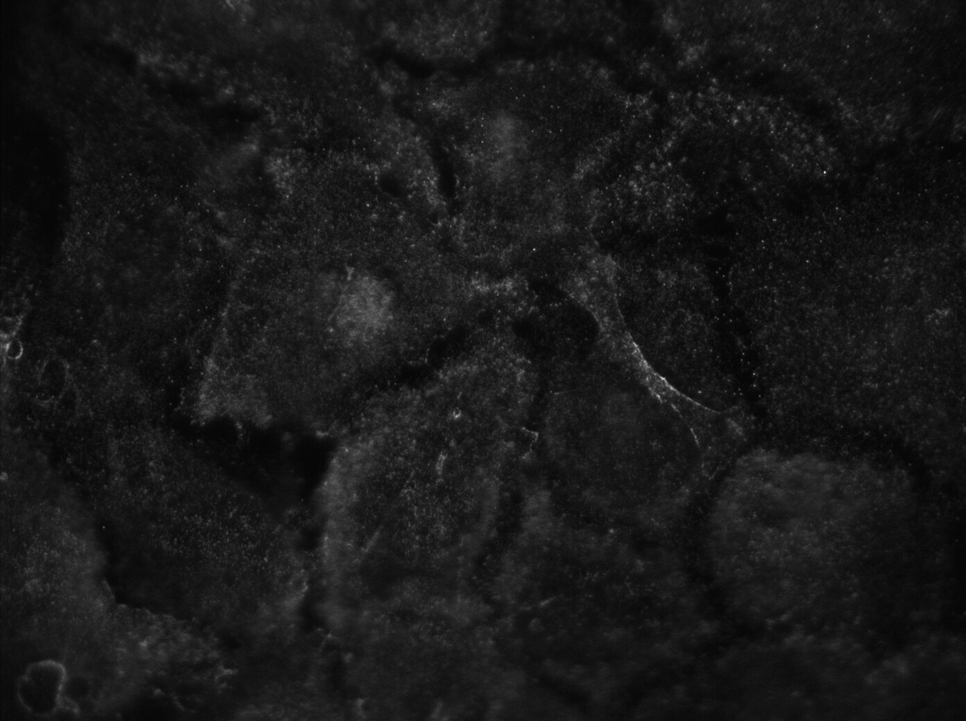
Immunocytochemistry/ Immunofluorescence: Septin-2 Antibody [NBP1-85212]
Immunocytochemistry/Immunofluorescence: Septin-2 Antibody [NBP1-85212] - Imaging of cultured rat corrtical astrocytes. Image submitted by a verified customer review.![Western Blot: Septin-2 Antibody [NBP1-85212]](https://resources.rndsystems.com/images/products/Septin-2-Antibody-Western-Blot-NBP1-85212-img0009.jpg "Western Blot: Septin-2 Antibody [NBP1-85212]")
Western Blot: Septin-2 Antibody [NBP1-85212]
Western Blot: Septin-2 Antibody [NBP1-85212] - Analysis in mouse cell line NIH-3T3 and rat cell line NBT-II.![Immunocytochemistry/ Immunofluorescence: Septin-2 Antibody [NBP1-85212]](https://resources.rndsystems.com/images/products/Septin-2-Antibody-Immunocytochemistry-Immunofluorescence-NBP1-85212-img0011.jpg "Immunocytochemistry/ Immunofluorescence: Septin-2 Antibody [NBP1-85212]")
Immunocytochemistry/ Immunofluorescence: Septin-2 Antibody [NBP1-85212]
Immunocytochemistry/Immunofluorescence: Septin-2 Antibody [NBP1-85212] - Staining of human cell line U-2 OS shows localization to nucleus, nucleoli & actin filaments. Antibody staining is shown in green.![Septin-2 Antibody - BSA Free Immunohistochemistry-Paraffin: Septin-2 Antibody [NBP1-85212]](https://resources.rndsystems.com/images/products/nbp1-85212_-immunohistochemistry-paraffin-639173123319920624.jpg "Immunohistochemistry-Paraffin: Septin-2 Antibody [NBP1-85212]")
Immunohistochemistry-Paraffin: Septin-2 Antibody [NBP1-85212]
Staining of human small intestine shows weak cytoplasmic positivity in glandular cells.![Septin-2 Antibody - BSA Free Immunohistochemistry-Paraffin: Septin-2 Antibody [NBP1-85212]](https://resources.rndsystems.com/images/products/nbp1-85212_-immunohistochemistry-paraffin-639173123524732871.jpg "Immunohistochemistry-Paraffin: Septin-2 Antibody [NBP1-85212]")
Immunohistochemistry-Paraffin: Septin-2 Antibody [NBP1-85212]
Staining of human testis shows strong membranous/cytoplasmic positivity in cells in seminiferous ducts.![Septin-2 Antibody - BSA Free Immunohistochemistry-Paraffin: Septin-2 Antibody [NBP1-85212]](https://resources.rndsystems.com/images/products/nbp1-85212_-immunohistochemistry-paraffin-639173132485736163.jpg "Immunohistochemistry-Paraffin: Septin-2 Antibody [NBP1-85212]")
Immunohistochemistry-Paraffin: Septin-2 Antibody [NBP1-85212]
Staining of human cerebral cortex shows strong cytoplasmic positivity in glial cells.![Septin-2 Antibody - BSA Free Immunohistochemistry-Paraffin: Septin-2 Antibody [NBP1-85212]](https://resources.rndsystems.com/images/products/nbp1-85212_-immunohistochemistry-paraffin-639173132674909254.jpg "Immunohistochemistry-Paraffin: Septin-2 Antibody [NBP1-85212]")
Immunohistochemistry-Paraffin: Septin-2 Antibody [NBP1-85212]
Staining of human endometrium shows moderate cytoplasmic positivity in glandular cells.Applications for Septin-2 Antibody - BSA Free
Application
Recommended Usage
Immunocytochemistry/ Immunofluorescence
0.25-2 ug/ml
Immunohistochemistry-Paraffin
1:200 - 1:500
Western Blot
0.04-0.4 ug/ml
Application Notes
For IHC-Paraffin, HIER pH 6 retrieval is recommended. ICC/IF Fixation Permeabilization: Use PFA/Triton X-100.
Reviewed Applications
Read 1 review rated 5 using NBP1-85212 in the following applications:
Formulation, Preparation, and Storage
Purification
Affinity purified
Formulation
PBS (pH 7.2) and 40% Glycerol
Format
BSA Free
Preservative
0.02% Sodium Azide
Concentration
Concentrations vary lot to lot. See vial label for concentration. If unlisted please contact technical services.
Shipping
The product is shipped with polar packs. Upon receipt, store it immediately at the temperature recommended below.
Stability & Storage
Store at 4C short term. Aliquot and store at -20C long term. Avoid freeze-thaw cycles.
Background: Septin-2
Alternate Names
DIFF6NEDD-5, hNedd5, KIAA0158Neural precursor cell expressed developmentally down-regulated protein 5, NEDD5Pnutl3, neural precursor cell expressed, developmentally down-regulated 5, septin 2, septin-2
Entrez Gene IDs
4735 (Human)
Gene Symbol
SEPTIN2
UniProt
Additional Septin-2 Products
Product Documents for Septin-2 Antibody - BSA Free
Certificate of Analysis
To download a Certificate of Analysis, please enter a lot or batch number in the search box below.
Product Specific Notices for Septin-2 Antibody - BSA Free
This product is for research use only and is not approved for use in humans or in clinical diagnosis. Primary Antibodies are guaranteed for 1 year from date of receipt.
Citations for Septin-2 Antibody - BSA Free
 Powered by Bioz
Customer Reviews for Septin-2 Antibody - BSA Free (1)
5 out of 5
1 Customer Rating
Have you used Septin-2 Antibody - BSA Free?
Submit a review and receive an Amazon gift card!
$25/€18/£15/$25CAN/¥2500 Yen for a review with an image
$10/€7/£6/$10CAN/¥1110 Yen for a review without an image
Submit a review
Customer Images
Showing
1
-
1 of
1 review
Showing All
Filter By:
-
Application: ImmunocytochemistrySample Tested: Rat cortical astrocytesSpecies: RatVerified Customer | Posted 06/27/2017The antibody labels the cultured astrocytes - some labelling is found under the membrane.Fixation Solution and Conditions: 4% paraformaldehyde in PBS, 15 minutes RT Blocking Solution & Duration: 1% BSA in PBS with 0.1% triton, 1 hour RT Primary Antibody Diluent and Dilutions Tested: 1:1000 in 1% BSA in PBS, over night 4°C Secondary Antibody Manufacturer, Host Species, Dilution, & Diluent: Thermo donkey anti-rabbit Alexa647, 1:2000 in PBS, 2 hours, RT

There are no reviews that match your criteria.
Protocols
Find general support by application which include: protocols, troubleshooting, illustrated assays, videos and webinars.
- Antigen Retrieval Protocol (PIER)
- Antigen Retrieval for Frozen Sections Protocol
- Appropriate Fixation of IHC/ICC Samples
- Cellular Response to Hypoxia Protocols
- Chromogenic IHC Staining of Formalin-Fixed Paraffin-Embedded (FFPE) Tissue Protocol
- Chromogenic Immunohistochemistry Staining of Frozen Tissue
- ClariTSA™ Fluorophore Kits
- Detection & Visualization of Antibody Binding
- Fluorescent IHC Staining of Frozen Tissue Protocol
- Graphic Protocol for Heat-induced Epitope Retrieval
- Graphic Protocol for the Preparation and Fluorescent IHC Staining of Frozen Tissue Sections
- Graphic Protocol for the Preparation and Fluorescent IHC Staining of Paraffin-embedded Tissue Sections
- Graphic Protocol for the Preparation of Gelatin-coated Slides for Histological Tissue Sections
- ICC Cell Smear Protocol for Suspension Cells
- ICC Immunocytochemistry Protocol Videos
- ICC for Adherent Cells
- IHC Sample Preparation (Frozen sections vs Paraffin)
- Immunocytochemistry (ICC) Protocol
- Immunocytochemistry Troubleshooting
- Immunofluorescence of Organoids Embedded in Cultrex Basement Membrane Extract
- Immunofluorescent IHC Staining of Formalin-Fixed Paraffin-Embedded (FFPE) Tissue Protocol
- Immunohistochemistry (IHC) and Immunocytochemistry (ICC) Protocols
- Immunohistochemistry Frozen Troubleshooting
- Immunohistochemistry Paraffin Troubleshooting
- Preparing Samples for IHC/ICC Experiments
- Preventing Non-Specific Staining (Non-Specific Binding)
- Primary Antibody Selection & Optimization
- Protocol for Heat-Induced Epitope Retrieval (HIER)
- Protocol for Making a 4% Formaldehyde Solution in PBS
- Protocol for VisUCyte™ HRP Polymer Detection Reagent
- Protocol for the Fluorescent ICC Staining of Cell Smears - Graphic
- Protocol for the Fluorescent ICC Staining of Cultured Cells on Coverslips - Graphic
- Protocol for the Preparation & Fixation of Cells on Coverslips
- Protocol for the Preparation and Chromogenic IHC Staining of Frozen Tissue Sections
- Protocol for the Preparation and Chromogenic IHC Staining of Frozen Tissue Sections - Graphic
- Protocol for the Preparation and Chromogenic IHC Staining of Paraffin-embedded Tissue Sections
- Protocol for the Preparation and Chromogenic IHC Staining of Paraffin-embedded Tissue Sections - Graphic
- Protocol for the Preparation and Fluorescent ICC Staining of Cells on Coverslips
- Protocol for the Preparation and Fluorescent ICC Staining of Non-adherent Cells
- Protocol for the Preparation and Fluorescent ICC Staining of Stem Cells on Coverslips
- Protocol for the Preparation and Fluorescent IHC Staining of Frozen Tissue Sections
- Protocol for the Preparation and Fluorescent IHC Staining of Paraffin-embedded Tissue Sections
- Protocol for the Preparation of Gelatin-coated Slides for Histological Tissue Sections
- Protocol for the Preparation of a Cell Smear for Non-adherent Cell ICC - Graphic
- R&D Systems Quality Control Western Blot Protocol
- TUNEL and Active Caspase-3 Detection by IHC/ICC Protocol
- The Importance of IHC/ICC Controls
- Troubleshooting Guide: Immunohistochemistry
- Troubleshooting Guide: Western Blot Figures
- Western Blot Conditions
- Western Blot Protocol
- Western Blot Protocol for Cell Lysates
- Western Blot Troubleshooting
- Western Blot Troubleshooting Guide
- View all Protocols, Troubleshooting, Illustrated assays and Webinars
Loading...