Septin-9 Antibody - BSA Free
Novus Biologicals | Catalog # NBP1-28711
![Western Blot: Septin-9 Antibody [NBP1-28711]](https://resources.rndsystems.com/images/products/Septin-9-Antibody-Western-Blot-NBP1-28711-img0003.jpg "Western Blot: Septin-9 Antibody [NBP1-28711]")
Key Product Details
Validated by
Biological Validation
Species Reactivity
Validated:
Human
Cited:
Human
Applications
Validated:
Immunohistochemistry, Immunohistochemistry-Paraffin, Western Blot, Immunocytochemistry/ Immunofluorescence, Immunoprecipitation
Cited:
Immunocytochemistry/ Immunofluorescence
Label
Unconjugated
Antibody Source
Polyclonal Rabbit IgG
Format
BSA Free
Loading...
Product Specifications
Immunogen
The immunogen recognized by this antibody maps to a region between residue 536 and 586 of human septin 9 using the numbering given in entry NP_001106963.1 (GeneID 10801).
Clonality
Polyclonal
Host
Rabbit
Isotype
IgG
Scientific Data Images for Septin-9 Antibody - BSA Free
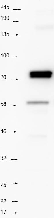
Western Blot: Septin-9 Antibody [NBP1-28711]
Western Blot: Septin-9 Antibody [NBP1-28711] - Lane 1: marker. Lane 2: 10mg of HeLa whole cell lysate probed with NBP1-28711 antibody 1:1000 in PBST. Blot blocked with 5% milk in PBS with 0.1% tween 20 probed overnight at 4C with NBP1-28711 at 1:1000 washed 3X PBST incubated with anti rabbit HRP secondary antiboy, washed 3X PBST, developed with ECL prime kit GE healthcare. Image from verified customer review.![Immunocytochemistry/ Immunofluorescence: Septin-9 Antibody [NBP1-28711]](https://resources.rndsystems.com/images/products/Septin-9-Antibody-Immunocytochemistry-Immunofluorescence-NBP1-28711-img0004.jpg "Immunocytochemistry/ Immunofluorescence: Septin-9 Antibody [NBP1-28711]")
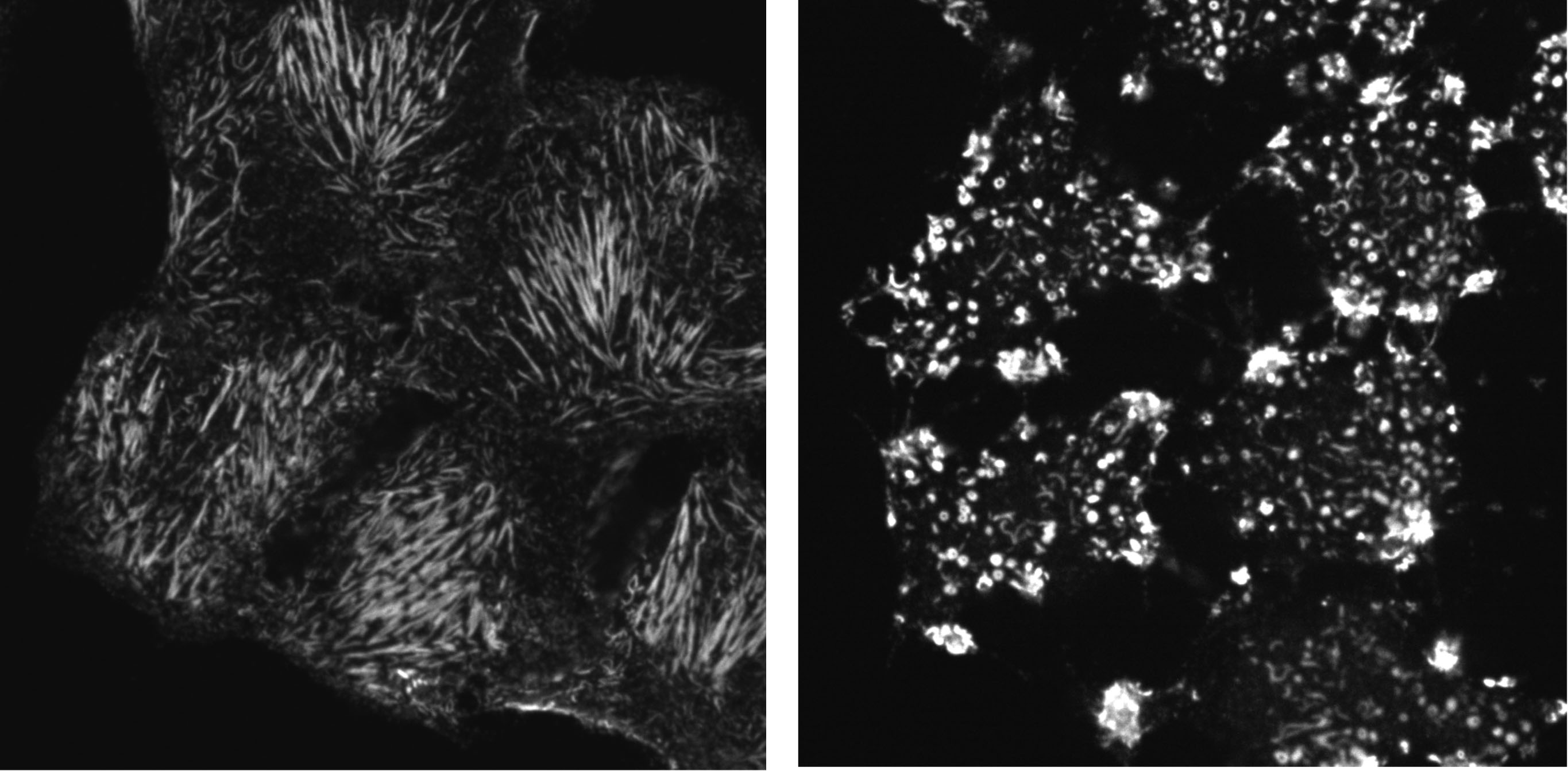
Immunocytochemistry/ Immunofluorescence: Septin-9 Antibody [NBP1-28711]
Immunocytochemistry/Immunofluorescence: Septin-9 Antibody [NBP1-28711] - HeLa cells fixed with PFA (4%, 20 min, RT) with anti septin 9 antibody NBP1-28711 at 1:1000. Left panel: Control localisation to sub nuclear filaments. Right panel: Cells treated with cytochalasin D to depolymerize F-actin. Note localization to ring like structures. Image from verified customer review.![Immunohistochemistry-Paraffin: Septin-9 Antibody [NBP1-28711]](https://resources.rndsystems.com/images/products/Septin-9-Antibody-Immunohistochemistry-Paraffin-NBP1-28711-img0005.jpg "Immunohistochemistry-Paraffin: Septin-9 Antibody [NBP1-28711]")
Immunohistochemistry-Paraffin: Septin-9 Antibody [NBP1-28711]
Immunohistochemistry-Paraffin: Septin-9 Antibody [NBP1-28711] - Section of human breast carcinoma. Antibody: Affinity purified rabbit anti- Septin 9 used at a dilution of 1:1,000 (1ug/ml). Detection: Red-fluorescent goat anti-rabbit IgG-heavy and light chain cross-adsorbed Antibody DyLight® 594 Conjugated used at a dilution of 1:100.![Immunoprecipitation: Septin-9 Antibody [NBP1-28711]](https://resources.rndsystems.com/images/products/Septin-9-Antibody-Immunoprecipitation-NBP1-28711-img0001.jpg "Immunoprecipitation: Septin-9 Antibody [NBP1-28711]")
Immunoprecipitation: Septin-9 Antibody [NBP1-28711]
Immunoprecipitation: Septin-9 Antibody [NBP1-28711] - Samples: Whole cell lysate (1 mg for IP, 20% of IP loaded) from HeLa cells. Antibodies: Affinity purified rabbit anti-Septin 9 antibody NBP1-28711 used for IP at 3 ug/mg lysate. Septin 9 was also immunoprecipitated by rabbit anti-Septin 9 antibody NBP1-28764, which recognizes an upstream epitope. For blotting immunoprecipitated Septin 9, NBP1-28764 was used at 1 ug/ml. Detection: Chemiluminescence with an exposure time of 1 second.Applications for Septin-9 Antibody - BSA Free
Application
Recommended Usage
Immunocytochemistry/ Immunofluorescence
1:1000 - 1:5000
Immunoprecipitation
2-5 ug/mg lysate
Application Notes
Septin-9 antibody validated for WB, ICC/IF from verified customer reviews.
Reviewed Applications
Read 2 reviews rated 4.5 using NBP1-28711 in the following applications:
Formulation, Preparation, and Storage
Purification
Immunogen affinity purified
Formulation
Tris-Citrate/Phosphate (pH 7.0 - 8.0)
Format
BSA Free
Preservative
0.09% Sodium Azide
Concentration
1.0 mg/ml
Shipping
The product is shipped with polar packs. Upon receipt, store it immediately at the temperature recommended below.
Stability & Storage
Store at 4C. Do not freeze.
Background: Septin-9
Alternate Names
AF17q25, FLJ75490, KIAA0991Ov/Br septin, MLL septin-like fusion protein, MLL septin-like fusion protein MSF-A, MSF1NAPB, MSFMLL septin-like fusion, Ovarian/Breast septin, PNUTL4, SeptD1, septin 9, Septin D1, septin-9, SINT1
Entrez Gene IDs
10801 (Human)
Gene Symbol
SEPTIN9
UniProt
Additional Septin-9 Products
Product Documents for Septin-9 Antibody - BSA Free
Certificate of Analysis
To download a Certificate of Analysis, please enter a lot or batch number in the search box below.
Product Specific Notices for Septin-9 Antibody - BSA Free
This product is for research use only and is not approved for use in humans or in clinical diagnosis. Primary Antibodies are guaranteed for 1 year from date of receipt.
Citations for Septin-9 Antibody - BSA Free
 Powered by Bioz
Customer Reviews for Septin-9 Antibody - BSA Free (2)
4.5 out of 5
2 Customer Ratings
Have you used Septin-9 Antibody - BSA Free?
Submit a review and receive an Amazon gift card!
$25/€18/£15/$25CAN/¥2500 Yen for a review with an image
$10/€7/£6/$10CAN/¥1110 Yen for a review without an image
Submit a review
Customer Images
Showing
1
-
2 of
2 reviews
Showing All
Filter By:
-
Application: ImmunocytochemistrySample Tested: hela cellSpecies: HumanVerified Customer | Posted 10/25/2018IF images of HeLa cells fixed with PFA with anti septin 9 antibody NBP1-28711 1:1000. Left Control localisation to sub nuclear filaments. Right cells treated with cytochalasin D to depolymerize F-actin. Note localization to ring like structures.Cells fixed with 4% PFA for 20 minutes room temperature. Fixed cells incubated with with 0.5% triton x100 in 1xPBS for 5 minutes room. Washed twice in blocking buffer (1% BSA 1xPBS, 20mM glycine ). Washed once with blocking buffer left for 1hour room temperature. incubated with Anti septin 9 antibody in blocking buffer 1:1000 for 20 hours at 4 degrees. Washed 3 x 5 minutes in blocking buffer Incubated with Anti rabbit-Alexa 594 conjugated antibody (Jackson immunoscience) for 2 hours room temperature. Washed 3 x 5 minutes in blocking buffer Washed 1 x 5 minutes in PBS Imaged with 60X oil objective on Nikon Eclipse Ti Inverted Fast confocal - Equipped with a Yokogawa CSU-X1 spinning disk unit and okogawa CSU-1 disk head and Andor Neo sCMOS camera.

-
Application: Western BlotSample Tested: Hela whole cell lysateSpecies: HumanVerified Customer | Posted 10/22/2018lane 1 marker lane 2 10mg of HeLa whole cell lysate probed with NBP1-28711 antibody 1:1000 in PBST10mg of HeLa whole cell lysate was loaded run on a 4-12% bi-Tris acrylamide gell run in 1X mops buffer. Blot blocked with 5%milk in PBS with 0.1% tween 20 probed overnight at 4 degrees C with NBP1-28711 1:1000 washed 3X PBST incubated with anti rabbit HRP secondary washed 3X PBST, developed with ECL prime kit GE healthcare.
There are no reviews that match your criteria.
Protocols
Find general support by application which include: protocols, troubleshooting, illustrated assays, videos and webinars.
- Antigen Retrieval Protocol (PIER)
- Antigen Retrieval for Frozen Sections Protocol
- Appropriate Fixation of IHC/ICC Samples
- Cellular Response to Hypoxia Protocols
- Chromogenic IHC Staining of Formalin-Fixed Paraffin-Embedded (FFPE) Tissue Protocol
- Chromogenic Immunohistochemistry Staining of Frozen Tissue
- ClariTSA™ Fluorophore Kits
- Detection & Visualization of Antibody Binding
- Fluorescent IHC Staining of Frozen Tissue Protocol
- Graphic Protocol for Heat-induced Epitope Retrieval
- Graphic Protocol for the Preparation and Fluorescent IHC Staining of Frozen Tissue Sections
- Graphic Protocol for the Preparation and Fluorescent IHC Staining of Paraffin-embedded Tissue Sections
- Graphic Protocol for the Preparation of Gelatin-coated Slides for Histological Tissue Sections
- ICC Cell Smear Protocol for Suspension Cells
- ICC Immunocytochemistry Protocol Videos
- ICC for Adherent Cells
- IHC Sample Preparation (Frozen sections vs Paraffin)
- Immunocytochemistry (ICC) Protocol
- Immunocytochemistry Troubleshooting
- Immunofluorescence of Organoids Embedded in Cultrex Basement Membrane Extract
- Immunofluorescent IHC Staining of Formalin-Fixed Paraffin-Embedded (FFPE) Tissue Protocol
- Immunohistochemistry (IHC) and Immunocytochemistry (ICC) Protocols
- Immunohistochemistry Frozen Troubleshooting
- Immunohistochemistry Paraffin Troubleshooting
- Immunoprecipitation Protocol
- Preparing Samples for IHC/ICC Experiments
- Preventing Non-Specific Staining (Non-Specific Binding)
- Primary Antibody Selection & Optimization
- Protocol for Heat-Induced Epitope Retrieval (HIER)
- Protocol for Making a 4% Formaldehyde Solution in PBS
- Protocol for VisUCyte™ HRP Polymer Detection Reagent
- Protocol for the Fluorescent ICC Staining of Cell Smears - Graphic
- Protocol for the Fluorescent ICC Staining of Cultured Cells on Coverslips - Graphic
- Protocol for the Preparation & Fixation of Cells on Coverslips
- Protocol for the Preparation and Chromogenic IHC Staining of Frozen Tissue Sections
- Protocol for the Preparation and Chromogenic IHC Staining of Frozen Tissue Sections - Graphic
- Protocol for the Preparation and Chromogenic IHC Staining of Paraffin-embedded Tissue Sections
- Protocol for the Preparation and Chromogenic IHC Staining of Paraffin-embedded Tissue Sections - Graphic
- Protocol for the Preparation and Fluorescent ICC Staining of Cells on Coverslips
- Protocol for the Preparation and Fluorescent ICC Staining of Non-adherent Cells
- Protocol for the Preparation and Fluorescent ICC Staining of Stem Cells on Coverslips
- Protocol for the Preparation and Fluorescent IHC Staining of Frozen Tissue Sections
- Protocol for the Preparation and Fluorescent IHC Staining of Paraffin-embedded Tissue Sections
- Protocol for the Preparation of Gelatin-coated Slides for Histological Tissue Sections
- Protocol for the Preparation of a Cell Smear for Non-adherent Cell ICC - Graphic
- R&D Systems Quality Control Western Blot Protocol
- TUNEL and Active Caspase-3 Detection by IHC/ICC Protocol
- The Importance of IHC/ICC Controls
- Troubleshooting Guide: Immunohistochemistry
- Troubleshooting Guide: Western Blot Figures
- Western Blot Conditions
- Western Blot Protocol
- Western Blot Protocol for Cell Lysates
- Western Blot Troubleshooting
- Western Blot Troubleshooting Guide
- View all Protocols, Troubleshooting, Illustrated assays and Webinars
Loading...