SHISA3 Antibody - BSA Free
Novus Biologicals | Catalog # NBP2-22340
![Western Blot: SHISA3 AntibodyBSA Free [NBP2-22340]](https://resources.rndsystems.com/images/products/SHISA3-Antibody-Western-Blot-NBP2-22340-img0002.jpg "Western Blot: SHISA3 AntibodyBSA Free [NBP2-22340]")
Key Product Details
Species Reactivity
Validated:
Human, Mouse, Rat
Cited:
Mouse
Applications
Validated:
Immunohistochemistry, Immunohistochemistry-Paraffin, Western Blot, ELISA, Immunocytochemistry/ Immunofluorescence
Cited:
IF/IHC
Label
Unconjugated
Antibody Source
Polyclonal Rabbit IgG
Format
BSA Free
Loading...
Product Specifications
Immunogen
Antibody was raised against a 15 amino acid peptide near the center of human SHISA3. The immunogen is located within amino acids 120 - 170 of SHISA3.
Specificity
SHISA3 antibody is predicted to not cross-react with other SHISA protein family members.
Clonality
Polyclonal
Host
Rabbit
Isotype
IgG
Theoretical MW
28 kDa.
Disclaimer note: The observed molecular weight of the protein may vary from the listed predicted molecular weight due to post translational modifications, post translation cleavages, relative charges, and other experimental factors.
Disclaimer note: The observed molecular weight of the protein may vary from the listed predicted molecular weight due to post translational modifications, post translation cleavages, relative charges, and other experimental factors.
Scientific Data Images for SHISA3 Antibody - BSA Free
Western Blot: SHISA3 AntibodyBSA Free [NBP2-22340]
Western Blot: SHISA3 Antibody [NBP2-22340] - Analysis SHISA3 in human small intestine Tissue lysate with SHISA3 antibody at 1 ug/mL in (A) the absence and (B) the presence of blocking peptide.![Immunohistochemistry: SHISA3 Antibody - BSA Free [NBP2-22340]](https://resources.rndsystems.com/images/products/SHISA3-Antibody-Immunohistochemistry-NBP2-22340-img0005.jpg "Immunohistochemistry: SHISA3 Antibody - BSA Free [NBP2-22340]")
Immunohistochemistry: SHISA3 Antibody - BSA Free [NBP2-22340]
Immunohistochemistry: SHISA3 Antibody [NBP2-22340] - SHISA3 in human small intestine tissue with SHISA3 antibody at 2.5 ug/ml.
Immunocytochemistry/ Immunofluorescence: SHISA3 Antibody - BSA Free [NBP2-22340] -
Immunocytochemistry/ Immunofluorescence: SHISA3 Antibody - BSA Free [NBP2-22340] - Immunofluorescence of SHISA3 in human small intestine tissue with SHISA3 antibody at 20 ug/ml.Applications for SHISA3 Antibody - BSA Free
Application
Recommended Usage
ELISA
1:100-1:2000
Immunocytochemistry/ Immunofluorescence
20 ug/ml
Immunohistochemistry
1:10-1:500
Immunohistochemistry-Paraffin
1:10-1:500
Western Blot
1 ug/ml
Reviewed Applications
Read 1 review rated 1 using NBP2-22340 in the following applications:
Formulation, Preparation, and Storage
Purification
Peptide affinity purified
Formulation
PBS
Format
BSA Free
Preservative
0.02% Sodium Azide
Concentration
1 mg/ml
Shipping
The product is shipped with polar packs. Upon receipt, store it immediately at the temperature recommended below.
Stability & Storage
Store at 4C short term. Aliquot and store at -20C long term. Avoid freeze-thaw cycles.
Background: SHISA3
Alternate Names
hShisa3, protein shisa-3 homolog, shisa homolog 3 (Xenopus laevis)
Gene Symbol
SHISA3
UniProt
Additional SHISA3 Products
Product Documents for SHISA3 Antibody - BSA Free
Certificate of Analysis
To download a Certificate of Analysis, please enter a lot or batch number in the search box below.
Product Specific Notices for SHISA3 Antibody - BSA Free
This product is for research use only and is not approved for use in humans or in clinical diagnosis. Primary Antibodies are guaranteed for 1 year from date of receipt.
Citations for SHISA3 Antibody - BSA Free
 Powered by Bioz
Customer Reviews for SHISA3 Antibody - BSA Free (1)
1 out of 5
1 Customer Rating
Have you used SHISA3 Antibody - BSA Free?
Submit a review and receive an Amazon gift card!
$25/€18/£15/$25CAN/¥2500 Yen for a review with an image
$10/€7/£6/$10CAN/¥1110 Yen for a review without an image
Submit a review
Customer Images
Showing
1
-
1 of
1 review
Showing All
Filter By:
-
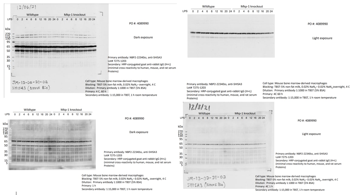
Application: Western BlotSample Tested: Bone marrow-derived macrophagesSpecies: MouseVerified Customer | Posted 12/07/2021Western blot analysis of wildtype and Mkp-1-/- BMDM treated with LPS for different times.Mouse bone-marrow-derived macrophages were stimulated with 100 ng/ml for 2, 3, 4, 6, 8, 12, 16, 20, and 24 h or left unstimulated as control, label as 0 h; in DMEM+10% FBS, and lysed in lysis buffer (20 mM HEPES, pH7.4, 50 mM beta-glycerol phosphate, 2 mM EGTA, 1 mM DTT, 10 mM NaF, 1 mM Sodium Orthovanadate, 1% Triton-100, 10% Glycerol, and various protease and phosphatase inhibitors). Lysates were resoled on 4-12% NuPAGE gel under denature condition. After transfering to nitrocellulose membrane, the blot was blocked in TBST containing 5% (w/v) Blotting Grade Blocker Non Fat Dry Milk (Bio-Rad) overnight at 4C. After blocking, the membrane was incubated with the primary antibody diluted to 1 ug/ml in TBST containing 5% BSA and 0.02% sodium azide for 1 h at room temperature or 60 h at 4 C. After 3 washes with TBST (5', 10' and 15') the blot was incubated with HRP-conjugated goat anti-rabbit secondary antibody (1:15,000) for 1 h at room temperature. After washing 3 times with TBST, the image was developed using ECL.
 Bio-Techne ResponseThank you for reviewing our product. We are sorry to hear that this product did not perform as expected. We have been in touch with the customer to resolve this issue according to our Product Guarantee and to the customer’s satisfaction.
There are no reviews that match your criteria.
Protocols
Find general support by application which include: protocols, troubleshooting, illustrated assays, videos and webinars.
- Antigen Retrieval Protocol (PIER)
- Antigen Retrieval for Frozen Sections Protocol
- Appropriate Fixation of IHC/ICC Samples
- Cellular Response to Hypoxia Protocols
- Chromogenic IHC Staining of Formalin-Fixed Paraffin-Embedded (FFPE) Tissue Protocol
- Chromogenic Immunohistochemistry Staining of Frozen Tissue
- ClariTSA™ Fluorophore Kits
- Detection & Visualization of Antibody Binding
- ELISA Sample Preparation & Collection Guide
- ELISA Troubleshooting Guide
- Fluorescent IHC Staining of Frozen Tissue Protocol
- Graphic Protocol for Heat-induced Epitope Retrieval
- Graphic Protocol for the Preparation and Fluorescent IHC Staining of Frozen Tissue Sections
- Graphic Protocol for the Preparation and Fluorescent IHC Staining of Paraffin-embedded Tissue Sections
- Graphic Protocol for the Preparation of Gelatin-coated Slides for Histological Tissue Sections
- How to Run an R&D Systems DuoSet ELISA
- How to Run an R&D Systems Quantikine ELISA
- How to Run an R&D Systems Quantikine™ QuicKit™ ELISA
- ICC Cell Smear Protocol for Suspension Cells
- ICC Immunocytochemistry Protocol Videos
- ICC for Adherent Cells
- IHC Sample Preparation (Frozen sections vs Paraffin)
- Immunocytochemistry (ICC) Protocol
- Immunocytochemistry Troubleshooting
- Immunofluorescence of Organoids Embedded in Cultrex Basement Membrane Extract
- Immunofluorescent IHC Staining of Formalin-Fixed Paraffin-Embedded (FFPE) Tissue Protocol
- Immunohistochemistry (IHC) and Immunocytochemistry (ICC) Protocols
- Immunohistochemistry Frozen Troubleshooting
- Immunohistochemistry Paraffin Troubleshooting
- Preparing Samples for IHC/ICC Experiments
- Preventing Non-Specific Staining (Non-Specific Binding)
- Primary Antibody Selection & Optimization
- Protocol for Heat-Induced Epitope Retrieval (HIER)
- Protocol for Making a 4% Formaldehyde Solution in PBS
- Protocol for VisUCyte™ HRP Polymer Detection Reagent
- Protocol for the Fluorescent ICC Staining of Cell Smears - Graphic
- Protocol for the Fluorescent ICC Staining of Cultured Cells on Coverslips - Graphic
- Protocol for the Preparation & Fixation of Cells on Coverslips
- Protocol for the Preparation and Chromogenic IHC Staining of Frozen Tissue Sections
- Protocol for the Preparation and Chromogenic IHC Staining of Frozen Tissue Sections - Graphic
- Protocol for the Preparation and Chromogenic IHC Staining of Paraffin-embedded Tissue Sections
- Protocol for the Preparation and Chromogenic IHC Staining of Paraffin-embedded Tissue Sections - Graphic
- Protocol for the Preparation and Fluorescent ICC Staining of Cells on Coverslips
- Protocol for the Preparation and Fluorescent ICC Staining of Non-adherent Cells
- Protocol for the Preparation and Fluorescent ICC Staining of Stem Cells on Coverslips
- Protocol for the Preparation and Fluorescent IHC Staining of Frozen Tissue Sections
- Protocol for the Preparation and Fluorescent IHC Staining of Paraffin-embedded Tissue Sections
- Protocol for the Preparation of Gelatin-coated Slides for Histological Tissue Sections
- Protocol for the Preparation of a Cell Smear for Non-adherent Cell ICC - Graphic
- Quantikine HS ELISA Kit Assay Principle, Alkaline Phosphatase
- Quantikine HS ELISA Kit Principle, Streptavidin-HRP Polymer
- R&D Systems Quality Control Western Blot Protocol
- Sandwich ELISA (Colorimetric) – Biotin/Streptavidin Detection Protocol
- Sandwich ELISA (Colorimetric) – Direct Detection Protocol
- TUNEL and Active Caspase-3 Detection by IHC/ICC Protocol
- The Importance of IHC/ICC Controls
- Troubleshooting Guide: ELISA
- Troubleshooting Guide: Immunohistochemistry
- Troubleshooting Guide: Western Blot Figures
- Western Blot Conditions
- Western Blot Protocol
- Western Blot Protocol for Cell Lysates
- Western Blot Troubleshooting
- Western Blot Troubleshooting Guide
- View all Protocols, Troubleshooting, Illustrated assays and Webinars
Loading...