Siglec-1/CD169 Antibody (HSn 7D2) - BSA Free
Novus Biologicals | Catalog # NB600-534
Key Product Details
Species Reactivity
Validated:
Human, Mouse, Porcine, Virus
Cited:
Human, Mouse, Porcine, Virus
Applications
Validated:
Immunohistochemistry, Immunohistochemistry-Paraffin, Immunohistochemistry-Frozen, Western Blot, ELISA, Flow Cytometry, Immunocytochemistry/ Immunofluorescence, In vitro assay, Radioimmunoassay, In vivo assay
Cited:
Immunohistochemistry-Paraffin, Immunohistochemistry-Frozen, Western Blot, Flow Cytometry, Immunocytochemistry/ Immunofluorescence, In vitro assay, IF/IHC
Label
Unconjugated
Antibody Source
Monoclonal Mouse IgG1 Clone # HSn 7D2
Format
BSA Free
Loading...
Product Specifications
Immunogen
Full-length purified human Sialoadhesin (d1-4)Fc protein. [Swiss-Prot# Q9BZZ2]
Epitope
Within domains 1-4.
Reactivity Notes
Porcine reactivity reported in scientific literature (PMID: 27441395). Mouse reactivity reported in scientific literature. Not yet tested in other species. Use in Virus reported in scientific literature (PMID:27487279).
Localization
Isoform 1: Cell membrane; Single-pass type I membrane protein. Isoform 2: Secreted.
Clonality
Monoclonal
Host
Mouse
Isotype
IgG1
Scientific Data Images for Siglec-1/CD169 Antibody (HSn 7D2) - BSA Free
![Immunohistochemistry-Frozen: Siglec-1/CD169 Antibody (HSn 7D2) - BSA Free [NB600-534]](https://resources.rndsystems.com/images/products/Siglec-1-CD169-Antibody-HSn-7D2-Immunohistochemistry-Frozen-NB600-534-img0003.jpg "Immunohistochemistry-Frozen: Siglec-1/CD169 Antibody (HSn 7D2) - BSA Free [NB600-534]")
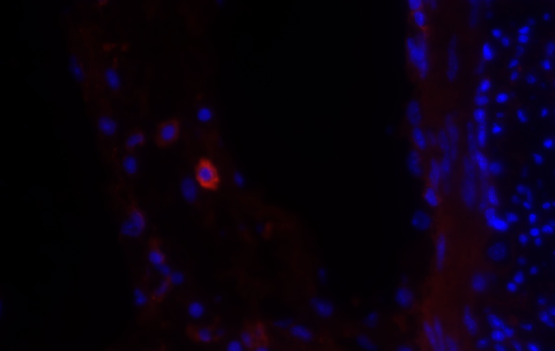
Immunohistochemistry-Frozen: Siglec-1/CD169 Antibody (HSn 7D2) - BSA Free [NB600-534]
Immunohistochemistry-Frozen: Siglec-1/CD169 Antibody (HSn 7D2) [NB600-534] - Adult mouse small intestine. NB600-534 Siglec-1 was used at 1:500 and labeled with Alexa Fluor 568 conjugated secondary antibody (Red). DAPI shown as blue. Image from verified customer review.![Flow Cytometry: Siglec-1/CD169 Antibody (HSn 7D2) - BSA Free [NB600-534]](https://resources.rndsystems.com/images/products/Siglec-1-CD169-Antibody-HSn-7D2-Flow-Cytometry-NB600-534-img0002.jpg "Flow Cytometry: Siglec-1/CD169 Antibody (HSn 7D2) - BSA Free [NB600-534]")
Flow Cytometry: Siglec-1/CD169 Antibody (HSn 7D2) - BSA Free [NB600-534]
Flow Cytometry: Siglec-1/CD169 Antibody (HSn 7D2) [NB600-534] - Human CD14+ PBMC differentiated to M1 macrophages with rhGM-CSF were stained with Mouse Anti-Siglec-1/CD169 Monoclonal Antibody (NB600-534, filled histogram), or Mouse IgG1 isotype control (MAB002, open histogram) followed by APC-conjugated Anti-Mouse IgG Secondary Antibody (F0101B).![Immunohistochemistry-Paraffin: Siglec-1/CD169 Antibody (HSn 7D2) - BSA Free [NB600-534]](https://resources.rndsystems.com/images/products/Sialoadhesin-Antibody-HSn-7D2-Immunohistochemistry-Paraffin-NB600-534-img0001.jpg "Immunohistochemistry-Paraffin: Siglec-1/CD169 Antibody (HSn 7D2) - BSA Free [NB600-534]")
Immunohistochemistry-Paraffin: Siglec-1/CD169 Antibody (HSn 7D2) - BSA Free [NB600-534]
Immunohistochemistry-Paraffin: Siglec-1/CD169 Antibody (HSn 7D2) [NB600-534] - Staining of Sialoadhesin in human spleen using DAB with hematoxylin counterstain.Applications for Siglec-1/CD169 Antibody (HSn 7D2) - BSA Free
Application
Recommended Usage
ELISA
1:100-1:2000
Flow Cytometry
1:10-1:1000
Immunocytochemistry/ Immunofluorescence
reported in scientific literature (PMID 28794041)
Immunohistochemistry
1:10-1:500. Use reported in scientific literature (PMID 26106049)
Immunohistochemistry-Frozen
1:10-1:500
Immunohistochemistry-Paraffin
1:10-1:500
In vivo assay
reported in scientific literature (PMID 18414664)
Western Blot
reported in scientific literature
Application Notes
This Sialoadhesin Antibody (HSn 7D2) is useful for Immunohistochemistry (paraffin-embedded, acetone-fixed, and frozen sections), Flow Cytometry, Radioimmunoassay and ELISA.
Reviewed Applications
Read 1 review rated 5 using NB600-534 in the following applications:
Flow Cytometry Panel Builder
Bio-Techne Knows Flow Cytometry
Save time and reduce costly mistakes by quickly finding compatible reagents using the Panel Builder Tool.
Advanced Features
- Spectra Viewer - Custom analysis of spectra from multiple fluorochromes
- Spillover Popups - Visualize the spectra of individual fluorochromes
- Antigen Density Selector - Match fluorochrome brightness with antigen density

Formulation, Preparation, and Storage
Purification
Protein G purified
Formulation
PBS
Format
BSA Free
Preservative
0.05% Sodium Azide
Concentration
1.0 mg/ml
Shipping
The product is shipped with polar packs. Upon receipt, store it immediately at the temperature recommended below.
Stability & Storage
Store at 4C short term. Aliquot and store at -20C long term. Avoid freeze-thaw cycles.
Background: Siglec-1/CD169
Long Name
Sialic Acid Binding Ig-like Lectin 1
Alternate Names
CD169, Siglec1
Entrez Gene IDs
6614 (Human)
Gene Symbol
SIGLEC1
UniProt
Additional Siglec-1/CD169 Products
Product Documents for Siglec-1/CD169 Antibody (HSn 7D2) - BSA Free
Certificate of Analysis
To download a Certificate of Analysis, please enter a lot or batch number in the search box below.
Product Specific Notices for Siglec-1/CD169 Antibody (HSn 7D2) - BSA Free
This product is for research use only and is not approved for use in humans or in clinical diagnosis. Primary Antibodies are guaranteed for 1 year from date of receipt.
Related Research Areas
Citations for Siglec-1/CD169 Antibody (HSn 7D2) - BSA Free
 Powered by Bioz
Customer Reviews for Siglec-1/CD169 Antibody (HSn 7D2) - BSA Free (1)
5 out of 5
1 Customer Rating
Have you used Siglec-1/CD169 Antibody (HSn 7D2) - BSA Free?
Submit a review and receive an Amazon gift card!
$25/€18/£15/$25CAN/¥2500 Yen for a review with an image
$10/€7/£6/$10CAN/¥1110 Yen for a review without an image
Submit a review
Customer Images
Showing
1
-
1 of
1 review
Showing All
Filter By:
-
Application: Immunohistochemistry-FrozenSample Tested: Adult small intestineSpecies: MouseVerified Customer | Posted 12/16/2018NB600-534 Siglec-1 was used at 1:500 and labelled with Alexa Fluor 568 conjugated secondary antibody - red. DAPI shown as blue.

There are no reviews that match your criteria.
Protocols
View specific protocols for Siglec-1/CD169 Antibody (HSn 7D2) - BSA Free (NB600-534):
Immunohistochemistry-Paraffin Embedded Sections
Antigen Unmasking:
Bring slides to a boil in 10 mM sodium citrate buffer (pH 6.0) then maintain at a sub-boiling temperature for 10 minutes. Cool slides on bench-top for 30 minutes.
Staining:
1. Wash sections in deionized water three times for 5 minutes each.
2. Wash sections in wash buffer for 5 minutes.
3. Block each section with 100-400 ul blocking solution for 1 hour at room temperature.
4. Remove blocking solution and add 100-400 ul diluted primary antibody. Incubate overnight at 4 C.
5. Remove antibody solution and wash sections in wash buffer three times for 5 minutes each.
6. Add 100-400 ul biotinylated diluted secondary antibody. Incubate 30 minutes at room temperature.
7. Remove secondary antibody solution and wash sections three times with wash buffer for 5 minutes each.
8. Add 100-400 ul Streptavidin-HRP reagent to each section and incubate for 30 minutes at room temperature.
9. Wash sections three times in wash buffer for 5 minutes each.
10. Add 100-400 ul DAB substrate to each section and monitor staining closely.
11. As soon as the sections develop, immerse slides in deionized water.
12. Counterstain sections in hematoxylin.
13. Wash sections in deionized water two times for 5 minutes each.
14. Dehydrate sections.
15. Mount coverslips.
*The above information is only intended as a guide. The researcher should determine what protocol best meets their needs. Please follow safe laboratory procedures.
Find general support by application which include: protocols, troubleshooting, illustrated assays, videos and webinars.
- 7-Amino Actinomycin D (7-AAD) Cell Viability Flow Cytometry Protocol
- Antigen Retrieval Protocol (PIER)
- Antigen Retrieval for Frozen Sections Protocol
- Appropriate Fixation of IHC/ICC Samples
- Cellular Response to Hypoxia Protocols
- Chromogenic IHC Staining of Formalin-Fixed Paraffin-Embedded (FFPE) Tissue Protocol
- Chromogenic Immunohistochemistry Staining of Frozen Tissue
- ClariTSA™ Fluorophore Kits
- Detection & Visualization of Antibody Binding
- ELISA Sample Preparation & Collection Guide
- ELISA Troubleshooting Guide
- Extracellular Membrane Flow Cytometry Protocol
- Flow Cytometry Protocol for Cell Surface Markers
- Flow Cytometry Protocol for Staining Membrane Associated Proteins
- Flow Cytometry Staining Protocols
- Flow Cytometry Troubleshooting Guide
- Fluorescent IHC Staining of Frozen Tissue Protocol
- Graphic Protocol for Heat-induced Epitope Retrieval
- Graphic Protocol for the Preparation and Fluorescent IHC Staining of Frozen Tissue Sections
- Graphic Protocol for the Preparation and Fluorescent IHC Staining of Paraffin-embedded Tissue Sections
- Graphic Protocol for the Preparation of Gelatin-coated Slides for Histological Tissue Sections
- How to Run an R&D Systems DuoSet ELISA
- How to Run an R&D Systems Quantikine ELISA
- How to Run an R&D Systems Quantikine™ QuicKit™ ELISA
- ICC Cell Smear Protocol for Suspension Cells
- ICC Immunocytochemistry Protocol Videos
- ICC for Adherent Cells
- IHC Sample Preparation (Frozen sections vs Paraffin)
- Immunocytochemistry (ICC) Protocol
- Immunocytochemistry Troubleshooting
- Immunofluorescence of Organoids Embedded in Cultrex Basement Membrane Extract
- Immunofluorescent IHC Staining of Formalin-Fixed Paraffin-Embedded (FFPE) Tissue Protocol
- Immunohistochemistry (IHC) and Immunocytochemistry (ICC) Protocols
- Immunohistochemistry Frozen Troubleshooting
- Immunohistochemistry Paraffin Troubleshooting
- Intracellular Flow Cytometry Protocol Using Alcohol (Methanol)
- Intracellular Flow Cytometry Protocol Using Detergents
- Intracellular Nuclear Staining Flow Cytometry Protocol Using Detergents
- Intracellular Staining Flow Cytometry Protocol Using Alcohol Permeabilization
- Intracellular Staining Flow Cytometry Protocol Using Detergents to Permeabilize Cells
- Preparing Samples for IHC/ICC Experiments
- Preventing Non-Specific Staining (Non-Specific Binding)
- Primary Antibody Selection & Optimization
- Propidium Iodide Cell Viability Flow Cytometry Protocol
- Protocol for Heat-Induced Epitope Retrieval (HIER)
- Protocol for Liperfluo
- Protocol for Making a 4% Formaldehyde Solution in PBS
- Protocol for VisUCyte™ HRP Polymer Detection Reagent
- Protocol for the Characterization of Human Th22 Cells
- Protocol for the Characterization of Human Th9 Cells
- Protocol for the Fluorescent ICC Staining of Cell Smears - Graphic
- Protocol for the Fluorescent ICC Staining of Cultured Cells on Coverslips - Graphic
- Protocol for the Preparation & Fixation of Cells on Coverslips
- Protocol for the Preparation and Chromogenic IHC Staining of Frozen Tissue Sections
- Protocol for the Preparation and Chromogenic IHC Staining of Frozen Tissue Sections - Graphic
- Protocol for the Preparation and Chromogenic IHC Staining of Paraffin-embedded Tissue Sections
- Protocol for the Preparation and Chromogenic IHC Staining of Paraffin-embedded Tissue Sections - Graphic
- Protocol for the Preparation and Fluorescent ICC Staining of Cells on Coverslips
- Protocol for the Preparation and Fluorescent ICC Staining of Non-adherent Cells
- Protocol for the Preparation and Fluorescent ICC Staining of Stem Cells on Coverslips
- Protocol for the Preparation and Fluorescent IHC Staining of Frozen Tissue Sections
- Protocol for the Preparation and Fluorescent IHC Staining of Paraffin-embedded Tissue Sections
- Protocol for the Preparation of Gelatin-coated Slides for Histological Tissue Sections
- Protocol for the Preparation of a Cell Smear for Non-adherent Cell ICC - Graphic
- Protocol: Annexin V and PI Staining by Flow Cytometry
- Protocol: Annexin V and PI Staining for Apoptosis by Flow Cytometry
- Quantikine HS ELISA Kit Assay Principle, Alkaline Phosphatase
- Quantikine HS ELISA Kit Principle, Streptavidin-HRP Polymer
- R&D Systems Quality Control Western Blot Protocol
- Sandwich ELISA (Colorimetric) – Biotin/Streptavidin Detection Protocol
- Sandwich ELISA (Colorimetric) – Direct Detection Protocol
- TUNEL and Active Caspase-3 Detection by IHC/ICC Protocol
- The Importance of IHC/ICC Controls
- Troubleshooting Guide: ELISA
- Troubleshooting Guide: Fluorokine Flow Cytometry Kits
- Troubleshooting Guide: Immunohistochemistry
- Troubleshooting Guide: Western Blot Figures
- Western Blot Conditions
- Western Blot Protocol
- Western Blot Protocol for Cell Lysates
- Western Blot Troubleshooting
- Western Blot Troubleshooting Guide
- View all Protocols, Troubleshooting, Illustrated assays and Webinars
Loading...