SR-BI Antibody - BSA Free
Novus Biologicals | Catalog # NB400-101
![Knockout Validated: SR-BI Antibody - BSA Free [NB400-101]](https://resources.rndsystems.com/images/products/SR-BI-Antibody-Knockout-Validated-NB400-101-img0018.jpg "Immunocytochemistry/ Immunofluorescence: SR-BI Antibody - BSA Free [NB400-101]")
Key Product Details
Validated by
Knockout/Knockdown
Species Reactivity
Validated:
Human, Mouse, Rat, Bovine, Chinese Hamster, Mustelid, Primate, S. japonicum
Cited:
Human, Mouse, Rat, Bovine, Hamster - Cricetulus (Chinese Hamster), Primate, S. japonicum
Applications
Validated:
Knockout Validated, Immunohistochemistry, Immunohistochemistry-Paraffin, Immunohistochemistry-Frozen, Western Blot, Immunoblotting, Block/Neutralize, Flow Cytometry, Flow (Intracellular), Immunocytochemistry/ Immunofluorescence, Simple Western, Immunoprecipitation
Cited:
Immunohistochemistry-Frozen, Western Blot, Immunoblotting, Block/Neutralize, Flow Cytometry, Immunocytochemistry/ Immunofluorescence, Immunoprecipitation, CyTof, IF/IHC
Label
Unconjugated
Antibody Source
Polyclonal Rabbit IgG
Format
BSA Free
Loading...
Product Specifications
Immunogen
A C-terminal peptide containing residues from mouse SR-BI (within residues 450-509). [Uniprot: Q61009]
Reactivity Notes
Bovine reactivity reported in scientific literature (PMID: 24196350).
Localization
Cell Membrane; multi-pass membrane protein
Clonality
Polyclonal
Host
Rabbit
Isotype
IgG
Theoretical MW
82 kDa.
Disclaimer note: The observed molecular weight of the protein may vary from the listed predicted molecular weight due to post translational modifications, post translation cleavages, relative charges, and other experimental factors.
Disclaimer note: The observed molecular weight of the protein may vary from the listed predicted molecular weight due to post translational modifications, post translation cleavages, relative charges, and other experimental factors.
Scientific Data Images for SR-BI Antibody - BSA Free
![Simple Western: SR-BI AntibodyBSA Free [NB400-101]](https://resources.rndsystems.com/images/products/SR-BI-Antibody-Simple-Western-NB400-101-img0007.jpg "Simple Western: SR-BI AntibodyBSA Free [NB400-101]")
Simple Western: SR-BI AntibodyBSA Free [NB400-101]
Simple Western: SR-BI Antibody [NB400-101] - Lane view shows a specific band for SR-BI in 0.5 mg/ml of HeLa lysate. This experiment was performed under reducing conditions using the 12-230 kDa separation system.![Knockout Validated: SR-BI Antibody - BSA Free [NB400-101]](https://resources.rndsystems.com/images/products/SR-BI-Antibody-Knockout-Validated-NB400-101-img0011.jpg "Immunohistochemistry: SR-BI Antibody - BSA Free [NB400-101]")
![Western Blot: SR-BI AntibodyBSA Free [NB400-101]](https://resources.rndsystems.com/images/products/SR-BI-Antibody-Western-Blot-NB400-101-img0005.jpg "Western Blot: SR-BI AntibodyBSA Free [NB400-101]")
Western Blot: SR-BI AntibodyBSA Free [NB400-101]
Western Blot: SR-BI Antibody [NB400-101] - Detection of SR-BI in mouse liver lysate (20 ug) using NB 400-101. ECL detection 5 seconds.![Flow (Intracellular): SR-BI Antibody - BSA Free [NB400-101]](https://resources.rndsystems.com/images/products/SR-BI-Antibody-Flow-Intracellular-NB400-101-img0010.jpg "Flow (Intracellular): SR-BI Antibody - BSA Free [NB400-101]")
Flow (Intracellular): SR-BI Antibody - BSA Free [NB400-101]
Flow (Intracellular): SR-BI Antibody [NB400-101] - An intracellular stain was performed on HeLa cells with NB400-101AF647 (blue) and a matched isotype control (orange). Cells were fixed with 4% PFA and then permeablized with 0.1% saponin. Cells were incubated in an antibody dilution of 2.5 ug/mL for 30 minutes at room temperature. Both antibodies were conjugated to Alexa Fluor 647.![Knockout Validated: SR-BI Antibody - BSA Free [NB400-101]](https://resources.rndsystems.com/images/products/SR-BI-Antibody-Knockout-Validated-NB400-101-img0014.jpg "Western Blot: SR-BI Antibody - BSA Free [NB400-101]")
![Immunocytochemistry/ Immunofluorescence: SR-BI Antibody - BSA Free [NB400-101]](https://resources.rndsystems.com/images/products/SR-BI-Antibody-Immunocytochemistry-Immunofluorescence-NB400-101-img0009.jpg "Immunocytochemistry/ Immunofluorescence: SR-BI Antibody - BSA Free [NB400-101]")
Immunocytochemistry/ Immunofluorescence: SR-BI Antibody - BSA Free [NB400-101]
Immunocytochemistry/Immunofluorescence: SR-BI Antibody [NB400-101] - HeLa cells were fixed and permeabilized for 10 minutes using -20C MeOH. The cells were incubated with anti-SR-BI at 2 ug/ml overnight at 4C and detected with an anti-rabbit DyLight 488 (Green) at a 1:500 dilution. Alpha tubulin (DM1A) NB100-690 was used as a co-stain at a 1:1000 dilution and detected with an anti-mouse DyLight 550 (Red) at a 1:500 dilution. Nuclei were counterstained with DAPI (Blue). Cells were imaged using a 40X objective.![Immunohistochemistry-Paraffin: SR-BI Antibody - BSA Free [NB400-101]](https://resources.rndsystems.com/images/products/SR-BI-Antibody-Immunohistochemistry-Paraffin-NB400-101-img0008.jpg "Immunohistochemistry-Paraffin: SR-BI Antibody - BSA Free [NB400-101]")
Immunohistochemistry-Paraffin: SR-BI Antibody - BSA Free [NB400-101]
Immunohistochemistry-Paraffin: SR-BI Antibody [NB400-101] - SR-B1 was detected in immersion fixed paraffin-embedded sections of human liver using rabbit anti-human antibody (Catalog # NB400-101) at 1:300 dilution overnight at 4C. Tissue was stained using the VisuCyte anti-rabbit HRP polymer detection reagent (Catalog # VC003) with DAB chromogen (brown) and counterstained with hematoxylin (blue). Images may not be copied, printed or otherwise disseminated without express written permission of Novus Biologicals a Bio-techne brand.![Western Blot: SR-BI AntibodyBSA Free [NB400-101]](https://resources.rndsystems.com/images/products/SR-BI-Antibody-Western-Blot-NB400-101-img0019.jpg "Western Blot: SR-BI AntibodyBSA Free [NB400-101]")
Western Blot: SR-BI AntibodyBSA Free [NB400-101]
SR-BI-Antibody-Western-Blot-NB400-101-img0019.jpg
Immunocytochemistry/ Immunofluorescence: SR-BI Antibody - BSA Free [NB400-101] -
Immunocytochemistry/ Immunofluorescence: SR-BI Antibody - BSA Free [NB400-101] - SR-BI distribution in polarized EC SVEC4-10 cells. SR-BI localization is presented in (a) as x-y image & orthogonal (x-z & y-z) views. (b) & (c) represent rendering of SR-BI staining (red) throughout the cells. Nuclei: blue. In (b) & (c) basolateral surface is on the top & the bottom, respectively. These results demonstrate the presence of SR-BI on both the apical & the basolateral sides of EC. The size of scale bar on (a) is 5 μm & on (b) & (c) 10 μm. Image collected & cropped by CiteAb from the following publication (https://pubmed.ncbi.nlm.nih.gov/26504816), licensed under a CC-BY license. Not internally tested by Novus Biologicals.
Immunocytochemistry/ Immunofluorescence: SR-BI Antibody - BSA Free [NB400-101] -
Immunocytochemistry/ Immunofluorescence: SR-BI Antibody - BSA Free [NB400-101] - Immunofluorescent analysis of SR-BI localization in aorta of Tie2-Scarb1 × Scarb1-KO & Scarb1-KO mice. Mouse aorta sections were stained for SR-BI (a & d, red) & CD31 (b & e, green). Colocalization of SR-BI & CD31 was presented on (c) & (f). Blue = DAPI. (a, b, & c) Immunohistological analysis of SR-BI localization in aortic sections of Tie2-Scarb1 × Scarb1-KO mice. Colocalization of SR-BI & CD31 was found in aortic endothelial cells. In several EC stained for SR-B1 red color was present on both apical & basolateral sides (a). (d, e, & f) Immunohistological analysis of SR-BI localization in aortic sections of Scarb1-KO mice. There is no red signal in aorta from Scarb1-KO mice. Image collected & cropped by CiteAb from the following publication (https://pubmed.ncbi.nlm.nih.gov/26504816), licensed under a CC-BY license. Not internally tested by Novus Biologicals.
Immunocytochemistry/ Immunofluorescence: SR-BI Antibody - BSA Free [NB400-101] -
Immunocytochemistry/ Immunofluorescence: SR-BI Antibody - BSA Free [NB400-101] - Immunofluorescent analysis of SR-BI localization in liver of normal, Tie2-Scarb1 × Scarb1-KO, & Scarb1-KO mice. Liver sections from normal (a, b) & Tie2-Scarb1 × Scarb1-KO mice (c) were stained for SR-BI (red) & cytokeratin 8–18 (green). Merge image for normal mouse (b) demonstrates strong presence of SR-BI in hepatocytes (b, yellow signal) & absence of detectable level of SR-BI protein in liver of Tie2-Scarb1 × Scarb1-KO mice (c). In Scarb1-KO mice there was no detectable level of SR-BI protein (d, staining for SR-BI). Blue = DAPI. Image collected & cropped by CiteAb from the following publication (https://pubmed.ncbi.nlm.nih.gov/26504816), licensed under a CC-BY license. Not internally tested by Novus Biologicals.
Immunocytochemistry/ Immunofluorescence: SR-BI Antibody - BSA Free [NB400-101] -
Immunocytochemistry/ Immunofluorescence: SR-BI Antibody - BSA Free [NB400-101] - Immunofluorescent analysis of SR-BI localization in aorta of Tie2-Scarb1 × Scarb1-KO & Scarb1-KO mice. Mouse aorta sections were stained for SR-BI (a & d, red) & CD31 (b & e, green). Colocalization of SR-BI & CD31 was presented on (c) & (f). Blue = DAPI. (a, b, & c) Immunohistological analysis of SR-BI localization in aortic sections of Tie2-Scarb1 × Scarb1-KO mice. Colocalization of SR-BI & CD31 was found in aortic endothelial cells. In several EC stained for SR-B1 red color was present on both apical & basolateral sides (a). (d, e, & f) Immunohistological analysis of SR-BI localization in aortic sections of Scarb1-KO mice. There is no red signal in aorta from Scarb1-KO mice. Image collected & cropped by CiteAb from the following publication (https://pubmed.ncbi.nlm.nih.gov/26504816), licensed under a CC-BY license. Not internally tested by Novus Biologicals.
Immunocytochemistry/ Immunofluorescence: SR-BI Antibody - BSA Free [NB400-101] -
Immunocytochemistry/ Immunofluorescence: SR-BI Antibody - BSA Free [NB400-101] - Immunofluorescent analysis of SR-BI localization in liver of normal, Tie2-Scarb1 × Scarb1-KO, & Scarb1-KO mice. Liver sections from normal (a, b) & Tie2-Scarb1 × Scarb1-KO mice (c) were stained for SR-BI (red) & cytokeratin 8–18 (green). Merge image for normal mouse (b) demonstrates strong presence of SR-BI in hepatocytes (b, yellow signal) & absence of detectable level of SR-BI protein in liver of Tie2-Scarb1 × Scarb1-KO mice (c). In Scarb1-KO mice there was no detectable level of SR-BI protein (d, staining for SR-BI). Blue = DAPI. Image collected & cropped by CiteAb from the following publication (https://pubmed.ncbi.nlm.nih.gov/26504816), licensed under a CC-BY license. Not internally tested by Novus Biologicals.
Immunocytochemistry/ Immunofluorescence: SR-BI Antibody - BSA Free [NB400-101] -
Immunocytochemistry/ Immunofluorescence: SR-BI Antibody - BSA Free [NB400-101] - SR-BI distribution in polarized EC SVEC4-10 cells. SR-BI localization is presented in (a) as x-y image & orthogonal (x-z & y-z) views. (b) & (c) represent rendering of SR-BI staining (red) throughout the cells. Nuclei: blue. In (b) & (c) basolateral surface is on the top & the bottom, respectively. These results demonstrate the presence of SR-BI on both the apical & the basolateral sides of EC. The size of scale bar on (a) is 5 μm & on (b) & (c) 10 μm. Image collected & cropped by CiteAb from the following publication (https://pubmed.ncbi.nlm.nih.gov/26504816), licensed under a CC-BY license. Not internally tested by Novus Biologicals.
Western Blot: SR-BI Antibody - BSA Free [NB400-101] -
Western Blot: SR-BI Antibody - BSA Free [NB400-101] - Western blot & Coomassie stain of membrane fractions isolated from livers of normal C57Bl/6N (lane 1), Scarb1-KO (lane 2), & LIV11-SCARB1 × Scarb1-KO (lane 3) mice. 20 μg of membrane protein was loaded into each lane. (a) Western blot (anti-SR-BI antibody). (b) Coomassie stain (loading control for Western blot) encompassing the same MW region as SR-BI. Aliquots from the same tube were loaded for the Western blot & for the Coomassie-stained gel, which shows comparable loading between the three samples. Image collected & cropped by CiteAb from the following publication (https://pubmed.ncbi.nlm.nih.gov/26504816), licensed under a CC-BY license. Not internally tested by Novus Biologicals.
Immunocytochemistry/ Immunofluorescence: SR-BI Antibody - BSA Free [NB400-101] -
Immunocytochemistry/ Immunofluorescence: SR-BI Antibody - BSA Free [NB400-101] - SR-BI distribution in polarized EC SVEC4-10 cells. SR-BI localization is presented in (a) as x-y image & orthogonal (x-z & y-z) views. (b) & (c) represent rendering of SR-BI staining (red) throughout the cells. Nuclei: blue. In (b) & (c) basolateral surface is on the top & the bottom, respectively. These results demonstrate the presence of SR-BI on both the apical & the basolateral sides of EC. The size of scale bar on (a) is 5 μm & on (b) & (c) 10 μm. Image collected & cropped by CiteAb from the following publication (https://pubmed.ncbi.nlm.nih.gov/26504816), licensed under a CC-BY license. Not internally tested by Novus Biologicals.
Immunocytochemistry/ Immunofluorescence: SR-BI Antibody - BSA Free [NB400-101] -
Immunocytochemistry/ Immunofluorescence: SR-BI Antibody - BSA Free [NB400-101] - Immunofluorescent analysis of SR-BI localization in liver of normal, Tie2-Scarb1 × Scarb1-KO, & Scarb1-KO mice. Liver sections from normal (a, b) & Tie2-Scarb1 × Scarb1-KO mice (c) were stained for SR-BI (red) & cytokeratin 8–18 (green). Merge image for normal mouse (b) demonstrates strong presence of SR-BI in hepatocytes (b, yellow signal) & absence of detectable level of SR-BI protein in liver of Tie2-Scarb1 × Scarb1-KO mice (c). In Scarb1-KO mice there was no detectable level of SR-BI protein (d, staining for SR-BI). Blue = DAPI. Image collected & cropped by CiteAb from the following publication (https://pubmed.ncbi.nlm.nih.gov/26504816), licensed under a CC-BY license. Not internally tested by Novus Biologicals.
Western Blot: SR-BI Antibody - BSA Free [NB400-101] -
Western Blot: SR-BI Antibody - BSA Free [NB400-101] - Liver mRNA & protein expression analysis of chow-fed Osbpl8KO mice.A: qPCR analysis of the quantity of the mRNAs identified at the bottom in chow-fed KO females (open bars) & males (closed bars). The mRNAs were quantified using ribosomal protein 36B4 message as a housekeeping reference. The data are expressed relative to quantity in littermate WT animals of the same gender, & represent mean ± s.e.m. (n = 6; *p<0.05, **p<0.01, T-test). B: Western blot analysis of ABCA1 & SR-B1 proteins in WT & KO mouse liver. The blots were probed with anti-beta -actin as a loading control. Densitometric quantification of the Western blot data is shown on the right. The results were normalized against beta -actin. The data represents mean ± s.e.m. (n = 4). Image collected & cropped by CiteAb from the following publication (https://dx.plos.org/10.1371/journal.pone.0058856), licensed under a CC-BY license. Not internally tested by Novus Biologicals.
Western Blot: SR-BI Antibody - BSA Free [NB400-101] -
Western Blot: SR-BI Antibody - BSA Free [NB400-101] - Western blot & Coomassie stain of membrane fractions isolated from livers of normal C57Bl/6N (lane 1), Scarb1-KO (lane 2), & LIV11-SCARB1 × Scarb1-KO (lane 3) mice. 20 μg of membrane protein was loaded into each lane. (a) Western blot (anti-SR-BI antibody). (b) Coomassie stain (loading control for Western blot) encompassing the same MW region as SR-BI. Aliquots from the same tube were loaded for the Western blot & for the Coomassie-stained gel, which shows comparable loading between the three samples. Image collected & cropped by CiteAb from the following publication (https://pubmed.ncbi.nlm.nih.gov/26504816), licensed under a CC-BY license. Not internally tested by Novus Biologicals.
Immunocytochemistry/ Immunofluorescence: SR-BI Antibody - BSA Free [NB400-101] -
Immunocytochemistry/ Immunofluorescence: SR-BI Antibody - BSA Free [NB400-101] - SR-BI distribution in polarized EC SVEC4-10 cells. SR-BI localization is presented in (a) as x-y image & orthogonal (x-z & y-z) views. (b) & (c) represent rendering of SR-BI staining (red) throughout the cells. Nuclei: blue. In (b) & (c) basolateral surface is on the top & the bottom, respectively. These results demonstrate the presence of SR-BI on both the apical & the basolateral sides of EC. The size of scale bar on (a) is 5 μm & on (b) & (c) 10 μm. Image collected & cropped by CiteAb from the following publication (https://pubmed.ncbi.nlm.nih.gov/26504816), licensed under a CC-BY license. Not internally tested by Novus Biologicals.
Immunocytochemistry/ Immunofluorescence: SR-BI Antibody - BSA Free [NB400-101] -
Immunocytochemistry/ Immunofluorescence: SR-BI Antibody - BSA Free [NB400-101] - Immunofluorescent analysis of SR-BI localization in aorta of Tie2-Scarb1 × Scarb1-KO & Scarb1-KO mice. Mouse aorta sections were stained for SR-BI (a & d, red) & CD31 (b & e, green). Colocalization of SR-BI & CD31 was presented on (c) & (f). Blue = DAPI. (a, b, & c) Immunohistological analysis of SR-BI localization in aortic sections of Tie2-Scarb1 × Scarb1-KO mice. Colocalization of SR-BI & CD31 was found in aortic endothelial cells. In several EC stained for SR-B1 red color was present on both apical & basolateral sides (a). (d, e, & f) Immunohistological analysis of SR-BI localization in aortic sections of Scarb1-KO mice. There is no red signal in aorta from Scarb1-KO mice. Image collected & cropped by CiteAb from the following publication (https://pubmed.ncbi.nlm.nih.gov/26504816), licensed under a CC-BY license. Not internally tested by Novus Biologicals.
Immunocytochemistry/ Immunofluorescence: SR-BI Antibody - BSA Free [NB400-101] -
Immunocytochemistry/ Immunofluorescence: SR-BI Antibody - BSA Free [NB400-101] - Immunofluorescent analysis of SR-BI localization in liver of normal, Tie2-Scarb1 × Scarb1-KO, & Scarb1-KO mice. Liver sections from normal (a, b) & Tie2-Scarb1 × Scarb1-KO mice (c) were stained for SR-BI (red) & cytokeratin 8–18 (green). Merge image for normal mouse (b) demonstrates strong presence of SR-BI in hepatocytes (b, yellow signal) & absence of detectable level of SR-BI protein in liver of Tie2-Scarb1 × Scarb1-KO mice (c). In Scarb1-KO mice there was no detectable level of SR-BI protein (d, staining for SR-BI). Blue = DAPI. Image collected & cropped by CiteAb from the following publication (https://pubmed.ncbi.nlm.nih.gov/26504816), licensed under a CC-BY license. Not internally tested by Novus Biologicals.Applications for SR-BI Antibody - BSA Free
Application
Recommended Usage
Block/Neutralize
reported in scientific literature (PMID 12119305; 26905520)
Flow Cytometry
1:400. Use reported in scientific literature (PMID 23029167)
Immunoblotting
reported in scientific literature (PMID 27599291)
Immunocytochemistry/ Immunofluorescence
1:50-1:1000
Immunohistochemistry
2.5-5 ug/ml
Immunohistochemistry-Frozen
reported in scientific literature (PMID 24244566)
Immunohistochemistry-Paraffin
2.5-5 ug/ml
Immunoprecipitation
1:10-1:500
Simple Western
1:100
Western Blot
1:1000-1:5000
Application Notes
In Western blot a band is observed at approx. 82 kDa in tissues that express SR-BI such as liver, ovary and adrenals and to a lesser extent testis, heart and mammary gland. In Simple Western only 10 - 15 uL of the recommended dilution is used per data point.
See Simple Western Antibody Database for Simple Western validation: Tested in HeLa lysate 0.5 mg/mL, separated by Size, antibody dilution of 1:100, apparent MW was 74 kDa. Separated by Size-Wes, Sally Sue/Peggy Sue.
See Simple Western Antibody Database for Simple Western validation: Tested in HeLa lysate 0.5 mg/mL, separated by Size, antibody dilution of 1:100, apparent MW was 74 kDa. Separated by Size-Wes, Sally Sue/Peggy Sue.
Reviewed Applications
Read 1 review rated 4 using NB400-101 in the following applications:
Flow Cytometry Panel Builder
Bio-Techne Knows Flow Cytometry
Save time and reduce costly mistakes by quickly finding compatible reagents using the Panel Builder Tool.
Advanced Features
- Spectra Viewer - Custom analysis of spectra from multiple fluorochromes
- Spillover Popups - Visualize the spectra of individual fluorochromes
- Antigen Density Selector - Match fluorochrome brightness with antigen density

Formulation, Preparation, and Storage
Purification
Immunogen affinity purified
Formulation
PBS
Format
BSA Free
Preservative
0.02% Sodium Azide
Concentration
1 mg/ml
Shipping
The product is shipped with polar packs. Upon receipt, store it immediately at the temperature recommended below.
Stability & Storage
Store at 4C short term. Aliquot and store at -20C long term. Avoid freeze-thaw cycles.
Background: SR-BI
At least 5 different human splice variants have been identified with theoretical molecular weights ranging from 60.9 kDa for the canonical sequence to 45 kDa for an N-terminally truncated variant (SR-BII) (3). The extracellular domain of human SR-BI (CLA-1) shares 80%, 80%, 89%, 86% and 84% aa sequence identity with mouse, rat, porcine, rabbit, and bovine SR-BI, respectively. SR-BI is highly glycosylated and removal of N-linked glycosylation reduces lipid transport.
References
1. Linton MF, Tao H, Linton EF, Yancey PG. (2017) SR-BI: A Multifunctional Receptor in Cholesterol Homeostasis and Atherosclerosis. Trends Endocrinol Metab. 28(6):461-472. PMID: 28259375
2. Hoekstra M, Sorci-Thomas M. (2017) Rediscovering scavenger receptor type BI: surprising new roles for the HDL receptor. Curr Opin Lipidol. 28(3):255-260. PMID: 28301373
3. Shen WJ, Asthana S, Kraemer FB, Azhar S. (2018) Scavenger receptor B type 1: expression, molecular regulation, and cholesterol transport function.
Long Name
Scavenger Receptor Class B, Member I
Alternate Names
CD36L1, CLA1, HDLQTL6, SCARB1, SR-B1, SRBI
Gene Symbol
SCARB1
UniProt
Additional SR-BI Products
Product Documents for SR-BI Antibody - BSA Free
Certificate of Analysis
To download a Certificate of Analysis, please enter a lot or batch number in the search box below.
Product Specific Notices for SR-BI Antibody - BSA Free
This product is for research use only and is not approved for use in humans or in clinical diagnosis. Primary Antibodies are guaranteed for 1 year from date of receipt.
Related Research Areas
Citations for SR-BI Antibody - BSA Free
 Powered by Bioz
Customer Reviews for SR-BI Antibody - BSA Free (1)
4 out of 5
1 Customer Rating
Have you used SR-BI Antibody - BSA Free?
Submit a review and receive an Amazon gift card!
$25/€18/£15/$25CAN/¥2500 Yen for a review with an image
$10/€7/£6/$10CAN/¥1110 Yen for a review without an image
Submit a review
Customer Images
Showing
1
-
1 of
1 review
Showing All
Filter By:
-
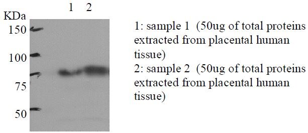
Application: Western BlotSample Tested:Species: HumanVerified Customer | Posted 04/09/2013Placental human tissue

There are no reviews that match your criteria.
Protocols
View specific protocols for SR-BI Antibody - BSA Free (NB400-101):
Protocol for Flow Cytometry Intracellular Staining
Sample Preparation.
1. Grow cells to 60-85% confluency. Flow cytometry requires between 2 x 10⁵ and 1 x 10⁶ cells for optimal performance.
2. If cells are adherent, harvest gently by washing once with staining buffer and then scraping. Avoid using trypsin as this can disrupt certain epitopes of interest. If enzymatic harvest is required, use Accutase, Collagenase, or TrypLE Express for a less damaging option.
3. Reserve 100 uL for counting, then transfer cell volume into a 50 mL conical tube and centrifuge for 8 minutes at 400 RCF.
a. Count cells using a hemocytometer and a 1:1 trypan blue exclusion stain to determine cell viability before starting the flow protocol. If cells appear blue, do not proceed.
4. Re-suspend cells to a concentration of 1 x 10⁶ cells/mL in staining buffer (NBP2-26247).
5. Aliquot out 100 uL samples in accordance with your experimental samples.
Tip: When cell surface and intracellular staining are required in the same sample, it is advisable that the cell surface staining be performed first since the fixation and permeabilization steps might reduce the availability of surface antigens.
Intracellular Staining.
Tip: When performing intracellular staining, it is important to use appropriate fixation and permeabilization reagents based upon the target and its subcellular location. Generally, our Intracellular Flow Assay Kit (NBP2-29450) is a good place to start as it contains an optimized combination of reagents for intracellular staining as well as an inhibitor of intracellular protein transport (necessary if staining secreted proteins). Certain targets may require more gentle or transient permeabilization protocols such as the commonly employed methanol or saponin-based methods.
Protocol for Cytoplasmic Targets:
1. Fix the cells by adding 100 uL fixation solution (such as 4% PFA) to each sample for 10-15 minutes.
2. Permeabilize cells by adding 100 uL of a permeabilization buffer to every 1 x 10⁶ cells present in the sample. Mix well and incubate at room temperature for 15 minutes.
a. For cytoplasmic targets, use a gentle permeabilization solution such as 1X PBS + 0.5% Saponin or 1X PBS + 0.5% Tween-20.
b. To maintain the permeabilized state throughout your experiment, use staining buffer + 0.1% of the permeabilization reagent (i.e. 0.1% Tween-20 or 0.1% Saponin).
3. Following the 15 minute incubation, add 2 mL of the staining buffer + 0.1% permeabilizer to each sample.
4. Centrifuge for 1 minute at 400 RCF.
5. Discard supernatant and re-suspend in 100 uL of staining buffer + 0.1% permeabilizer.
6. Add appropriate amount of each antibody (eg. 1 test or 1 ug per sample, as experimentally determined).
7. Mix well and incubate at room temperature for 30 minutes- 1 hour. Gently mix samples every 10-15 minutes.
8. Following the primary/conjugate incubation, add 1-2 mL/sample of staining buffer +0.1% permeabilizer and centrifuge for 1 minute at 400 RCF.
9. Wash twice by re-suspending cells in staining buffer (2 mL for tubes or 200 uL for wells) and centrifuging at 400 RCF for 5 minutes. Discard supernatant.
10. Add appropriate amount of secondary antibody (as experimentally determined) to each sample.
11. Incubate at room temperature in dark for 20 minutes.
12. Add 1-2 mL of staining buffer and centrifuge at 400 RCF for 1 minute and discard supernatant.
13. Wash twice by re-suspending cells in staining buffer (2 mL for tubes or 200 uL for wells) and centrifuging at 400 RCF for 5 minutes. Discard supernatant.
14. Resuspend in an appropriate volume of staining buffer (usually 500 uL per sample) and proceed with analysis on your flow cytometer.
Immunocytochemistry Protocol
Culture cells to appropriate density in 35 mm culture dishes or 6-well plates.
1. Remove culture medium and wash the cells briefly in PBS. Add 10% formalin to the dish and fix at room temperature for 10 minutes.
2. Remove the formalin and wash the cells in PBS.
3. Permeablize the cells with 0.1% Triton X100 or other suitable detergent for 10 min.
4. Remove the permeablization buffer and wash three times for 10 minutes each in PBS. Be sure to not let the specimen dry out.
5. To block nonspecific antibody binding, incubate in 10% normal goat serum from 1 hour to overnight at room temperature.
6. Add primary antibody at appropriate dilution and incubate overnight at 4C.
7. Remove primary antibody and replace with PBS. Wash three times for 10 minutes each.
8. Add secondary antibody at appropriate dilution. Incubate for 1 hour at room temperature.
9. Remove secondary antibody and replace with PBS. Wash three times for 10 minutes each.
10. Counter stain DNA with DAPi if required.
Immunohistochemistry-Paraffin Embedded Sections
Antigen Unmasking:
Bring slides to a boil in 10 mM sodium citrate buffer (pH 6.0) then maintain at a sub-boiling temperature for 10 minutes. Cool slides on bench-top for 30 minutes (keep slides in the sodium citrate buffer at all times).
Staining:
1. Wash sections in deionized water three times for 5 minutes each.
2. Wash sections in PBS for 5 minutes.
3. Block each section with 100-400 ul blocking solution (1% BSA in PBS) for 1 hour at room temperature.
4. Remove blocking solution and add 100-400 ul diluted primary antibody. Incubate overnight at 4 C.
5. Remove antibody solution and wash sections in wash buffer three times for 5 minutes each.
6. Add 100-400 ul HRP polymer conjugated secondary antibody. Incubate 30 minutes at room temperature.
7. Wash sections three times in wash buffer for 5 minutes each.
8. Add 100-400 ul DAB substrate to each section and monitor staining closely.
9. As soon as the sections develop, immerse slides in deionized water.
10. Counterstain sections in hematoxylin.
11. Wash sections in deionized water two times for 5 minutes each.
12. Dehydrate sections.
13. Mount coverslips.
Western Blot Protocol
1. Perform SDS-PAGE on samples to be analyzed, loading 10-25 ug of total protein per lane.
2. Transfer proteins to PVDF membrane according to the instructions provided by the manufacturer of the membrane and transfer apparatus.
3. Stain the membrane with Ponceau S (or similar product) to assess transfer success, and mark molecular weight standards where appropriate.
4. Rinse the blot TBS -0.05% Tween 20 (TBST).
5. Block the membrane in 5% Non-fat milk in TBST (blocking buffer) for at least 1 hour.
6. Wash the membrane in TBST three times for 10 minutes each.
7. Dilute primary antibody in blocking buffer and incubate overnight at 4C with gentle rocking.
8. Wash the membrane in TBST three times for 10 minutes each.
9. Incubate the membrane in diluted HRP conjugated secondary antibody in blocking buffer (as per manufacturer's instructions) for 1 hour at room temperature.
10. Wash the blot in TBST three times for 10 minutes each (this step can be repeated as required to reduce background).
11. Apply the detection reagent of choice in accordance with the manufacturer's instructions.
Find general support by application which include: protocols, troubleshooting, illustrated assays, videos and webinars.
- 7-Amino Actinomycin D (7-AAD) Cell Viability Flow Cytometry Protocol
- Antigen Retrieval Protocol (PIER)
- Antigen Retrieval for Frozen Sections Protocol
- Appropriate Fixation of IHC/ICC Samples
- Cellular Response to Hypoxia Protocols
- Chromogenic IHC Staining of Formalin-Fixed Paraffin-Embedded (FFPE) Tissue Protocol
- Chromogenic Immunohistochemistry Staining of Frozen Tissue
- ClariTSA™ Fluorophore Kits
- Detection & Visualization of Antibody Binding
- Extracellular Membrane Flow Cytometry Protocol
- Flow Cytometry Protocol for Cell Surface Markers
- Flow Cytometry Protocol for Staining Membrane Associated Proteins
- Flow Cytometry Staining Protocols
- Flow Cytometry Troubleshooting Guide
- Fluorescent IHC Staining of Frozen Tissue Protocol
- Graphic Protocol for Heat-induced Epitope Retrieval
- Graphic Protocol for the Preparation and Fluorescent IHC Staining of Frozen Tissue Sections
- Graphic Protocol for the Preparation and Fluorescent IHC Staining of Paraffin-embedded Tissue Sections
- Graphic Protocol for the Preparation of Gelatin-coated Slides for Histological Tissue Sections
- ICC Cell Smear Protocol for Suspension Cells
- ICC Immunocytochemistry Protocol Videos
- ICC for Adherent Cells
- IHC Sample Preparation (Frozen sections vs Paraffin)
- Immunocytochemistry (ICC) Protocol
- Immunocytochemistry Troubleshooting
- Immunofluorescence of Organoids Embedded in Cultrex Basement Membrane Extract
- Immunofluorescent IHC Staining of Formalin-Fixed Paraffin-Embedded (FFPE) Tissue Protocol
- Immunohistochemistry (IHC) and Immunocytochemistry (ICC) Protocols
- Immunohistochemistry Frozen Troubleshooting
- Immunohistochemistry Paraffin Troubleshooting
- Immunoprecipitation Protocol
- Intracellular Flow Cytometry Protocol Using Alcohol (Methanol)
- Intracellular Flow Cytometry Protocol Using Detergents
- Intracellular Nuclear Staining Flow Cytometry Protocol Using Detergents
- Intracellular Staining Flow Cytometry Protocol Using Alcohol Permeabilization
- Intracellular Staining Flow Cytometry Protocol Using Detergents to Permeabilize Cells
- Preparing Samples for IHC/ICC Experiments
- Preventing Non-Specific Staining (Non-Specific Binding)
- Primary Antibody Selection & Optimization
- Propidium Iodide Cell Viability Flow Cytometry Protocol
- Protocol for Heat-Induced Epitope Retrieval (HIER)
- Protocol for Liperfluo
- Protocol for Making a 4% Formaldehyde Solution in PBS
- Protocol for VisUCyte™ HRP Polymer Detection Reagent
- Protocol for the Characterization of Human Th22 Cells
- Protocol for the Characterization of Human Th9 Cells
- Protocol for the Fluorescent ICC Staining of Cell Smears - Graphic
- Protocol for the Fluorescent ICC Staining of Cultured Cells on Coverslips - Graphic
- Protocol for the Preparation & Fixation of Cells on Coverslips
- Protocol for the Preparation and Chromogenic IHC Staining of Frozen Tissue Sections
- Protocol for the Preparation and Chromogenic IHC Staining of Frozen Tissue Sections - Graphic
- Protocol for the Preparation and Chromogenic IHC Staining of Paraffin-embedded Tissue Sections
- Protocol for the Preparation and Chromogenic IHC Staining of Paraffin-embedded Tissue Sections - Graphic
- Protocol for the Preparation and Fluorescent ICC Staining of Cells on Coverslips
- Protocol for the Preparation and Fluorescent ICC Staining of Non-adherent Cells
- Protocol for the Preparation and Fluorescent ICC Staining of Stem Cells on Coverslips
- Protocol for the Preparation and Fluorescent IHC Staining of Frozen Tissue Sections
- Protocol for the Preparation and Fluorescent IHC Staining of Paraffin-embedded Tissue Sections
- Protocol for the Preparation of Gelatin-coated Slides for Histological Tissue Sections
- Protocol for the Preparation of a Cell Smear for Non-adherent Cell ICC - Graphic
- Protocol: Annexin V and PI Staining by Flow Cytometry
- Protocol: Annexin V and PI Staining for Apoptosis by Flow Cytometry
- R&D Systems Quality Control Western Blot Protocol
- TUNEL and Active Caspase-3 Detection by IHC/ICC Protocol
- The Importance of IHC/ICC Controls
- Troubleshooting Guide: Fluorokine Flow Cytometry Kits
- Troubleshooting Guide: Immunohistochemistry
- Troubleshooting Guide: Western Blot Figures
- Western Blot Conditions
- Western Blot Protocol
- Western Blot Protocol for Cell Lysates
- Western Blot Troubleshooting
- Western Blot Troubleshooting Guide
- View all Protocols, Troubleshooting, Illustrated assays and Webinars
FAQs for SR-BI Antibody - BSA Free
Showing
1
-
4 of
4 FAQs
Showing All
-
Q: Hi. I'm interested in scarb1 antibody. Can you tell me what is the difference between NB400-101 and 104?
A: SR-BI antibody NB400-101 has been validated for Block/Neutralize, while NB400-104 has not been.
-
Q: I did western blot for SR-BI using NB400-101 antibody. I just want to ask the denature condition when you test this antibody.
A: We denature all of our proteins using a standard protocol of 95 degrees for 10 minutes in 5% BME on all of our samples. Here is the rest of the Western Blot protocol used specifically for this antibody. 1. Run approximately 50 ug of protein on a 4-20% Tris-glycine mini-gel at 125V for 60 minutes. 2. Equilibrate gel, nitrocellulose membrane, Whatman paper, and blotting pads in transfer buffer for 15 minutes. 3. Transfer protein to the membrane at 25V for 90 minutes. 4. Allow membrane to air-dry. 5. Block membrane with 1XPBS/5% non-fat milk/0.1% Tween-20 for 1 hour at room temperature (approx. 23-27 degrees C). 6. Wash membrane twice, for 5 minutes each, with 1XPBS/0.05% Tween-20 (PBST). 7. Incubate membrane with 1:1,000 dilution of NB400-101 (anti-SR-BI), diluted in 1XPBS/1% BSA, for 1 hour at room temperature. 8. Wash membrane once for 15 minutes, then four times for 5 minutes each, with PBST. 9. Incubate membrane with 1:10,000 dilution of goat anti-rabbit IgG-HRP (BioRad), diluted in 1XPBS/1% BSA, for 1 hour at room temperature. 10. Wash membrane once for 15 minutes, then four times for 5 minutes each, with PBST. 11. Detect cross-reacting proteins using Renaissance Chemiluminescence Reagent Plus kit from NEN Life Sciences. NOTE: HL-60 whole cell extracts (NB800-PC3) were used as a positive control for this antibody.
-
Q: What is the difference between NB400-101 and NB400-104? The same IF image is shown on both product pages, so which antibody was really used?
A: Regarding your inquiry, NB400-101 and NB400-104 are the same antibodies and the reason why they have different catalog numbers is, in the first stage of production one of these antibodies was purified and the other was not but now both are purified and the same.
-
Q: Would you please help us to confirm the application of NB400-101? Can this antibody be used for IHC-P or IHC-Fr? If yes, what is the dilution for the two application? The customer finds that NB400-104 and NB400-101 have the same immunogen, but the molecular band is not the same, they are approx. 82kDa and approx. 75kDa separately. We are confused with this and would you please tell us the dilution for NB400-104 in IHC-P detection?
A: Both NB400-100 and NB400104 can be used with IHC paraffin embedded sections at 2.5-5 ug/ml. With regards to the WB image, since the markers are different on those gels, a 5 kDa difference between markers would not be out of the range of possibility but as far as the immunogen is concerned, both antibodies share the same region, meanwhile please keep in mind that NB400-104 can be used in human, mouse and rat tissues but NB400-101 can only be used in human and mouse.
-
Q: Hi. I'm interested in scarb1 antibody. Can you tell me what is the difference between NB400-101 and 104?
A: SR-BI antibody NB400-101 has been validated for Block/Neutralize, while NB400-104 has not been.
-
Q: I did western blot for SR-BI using NB400-101 antibody. I just want to ask the denature condition when you test this antibody.
A: We denature all of our proteins using a standard protocol of 95 degrees for 10 minutes in 5% BME on all of our samples. Here is the rest of the Western Blot protocol used specifically for this antibody. 1. Run approximately 50 ug of protein on a 4-20% Tris-glycine mini-gel at 125V for 60 minutes. 2. Equilibrate gel, nitrocellulose membrane, Whatman paper, and blotting pads in transfer buffer for 15 minutes. 3. Transfer protein to the membrane at 25V for 90 minutes. 4. Allow membrane to air-dry. 5. Block membrane with 1XPBS/5% non-fat milk/0.1% Tween-20 for 1 hour at room temperature (approx. 23-27 degrees C). 6. Wash membrane twice, for 5 minutes each, with 1XPBS/0.05% Tween-20 (PBST). 7. Incubate membrane with 1:1,000 dilution of NB400-101 (anti-SR-BI), diluted in 1XPBS/1% BSA, for 1 hour at room temperature. 8. Wash membrane once for 15 minutes, then four times for 5 minutes each, with PBST. 9. Incubate membrane with 1:10,000 dilution of goat anti-rabbit IgG-HRP (BioRad), diluted in 1XPBS/1% BSA, for 1 hour at room temperature. 10. Wash membrane once for 15 minutes, then four times for 5 minutes each, with PBST. 11. Detect cross-reacting proteins using Renaissance Chemiluminescence Reagent Plus kit from NEN Life Sciences. NOTE: HL-60 whole cell extracts (NB800-PC3) were used as a positive control for this antibody.
-
Q: What is the difference between NB400-101 and NB400-104? The same IF image is shown on both product pages, so which antibody was really used?
A: Regarding your inquiry, NB400-101 and NB400-104 are the same antibodies and the reason why they have different catalog numbers is, in the first stage of production one of these antibodies was purified and the other was not but now both are purified and the same.
-
Q: Would you please help us to confirm the application of NB400-101? Can this antibody be used for IHC-P or IHC-Fr? If yes, what is the dilution for the two application? The customer finds that NB400-104 and NB400-101 have the same immunogen, but the molecular band is not the same, they are approx. 82kDa and approx. 75kDa separately. We are confused with this and would you please tell us the dilution for NB400-104 in IHC-P detection?
A: Both NB400-100 and NB400104 can be used with IHC paraffin embedded sections at 2.5-5 ug/ml. With regards to the WB image, since the markers are different on those gels, a 5 kDa difference between markers would not be out of the range of possibility but as far as the immunogen is concerned, both antibodies share the same region, meanwhile please keep in mind that NB400-104 can be used in human, mouse and rat tissues but NB400-101 can only be used in human and mouse.
Loading...