Synapsin I Antibody - Azide Free
Novus Biologicals | Catalog # NB300-104
![Immunocytochemistry/ Immunofluorescence: Synapsin I Antibody [NB300-104]](https://resources.rndsystems.com/images/products/Synapsin-I-Antibody-Immunocytochemistry-Immunofluorescence-NB300-104-img0013.jpg "Immunocytochemistry/ Immunofluorescence: Synapsin I Antibody [NB300-104]")
Key Product Details
Validated by
Species Reactivity
Validated:
Cited:
Applications
Validated:
Cited:
Label
Antibody Source
Format
Product Specifications
Immunogen
Reactivity Notes
Localization
Marker
Specificity
Clonality
Host
Isotype
Theoretical MW
Disclaimer note: The observed molecular weight of the protein may vary from the listed predicted molecular weight due to post translational modifications, post translation cleavages, relative charges, and other experimental factors.
Description
Scientific Data Images for Synapsin I Antibody - Azide Free
Immunocytochemistry/ Immunofluorescence: Synapsin I Antibody [NB300-104]
Synapsin-I-Antibody-Immunocytochemistry-Immunofluorescence-NB300-104-img0013.jpg![Immunocytochemistry/ Immunofluorescence: Synapsin I Antibody [NB300-104]](https://resources.rndsystems.com/images/products/Synapsin-I-Antibody-Immunocytochemistry-Immunofluorescence-NB300-104-img0014.jpg "Immunocytochemistry/ Immunofluorescence: Synapsin I Antibody [NB300-104]")
![Western Blot: Synapsin I Antibody [NB300-104]](https://resources.rndsystems.com/images/products/Synapsin%20I%20Antibody-Western%20Blot-NB300-104-img0015.jpg "Western Blot: Synapsin I Antibody [NB300-104]")
Western Blot: Synapsin I Antibody [NB300-104]
Synapsin I Antibody-Western Blot-NB300-104-img0015.jpg![Immunohistochemistry-Paraffin: Synapsin I Antibody [NB300-104]](https://resources.rndsystems.com/images/products/Synapsin-I-Antibody-Immunohistochemistry-Paraffin-NB300-104-img0012.jpg "Immunohistochemistry-Paraffin: Synapsin I Antibody [NB300-104]")
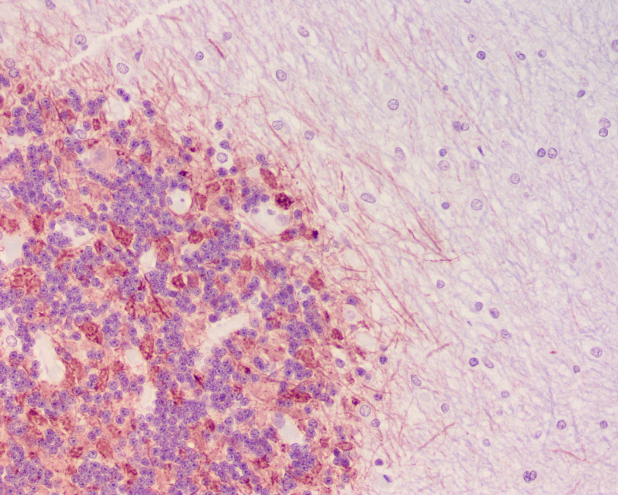
Immunohistochemistry-Paraffin: Synapsin I Antibody [NB300-104]
Immunohistochemistry-Paraffin: Synapsin I Antibody [NB300-104] - Human cerebellum section, formalin fixed paraffin embedded. Stained with Synpasin-1 at 1:250. Citrate antigen retrieval at pH 6. Image from verified customer review.![Western Blot: Synapsin I Antibody [NB300-104]](https://resources.rndsystems.com/images/products/Synapsin-I-Antibody-Western-Blot-NB300-104-img0007.jpg "Western Blot: Synapsin I Antibody [NB300-104]")
Western Blot: Synapsin I Antibody [NB300-104]
Western Blot: Synapsin I Antibody [NB300-104] - 10 ug of rat hippocampal (Hipp) lysate showing specific immunolabeling of the ~78k synapsin I doublet protein.![Western Blot: Synapsin I Antibody [NB300-104]](https://resources.rndsystems.com/images/products/Synapsin-I-Antibody-Western-Blot-NB300-104-img0008.jpg "Western Blot: Synapsin I Antibody [NB300-104]")
Western Blot: Synapsin I Antibody [NB300-104]
Western Blot: Synapsin I Antibody [NB300-104] - 10 ug of rat brain lysate showing specific immunolabeling of the ~78 kDa Synapsin I doublet.![Immunocytochemistry/ Immunofluorescence: Synapsin I Antibody [NB300-104]](https://resources.rndsystems.com/images/products/Synapsin-I-Antibody-Immunocytochemistry-Immunofluorescence-NB300-104-img0011.jpg "Immunocytochemistry/ Immunofluorescence: Synapsin I Antibody [NB300-104]")
Immunocytochemistry/ Immunofluorescence: Synapsin I Antibody [NB300-104]
Immunocytochemistry/Immunofluorescence: Synapsin I Antibody [NB300-104] - ICC-IF validation analysis of Synapsin 1 Antibody on cultured rat caudate neurons. This representative image shows the punctate distribution of Synapsin 1 (green) and MAP (red) proteins in a neuronal cell.
Immunocytochemistry/Immunofluorescence: Synapsin I Antibody [NB300-104] -
Characterization of WT and slow ESCs differentiated to neurons (related to Fig 4)Immunofluorescence staining for neuronal markers Map2, Syn1, and NeuN in neurons cultured on poly‐ornithine‐/laminin‐coated plates.Expression of neuronal markers in neurons cultured on poly‐ornithine‐/laminin‐coated plates from RNA‐seq analysis, n = 3.
Immunocytochemistry/ Immunofluorescence: Synapsin I Antibody [NB300-104] -
Immunocytochemistry/ Immunofluorescence: Synapsin I Antibody [NB300-104] - Characterization of WT & slow ESCs differentiated to neurons (related to Fig 4)Immunofluorescence staining for neuronal markers Map2, Syn1, & NeuN in neurons cultured on poly‐ornithine‐/laminin‐coated plates.Expression of neuronal markers in neurons cultured on poly‐ornithine‐/laminin‐coated plates from RNA‐seq analysis, n = 3. Image collected & cropped by CiteAb from the following publication (https://pubmed.ncbi.nlm.nih.gov/30988016), licensed under a CC-BY license. Not internally tested by Novus Biologicals.
Immunocytochemistry/ Immunofluorescence: Synapsin I Antibody [NB300-104] -
Immunocytochemistry/ Immunofluorescence: Synapsin I Antibody [NB300-104] - hPDLSCs-derived neural-like cells are connected by synapse-like interactions. hPDLSCs-derived neural-like cells connect to one another (a) through different types of synapses-like interactions, including dendrodendritic-like, axoaxonic-like & axodendritic-like synapses (b). (c) Synapse-associated proteins Cx43, Synaptophysin & Synapsin1 are found in the cell membrane of hPDLSCs-derived neural-like cells at the neurite contact areas. Scale bar: 25 μm. AA, axoaxonic-like synapse; AD, axodendritic-like synapse; DD, dendrodendritic-like synapse; LM, light microscopy; SEM, scanning electron microscopy. Image collected & cropped by CiteAb from the following publication (https://pubmed.ncbi.nlm.nih.gov/31792338), licensed under a CC-BY license. Not internally tested by Novus Biologicals.
Western Blot: Synapsin I Antibody [NB300-104] -
Western Blot: Synapsin I Antibody [NB300-104] - Synaptic localization of TA at the PC–PF synaptic contacts.Western blot of TA, V1, V2, VGAT proteins in cerebellar synaptosomal fractions (SYN) from P14 mice. Synaptosomal preparations are enriched of post synaptic density protein (PSD95) & Synapsin-I (Syn-I) in comparison to equal amount (ACT) of total cerebellar protein extract (LYS). TA is clearly present in synaptosomal enriched preparations. B) Cerebellar sagittal section immunostained for Cb (green), V1 (cyan) & TA (red). In the ML, anti-V1 specifically labels PF-terminals contacting PC-spines. C–E) The high magnification of the inset in B is reproduced in different merge images & by the relative split channels. C) Merge image of Cb & V1 staining plus the colocalization mask Cb/V1 (mCb/V1 in white); it represents the negative CTR (CTR-). D) Merge image of TA & Cb staining plus the colocalization mask TA/Cb (mTA/Cb in white), highlighting TA expression in PC-spines. E) Merge image of TA & V1 staining plus the colocalization mask TA/V1 (m TA/V1 in white), indicating the presence of TA also in this synaptic terminal. F) Quantitative colocalization analysis shows the mean overlap coefficients of TA/Cb (gray column) & TA/V1 (black column) significantly different from the negative CTR Cb/V1 (white column). The small insets (white boxes in C–D-E) are high magnifications of a representative PC-spine (green) contacted by a PF synaptic terminal (cyan) with the appropriate colocalization mask (white). * p < 0.05. Data are represented as mean ± SEM. Scale bars in B, 10 µm; in C–D-E, 5 µm; in F, 1 µm. Image collected & cropped by CiteAb from the following publication (https://pubmed.ncbi.nlm.nih.gov/23840813), licensed under a CC-BY license. Not internally tested by Novus Biologicals.
Western Blot: Synapsin I Antibody [NB300-104] -
Western Blot: Synapsin I Antibody [NB300-104] - Tau oligomers induce synaptic dysfunction. (A) Representative Western blot of mouse hippocampus homogenate. The levels of synaptophysin, synapsin-1, & septin-11 were measured by band quantification & normalized with the levels of tubulin. PBSo indicates representative bands of hippocampal area injected with PBS in mice also injected with tau oligomers, PBSf indicates PBS injection in mice also injected with tau fibrils, & PBSm indicates PBS injection in mice also injected with tau monomers. (B) Synaptophysin levels were significantly lower in the hemispheres injected with tau oligomers in comparison with the ones injected with fibrils, monomers, or PBS. (C) No significant differences in the levels of synapsin-1 were observed. (D) Only the hemisphere injected with tau oligomers presents a decrease in the level of septin-11. Data are represented as the mean ± SE. *p < 0.01, n = 6 Image collected & cropped by CiteAb from the following publication (https://pubmed.ncbi.nlm.nih.gov/21645391), licensed under a CC-BY license. Not internally tested by Novus Biologicals.Applications for Synapsin I Antibody - Azide Free
Immunocytochemistry/ Immunofluorescence
Immunohistochemistry
Immunohistochemistry-Frozen
Immunoprecipitation
Western Blot
Reviewed Applications
Read 1 review rated 5 using NB300-104 in the following applications:
Formulation, Preparation, and Storage
Purification
Formulation
Format
Preservative
Concentration
Shipping
Stability & Storage
Background: Synapsin I
Alternate Names
Gene Symbol
UniProt
Additional Synapsin I Products
Product Documents for Synapsin I Antibody - Azide Free
Certificate of Analysis
To download a Certificate of Analysis, please enter a lot or batch number in the search box below.
Product Specific Notices for Synapsin I Antibody - Azide Free
This product is for research use only and is not approved for use in humans or in clinical diagnosis. Primary Antibodies are guaranteed for 1 year from date of receipt.
Related Research Areas
Citations for Synapsin I Antibody - Azide Free
 Powered by Bioz
Customer Reviews for Synapsin I Antibody - Azide Free (1)
Have you used Synapsin I Antibody - Azide Free?
Submit a review and receive an Amazon gift card!
$25/€18/£15/$25CAN/¥2500 Yen for a review with an image
$10/€7/£6/$10CAN/¥1110 Yen for a review without an image
Submit a review
Customer Images
-
Application: Immunohistochemistry-ParaffinSample Tested: Brain (cerebellum) tissueSpecies: HumanVerified Customer | Posted 04/17/2018Human cerebellum section, formalin fixed paraffin embedded. Stained with Synpasin-1 at 1:250. Citrate antigen retrieval used.Citrate antigen retrieval, pH6 1 in 250 dilution

There are no reviews that match your criteria.
Protocols
Find general support by application which include: protocols, troubleshooting, illustrated assays, videos and webinars.
- Antigen Retrieval Protocol (PIER)
- Antigen Retrieval for Frozen Sections Protocol
- Appropriate Fixation of IHC/ICC Samples
- Cellular Response to Hypoxia Protocols
- Chromogenic IHC Staining of Formalin-Fixed Paraffin-Embedded (FFPE) Tissue Protocol
- Chromogenic Immunohistochemistry Staining of Frozen Tissue
- ClariTSA™ Fluorophore Kits
- Detection & Visualization of Antibody Binding
- Fluorescent IHC Staining of Frozen Tissue Protocol
- Graphic Protocol for Heat-induced Epitope Retrieval
- Graphic Protocol for the Preparation and Fluorescent IHC Staining of Frozen Tissue Sections
- Graphic Protocol for the Preparation and Fluorescent IHC Staining of Paraffin-embedded Tissue Sections
- Graphic Protocol for the Preparation of Gelatin-coated Slides for Histological Tissue Sections
- ICC Cell Smear Protocol for Suspension Cells
- ICC Immunocytochemistry Protocol Videos
- ICC for Adherent Cells
- IHC Sample Preparation (Frozen sections vs Paraffin)
- Immunocytochemistry (ICC) Protocol
- Immunocytochemistry Troubleshooting
- Immunofluorescence of Organoids Embedded in Cultrex Basement Membrane Extract
- Immunofluorescent IHC Staining of Formalin-Fixed Paraffin-Embedded (FFPE) Tissue Protocol
- Immunohistochemistry (IHC) and Immunocytochemistry (ICC) Protocols
- Immunohistochemistry Frozen Troubleshooting
- Immunohistochemistry Paraffin Troubleshooting
- Immunoprecipitation Protocol
- Preparing Samples for IHC/ICC Experiments
- Preventing Non-Specific Staining (Non-Specific Binding)
- Primary Antibody Selection & Optimization
- Protocol for Heat-Induced Epitope Retrieval (HIER)
- Protocol for Making a 4% Formaldehyde Solution in PBS
- Protocol for VisUCyte™ HRP Polymer Detection Reagent
- Protocol for the Fluorescent ICC Staining of Cell Smears - Graphic
- Protocol for the Fluorescent ICC Staining of Cultured Cells on Coverslips - Graphic
- Protocol for the Preparation & Fixation of Cells on Coverslips
- Protocol for the Preparation and Chromogenic IHC Staining of Frozen Tissue Sections
- Protocol for the Preparation and Chromogenic IHC Staining of Frozen Tissue Sections - Graphic
- Protocol for the Preparation and Chromogenic IHC Staining of Paraffin-embedded Tissue Sections
- Protocol for the Preparation and Chromogenic IHC Staining of Paraffin-embedded Tissue Sections - Graphic
- Protocol for the Preparation and Fluorescent ICC Staining of Cells on Coverslips
- Protocol for the Preparation and Fluorescent ICC Staining of Non-adherent Cells
- Protocol for the Preparation and Fluorescent ICC Staining of Stem Cells on Coverslips
- Protocol for the Preparation and Fluorescent IHC Staining of Frozen Tissue Sections
- Protocol for the Preparation and Fluorescent IHC Staining of Paraffin-embedded Tissue Sections
- Protocol for the Preparation of Gelatin-coated Slides for Histological Tissue Sections
- Protocol for the Preparation of a Cell Smear for Non-adherent Cell ICC - Graphic
- R&D Systems Quality Control Western Blot Protocol
- TUNEL and Active Caspase-3 Detection by IHC/ICC Protocol
- The Importance of IHC/ICC Controls
- Troubleshooting Guide: Immunohistochemistry
- Troubleshooting Guide: Western Blot Figures
- Western Blot Conditions
- Western Blot Protocol
- Western Blot Protocol for Cell Lysates
- Western Blot Troubleshooting
- Western Blot Troubleshooting Guide
- View all Protocols, Troubleshooting, Illustrated assays and Webinars
FAQs for Synapsin I Antibody - Azide Free
-
Q: How many days do you recommend culturing primary neurons to get a good Synapsin I signal? Can you please send the protocol used to validate this antibody in immunofluorescence?
A: It was validated in IF by a collaborator, so I do not have a specific protocol to offer. However I can tell you that the neurons were cultured for 14 days.