TAP1 Antibody (3D4) - Azide and BSA Free
Novus Biologicals | Catalog # H00006890-M04
![Immunocytochemistry/ Immunofluorescence: TAP1 Antibody (3D4) [H00006890-M04]](https://resources.rndsystems.com/images/products/TAP1-Antibody-3D4-Immunocytochemistry-Immunofluorescence-H00006890-M04-img0001.jpg "Immunocytochemistry/ Immunofluorescence: TAP1 Antibody (3D4) [H00006890-M04]")
Key Product Details
Validated by
Species Reactivity
Applications
Label
Antibody Source
Format
Product Specifications
Immunogen
Specificity
Clonality
Host
Isotype
Description
Scientific Data Images for TAP1 Antibody (3D4) - Azide and BSA Free
Immunocytochemistry/ Immunofluorescence: TAP1 Antibody (3D4) [H00006890-M04]
Immunocytochemistry/Immunofluorescence: TAP1 Antibody (3D4) [H00006890-M04] - Analysis of monoclonal antibody to TAP1 on HeLa cell. Antibody concentration 10 ug/ml![Flow Cytometry: TAP1 Antibody (3D4) [H00006890-M04]](https://resources.rndsystems.com/images/products/TAP1-Antibody-3D4-Flow-Cytometry-H00006890-M04-img0002.jpg "Flow Cytometry: TAP1 Antibody (3D4) [H00006890-M04]")
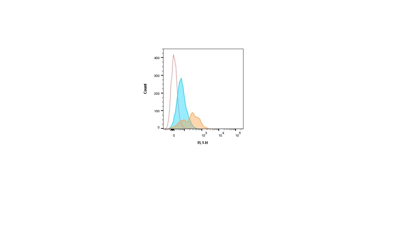
Flow Cytometry: TAP1 Antibody (3D4) [H00006890-M04]
Flow Cytometry: TAP1 Antibody (3D4) [H00006890-M04] - Intracellular flow cytometric analysis of TAP1 antibody H00006890-M04 (orange filled) on interferon gamma treated HCT116 cells. Red dotted line in histogram represents unstained and blue filled line represents isotype control. Flow cytometry image added from a verified customer review.Applications for TAP1 Antibody (3D4) - Azide and BSA Free
ELISA
Flow Cytometry
Immunocytochemistry/ Immunofluorescence
Western Blot
Reviewed Applications
Read 1 review rated 4 using H00006890-M04 in the following applications:
Flow Cytometry Panel Builder
Bio-Techne Knows Flow Cytometry
Save time and reduce costly mistakes by quickly finding compatible reagents using the Panel Builder Tool.
Advanced Features
- Spectra Viewer - Custom analysis of spectra from multiple fluorochromes
- Spillover Popups - Visualize the spectra of individual fluorochromes
- Antigen Density Selector - Match fluorochrome brightness with antigen density

Formulation, Preparation, and Storage
Purification
Formulation
Format
Preservative
Concentration
Shipping
Stability & Storage
Background: TAP1
Long Name
Alternate Names
Entrez Gene IDs
Gene Symbol
UniProt
Additional TAP1 Products
Product Documents for TAP1 Antibody (3D4) - Azide and BSA Free
Certificate of Analysis
To download a Certificate of Analysis, please enter a lot or batch number in the search box below.
Product Specific Notices for TAP1 Antibody (3D4) - Azide and BSA Free
This product is produced by and distributed for Abnova, a company based in Taiwan.
This product is for research use only and is not approved for use in humans or in clinical diagnosis. Primary Antibodies are guaranteed for 1 year from date of receipt.
Related Research Areas
Customer Reviews for TAP1 Antibody (3D4) - Azide and BSA Free (1)
Have you used TAP1 Antibody (3D4) - Azide and BSA Free?
Submit a review and receive an Amazon gift card!
$25/€18/£15/$25CAN/¥2500 Yen for a review with an image
$10/€7/£6/$10CAN/¥1110 Yen for a review without an image
Submit a review
Customer Images
-
Application: Flow CytometrySample Tested: HCT116 cellsSpecies: HumanVerified Customer | Posted 01/30/2020Intracellular flow cytometric analysis of TAP1 antibody H00006890-M04 (orange filled) on interferon gamma treated HCT116 cells. Red dotted line in histogram represents unstained and blue filled line represents isotype control.

There are no reviews that match your criteria.
Protocols
Find general support by application which include: protocols, troubleshooting, illustrated assays, videos and webinars.
- 7-Amino Actinomycin D (7-AAD) Cell Viability Flow Cytometry Protocol
- Appropriate Fixation of IHC/ICC Samples
- Cellular Response to Hypoxia Protocols
- ClariTSA™ Fluorophore Kits
- Detection & Visualization of Antibody Binding
- ELISA Sample Preparation & Collection Guide
- ELISA Troubleshooting Guide
- Extracellular Membrane Flow Cytometry Protocol
- Flow Cytometry Protocol for Cell Surface Markers
- Flow Cytometry Protocol for Staining Membrane Associated Proteins
- Flow Cytometry Staining Protocols
- Flow Cytometry Troubleshooting Guide
- How to Run an R&D Systems DuoSet ELISA
- How to Run an R&D Systems Quantikine ELISA
- How to Run an R&D Systems Quantikine™ QuicKit™ ELISA
- ICC Cell Smear Protocol for Suspension Cells
- ICC Immunocytochemistry Protocol Videos
- ICC for Adherent Cells
- Immunocytochemistry (ICC) Protocol
- Immunocytochemistry Troubleshooting
- Immunofluorescence of Organoids Embedded in Cultrex Basement Membrane Extract
- Immunohistochemistry (IHC) and Immunocytochemistry (ICC) Protocols
- Intracellular Flow Cytometry Protocol Using Alcohol (Methanol)
- Intracellular Flow Cytometry Protocol Using Detergents
- Intracellular Nuclear Staining Flow Cytometry Protocol Using Detergents
- Intracellular Staining Flow Cytometry Protocol Using Alcohol Permeabilization
- Intracellular Staining Flow Cytometry Protocol Using Detergents to Permeabilize Cells
- Preparing Samples for IHC/ICC Experiments
- Preventing Non-Specific Staining (Non-Specific Binding)
- Primary Antibody Selection & Optimization
- Propidium Iodide Cell Viability Flow Cytometry Protocol
- Protocol for Liperfluo
- Protocol for VisUCyte™ HRP Polymer Detection Reagent
- Protocol for the Characterization of Human Th22 Cells
- Protocol for the Characterization of Human Th9 Cells
- Protocol for the Fluorescent ICC Staining of Cell Smears - Graphic
- Protocol for the Fluorescent ICC Staining of Cultured Cells on Coverslips - Graphic
- Protocol for the Preparation and Fluorescent ICC Staining of Cells on Coverslips
- Protocol for the Preparation and Fluorescent ICC Staining of Non-adherent Cells
- Protocol for the Preparation and Fluorescent ICC Staining of Stem Cells on Coverslips
- Protocol for the Preparation of a Cell Smear for Non-adherent Cell ICC - Graphic
- Protocol: Annexin V and PI Staining by Flow Cytometry
- Protocol: Annexin V and PI Staining for Apoptosis by Flow Cytometry
- Quantikine HS ELISA Kit Assay Principle, Alkaline Phosphatase
- Quantikine HS ELISA Kit Principle, Streptavidin-HRP Polymer
- R&D Systems Quality Control Western Blot Protocol
- Sandwich ELISA (Colorimetric) – Biotin/Streptavidin Detection Protocol
- Sandwich ELISA (Colorimetric) – Direct Detection Protocol
- TUNEL and Active Caspase-3 Detection by IHC/ICC Protocol
- The Importance of IHC/ICC Controls
- Troubleshooting Guide: ELISA
- Troubleshooting Guide: Fluorokine Flow Cytometry Kits
- Troubleshooting Guide: Western Blot Figures
- Western Blot Conditions
- Western Blot Protocol
- Western Blot Protocol for Cell Lysates
- Western Blot Troubleshooting
- Western Blot Troubleshooting Guide
- View all Protocols, Troubleshooting, Illustrated assays and Webinars